Collection of materials relating to neuro-ophthalmology as part of the Neuro-Ophthalmology Virtual Education Library.
NOVEL: https://novel.utah.edu/
TO
- NOVEL966
Filters: Collection: "ehsl_novel_novel"
| Title | Creator | Description | Subject | ||
|---|---|---|---|---|---|
| 51 |
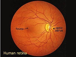 |
Simple Anatomy of the Retina (Webvision) | Helga Kolb, MD | Description of the anatomy of the retina with diagrams. | Retina Anatomy |
| 52 |
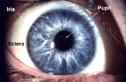 |
Gross Anatomy of the Eye (Webvision) | Helga Kolb, MD | Description of the gross anatomy of the eye, with diagrams. | Gross Anatomy Eye |
| 53 |
 |
Optic Atrophy (PowerPoint) | William F. Hoyt, PhD | a) Evolution of optic disc pallor after optic nerve transection. Normal Right eye. Photo taken December 9, 1978. b) Injury on December 8, 1978. Evolution of optic disc pallor after optic nerve transection. Woman having rhinoplasty suffered optic nerve transection. One day after nerve transection. N... | Optic Disc Atrophy from Retrobulbar Causes (Retrograde Optic Nerve Degeneration); Severe Atrophy; Optic Atrophic |
| 54 |
 |
Temporal Atrophy (PowerPoint) | William F. Hoyt, PhD | Segmental Atrophy - Temporal pallor - Nutritional amblyopia (alcoholic). 1985. | Temporal Atrophy; Optic Disc Atrophy from Retrobulbar Causes (Retrograde Optic Nerve Degeneration); Severe Atrophy; Alcohol |
| 55 |
 |
Evolution of Optociliary Veins with Perioptic Nerve Sheath | William F. Hoyt, PhD | Series of images showing progression of disc swelling and macular degeneration. Pathology: Optociliary Vein. Disease/Diagnosis: Perioptic nerve sheath meningioma evolution. Clinical notes: Visual Loss. | Optic Disc Atrophy with Special Features; Optociliary Veins; Shunt Vessels (Meningioma) |
| 56 |
 |
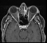
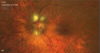
Exposed Drusen (PowerPoint) | William F. Hoyt, PhD | PP25a: Left eye: Severe visual field defect. PP25b: right eye with exposed drusen and field loss: visual field defects; PP25c: right eye visual field PP25d: left eye visual field. | Pseudopapilledema; Exposed Drusen |
| 57 |
 |
Curtain Sign (Enhanced Ptosis) - Associated Image 2 | Bashaer Aldhahwani, MD; Hong Jiang, MD, PhD | This is a 78-year-old male patient who presented with diplopia, right eyelid ptosis, and ophthalmoplegia. He had severe ptosis OD and pseudo-proptosis (lid retraction) OS at baseline, but when the right eyelid was manually elevated, there was marked enhanced ptosis of the left eyelid (Video). He was... | Myasthenia GravIs; Clinical Signs |
| 58 |
 |
Myelinated Retinal Nerve Fiber Layer | Bashaer Aldhahwani, MD; Hong Jiang, MD, PhD | A 78 YOF with no visual symptoms has an incidental finding of yellow-white well-demarcated patches with ragged borders at the peripapillary area of her left eye (see the fundus photo). | Myelinated Retinal Nerve Fiber Layer |
| 59 |
 |
Aneurysms | AAO/NANOS - American Academy of Ophthalmology / North American Neuro-Ophthalmology Society | Aneurisms may result in neuro-ophthalmologic sign and symptoms by direct compression of the afferent or efferent systems or by the secondary effects of hemorrhage. Basilar aneurisms may result in ocular motor deficits such as a unilateral or bilateral third nerve palsy. | Aneurysm |
| 60 |
 |
Voluntary Nystagmus | Sangeeta Khanna, MD | A short presentation on the phenomena of voluntary nystagmus. | Nystagmus; Voluntary Nystagmus |
| 61 |
 |
Paediatric Neuro-ophthalmology: Visual Acuity Assessment Strategies | Anat Bachar Zipori, MD; Nasrin Najm-Tehrani, FRCS Ed (Ophth), FRCSC | Assessing the visual function of a child can be challenging at times. When approaching a child one must understand visual development and accommodate to the child's capabilities, level of development and communication skills. The examining physician may need to apply more than one method to assess t... | Visual Acuity Assessment; Pediatric Visual Acuity Tests |
| 62 |
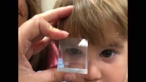 |
Pediatric Visual Acuity Strategies: Induced Tropia Test | Anat Bachar Zipori, MD; Nasrin Najm-Tehrani, FRCS Ed (Ophth), FRCSC | Induced Tropia Test. Using a 10-20 base-down prism, this exam demonstrates equal vision in both eyes. If there is evidence of ocular misalignment, prisms can be used to neutralize the movement and thereby measure the deviation, whether it is a heterotropia or heterophoria. Prisms are placed in front... | Induced Tropia; Prism Test |
| 63 |
 |
Vertical optokinetic nystagmus | Anat Bachar Zipori, MD; Nasrin Najm-Tehrani, FRCS Ed (Ophth), FRCSC | Visual depiction of the vertical OKN drum test. The vertical optokinetic drum test is an exam used to elicit vertical optokinetic nystagmus (OKN) when there is horizontal nystagmus. The hand-held "optokinetic" drums or tapes that are used to elicit smooth movements primarily test the pursuit system.... | Vertical Optokinetic Nystagmus; OKN Drum Test |
| 64 |
 |
Central Retinal Artery Occlusion | Natasha Nayak, MD; Rudrani Banik, MD | Power point of case presentation of acute central retinal artery occlusion (CRAO) treated with tPA. Risk factors for stroke and results of EAGLE study reviewed. Imaging: Number of Figures and legend for each: 12 Slide 3: Figure 1: Table 1: Exam Findings Slide 3: Figure 2: Table 2: Exam Findings Cont... | Central Retinal Artery Occlusion; Stroke; Tissue Plasminogen Activator; EAGLE Study |
| 65 |
 |
Non-Surgical Management of Strabismus | Alex Christoff, MD | An overview of non-surgical treatment of strabismus. | Strabismus |
| 66 |
 |
Carotid Cavernous Fistula | Adam Botwinick, MD; Rudrani Banik, MD | Power point of case presentation of 66-year-old female with chronic red eye OU x 2 months, misdiagnosed as conjunctivitis. Exam showed dilated, tortuous episcleral vessels OU with proptosis OU and elevated intraocular pressure. MRI showed suspicion of carotid cavernous fistula (CCF), confirmed by ... | Carotid Cavernous Fistula; Dural CCF; Chemosis; Corkscrew Vessels; Proptosis; Embolization; Neurointerventional Radiology |
| 67 |
 |
Optic Neuropathy: A Recipe for Blindness | Karim Kozhaya, MD; Alaa Bou Ghannam, MD; Alfredo Sadun, MD, PhD | An epidemic of blindness and peripheral neuropathy struck Cuba in the early 90s. By the end of 1993, 7% of the population was affected. Most patients were men and presented with sub-acute, painless, bilateral loss of vision. The etiology of the disease pondered local and international scientists, es... | Cuban Epidemic Optic Neuropathy; Leber's Hereditary Optic Neuropathy; Mitochondrial Insufficiency; Nutritional Optic Neuropathy; Pale Optic Nerve |
| 68 |
 |
Bitemporal Hemianopia | Julia Mathew Padiyedathu, MD; Rudrani Banik, MD | Power point of case presentation of patient with painless progressive vision loss, optic nerve cupping with pallor and history of significant alcohol and tobacco use. Patient initially diagnosed at outside institution with normal tension glaucoma and toxic optic neuropathy. Exam suggests bitempora... | Ditemporal Visual Field Defect; Toxic Optic Neuropathy; Pituitary Adenoma; Compressive Optic Neuropathy |
| 69 |
 |
Pupillary reflex and the APD | Wade Crow, MD | Illustrations describing pupillary reflex. | Pupillary Reflex, APD |
| 70 |
 |
Eyebrow Spasm (Guest Lecture) | Shirley H. Wray, MD, PhD, FRCP | This case is published courtesy of Daniel J. Costello, M.D., Department of Neurology, Massachusetts General Hospital, Boston. The patient is a 32-year-old right-handed man with an established diagnosis of Tuberous Sclerosis Complex characterized by: -medically intractable epilepsy -developmental del... | Rhythmic Eyebrow Spasm; Torsional Nystagmus; Primary Position Left Beating Nystagmus; Epileptic Seizures; Tuberous Sclerosis Complex (TSC-2 DNA sequence variant); Bipolar Affective Disorder |
| 71 |
 |
Optic Nerve Hypoplasia (ONH) - Double Ring Sign | Bashaer Aldhahwani, MD; Joshua Pasol, MD | Optic nerve hypoplasia (ONH) is characterized by a decreased number of optic nerve axons. It can present unilaterally or bilaterally, Isolated or associated with midline cerebral structural defects, such as septum pellucidum absence, agenesis of corpus callosum, cerebral hemisphere abnormalities, or... | Optic Nerve Hypoplasia (ONH) |
| 72 |
 |
Horizontal Right Optokinetic Nystagmus | Anat Bachar Zipori, MD; Nasrin Najm-Tehrani, FRCS Ed (Ophth), FRCSC | Visual depiction of the horizontal OKN drum test. The hand-held "optokinetic" drums or tapes that are used to elicit smooth movements primarily test the pursuit system. This video supplements Paediatric Neuro-ophthalmology: Visual Acuity Assessment Strategies: https://collections.lib.utah.edu/ark:/... | Horizontal Right Optokinetic Nystagmus; OKN Drum Test |
| 73 |
 |
Supranuclear and Infranuclear Motility Disorder | Brittany Lin, MD; Rudrani Banik, MD | Power point of case presentation of patient with supranuclear left gaze preference from frontotemporal CVA (overcome by Doll's head), as well as right sixth nerve palsy with incomitant esotropia from pontine CVA. | Supranuclear Gaze Palsy; Sixth Nerve Palsy; Esotropia; Gaze Preference; Stroke |
| 74 |
 |
Melanoma Associated Retinopathy (MAR) | James O'Brien, MD; Brian Firestone, MD | Grand rounds PowerPoint presentation slides regarding a case of MAR diagnosed at our institution. | Paraneoplastic Syndrome; Melanoma; Retinopathy |
| 75 |
 |
Fundus Fluorescein Angiography: What Is It and When Is It Useful for Neuro-Ophthalmology? | Clare L. Fraser, MBBS; Elisa E. Cornish, PhD | An introduction to the use of fluorescein angiography. | Fluorescein Angiography; Visual Exam |