John A. Moran Eye Center Neuro-Ophthalmology Collection: A variety of lectures, videos and images relating to topics in Neuro-Ophthalmology created by faculty at the Moran Eye Center, University of Utah, in Salt Lake City.
NOVEL: https://novel.utah.edu/
TO
Filters: Collection: "ehsl_novel_jmec"
| Title | Description | Type | ||
|---|---|---|---|---|
| 51 |
 |
Retinitis Pigmentosa Disease of Rods | Discussion of retinitis pigmentosa which is a retinal/choroidal degeneration caused by various genetic defects. | Text |
| 52 |
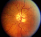 |
Shunt Vessel Meningioma | RETINO-CHOROIDAL (OPTO-CILIARY) COLLATERAL VESSELS: (also known as Retinal-choroidal venous collaterals, opticociliary veins or ciliary shunt vessels) Retino-choroidal collaterals are potential telangiectatic connections between the retina and choroidal circulation. Although sometimes called "shunts... | Image |
| 53 |
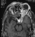 |
Shunt Vessel Meningioma - MRI | Meningiomas block venous egress and open potential venous channels known as retinochoroidal (optociliary) collateral vein. This meningioma extends from the back of the globe through the optic canal. | Image |
| 54 |
 |
Stage 2 - Papilledema | Image | |
| 55 |
 |
Stargardt's Disease | Discussion of Stargardt's disease, an inherited maculopathy which frequently presents with a loss of central vision. | Text |
| 56 |
 |
Tangent Screen Recording Chart | The tangent screen recording chart. | |
| 57 |
 |
Tangent Screen Testing Visual Field | Description of tangent screen testing. | |
| 58 |
 |
Third Nerve Palsy | Patient with third nerve palsy (no audio) | Image/MovingImage |
| 59 |
 |
Tilted Discs | Short PowerPoint discussion of tilted discs with illustrations and images. | |
| 60 |
 |
Trigeminal Nerve Exam | Explanation of a trigeminal nerve exam. | |
| 61 |
 |
Tunnel Vision on Tangent Screen Testing | Description of tunnel vision and tangent screen testing. | |
| 62 |
 |
Vestibular Nystagmus | Example of patient with vestibular nystagmus. Patient is led through instructions for direction of gaze. Shown also with Frenzel goggles. | Image/MovingImage |
| 63 |
 |
Abducting (Dissociated) Nystagmus | Example of a patient with abducting (dissociated) nystagmus. Patient has a subtle internuclear ophthalmoplegia. Right eye has right-beating jerk nystagmus, with smaller oscillations in the left eye. Disease/Diagnosis: Abducting Nystagmus | Image/MovingImage |
| 64 |
 |
Before Tensilon | Example of patient with myasthenia gravis. Demonstration of baseline examination, followed by administration of 2mg of tensilon, which is a test dose. Procedure for administration of tensilon test is described, including variations. Patient is then shown after being given 4mg of tensilon, with very ... | Image/MovingImage |
| 65 |
 |
Congenital Ocular Motor Apraxia | Two examples of congenital ocular motor apraxia. Patients have trouble initiating saccades, and compensate with head movement. Discussion of how to distinguish this condition from simply not seeing well. | Image/MovingImage |
| 66 |
 |
Dissociated Nystagmus | Example of a patient with dissociated nystagmus. Demonstrates difference in movements between each eye. | Image/MovingImage |
| 67 |
 |
Internuclear Ophthalmoplegia (2 Examples) | Two examples of patients with internuclear ophthalmoplegia. First patient has a right internuclear ophthalmoplegia. Patient had subacute bacterial endocarditis with a bacterial abscess in the brain stem. Ductions and gaze to the right look good, but when gazing to the left, the right eye does not ad... | Image/MovingImage |
| 68 |
 |
Latent Nystagmus | Example of a patient with latent nystagmus. Demonstrates a lack of oscillations in forward gaze, followed by the occlusion of each eye, showing how this generates a jerking oscillation in the non-occluded eye away from the occluded eye. | Image/MovingImage |
| 69 |
 |
Opsoclonus | Example of patients with opsoclonus, a saccadic abnormality. | Image/MovingImage |
| 70 |
 |
Paradoxical Constriction of Pupils to Darkness (Flynn Phenomenon) | Example of patients both with and without paradoxical constriction of pupils. Observed in many congenital retinal disorders, such as achromatopsia, congenital stationary night-blindness, and Leber's congenital amaurosis. Sometimes seen in optic nerve disorders, such as dominant optic atrophy. | Image/MovingImage |
| 71 |
 |
Parinaud's Syndrome | Two examples of patients with Parinaud's syndrome, a dorsal midbrain syndrome. Discussion of hallmarks of this syndrome, including convergence retraction nystagmus, vertical gaze palsies, light-near dissociation, and Collier's Sign. Discussion of age-dependent disorders associated with this syndrome... | Image/MovingImage |
| 72 |
 |
Physiologic (End-Gaze) Nystagmus | Demonstration of physiological nystagmus, where oscillations do not represent pathology, but occur when the patient's gaze is drawn too far laterally. | Image/MovingImage |
| 73 |
|
Progressive Supranuclear Palsy | Example of patient with progressive supranuclear palsy. Discussion of difference between saccadic movement in supranuclear palsy and nystagmus. Shows saccadic intrusions in forward gaze, pursuit, saccades, and doll's head maneuver. | Image/MovingImage |
| 74 |
 |
Spasm of the Near Reflex | Example of patient with spasm of the near reflex and voluntary nystagmus. Discussion of similar-looking conditions (e.g. six nerve palsy, limitation of abduction, lateral rectus muscle problems) and how to tell them apart from spasm of the near reflex by observing the myosis evoked by the near respo... | Image/MovingImage |
| 75 |
|
Transillumination Ocular Melanoma | Video describing condition. | Image/MovingImage |