John A. Moran Eye Center Neuro-Ophthalmology Collection: A variety of lectures, videos and images relating to topics in Neuro-Ophthalmology created by faculty at the Moran Eye Center, University of Utah, in Salt Lake City.
NOVEL: https://novel.utah.edu/
TO
Filters: Collection: ehsl_novel_jmec
| Identifier | Title | Description | Subject | ||
|---|---|---|---|---|---|
| 26 |
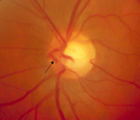 |
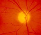
3-65 | 3-65 - Shunt Vessels (Glaucoma) | Chronic end-stage glaucoma produces high pressure that interferes with venous drainage from the disc and broad smooth venous collaterals drain the disc centrifugally to the disc margin where they drain. | Shunt Vessels (Glaucoma) |
| 27 |
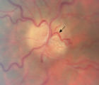 |
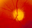
3-66a | 3-66a - Shunt Vessels (Post-papilledema) | The retino-choroidal collaterals seen with chronic papilledema begin with a "Hairnet" of telangiectasias that gradually winnow down to one or more large collateral tortuous draining channel. The presence of these vessels is evidence of long standing disc swelling. When the CSF pressure is lowered, t... | Shunt Vessels (Post-papilledema) |
| 28 |
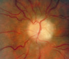 |
3-66d | 3-66d - Shunt Vessels (Post-papilledema) | The retino-choroidal collaterals seen with chronic papilledema begin with a "Hairnet" of telangiectasias that gradually winnow down to one or more large collateral tortuous draining channel. The presence of these vessels is evidence of long standing disc swelling. When the CSF pressure is lowered, t... | Shunt Vessels (Post-papilledema) |
| 29 |
 |
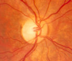
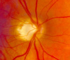
4-35 | 4-35 - Cupped Optic Nerve | Atrophic Glaucoma Atrophic glaucomatous discs show thinning of the neuro-retinal rim, "saucerization" (which is shallow cupping), evidence of peripapillary atrophy, and pallor of the very narrow neuroretinal rim. Notice that there is severe atrophy of the nerve fiber layer. | Cupped Optic Nerve |
| 30 |
 |
4-52b | 4-52b - Dominant Optic Neuropathy | A son presented with bilateral optic atrophy of unknown etiology after he failed a school visual exam. When looking for dominant optic atrophy, look at the parents. Mother was examined to find similar kind of atrophy. 4-52a mother, 4-52b son. | Dominant Optic Neuropathy |
| 31 |
 |
4-54a | 4-54a -Optic Neuropathy, Ischemic: Posterior | Optic Neuropathy, Ischemic; Posterior Ischemic Optic Neuropathy | |
| 32 |
 |
4-54b | 4-54b - Optic Neuropathy, Ischemic: Posterior | Optic Neuropathy, Ischemic; Posterior Ischemic Optic Neuropathy | |
| 33 |
 |
4-60a | 4-60a - Dominant Optic Neuropathy | A son presented with bilateral optic atrophy of unknown etiology after he failed a school visual exam. When looking for dominant optic atrophy, look at the parents. Mother was examined to find similar kind of atrophy. 4-60a mother, 4-60b son. | Dominant Optic Neuropathy |
| 34 |
 |
A-scan_technique | A-scan Technique | This video describes and demonstrates the A-scan examination technique for examination of the eye using ultrasonography. | Ultrasonography Eye Examination Techniques |
| 35 |
 |
2-7 | Abducting (Dissociated) Nystagmus | Example of a patient with abducting (dissociated) nystagmus. Patient has a subtle internuclear ophthalmoplegia. Right eye has right-beating jerk nystagmus, with smaller oscillations in the left eye. Disease/Diagnosis: Abducting Nystagmus | Abducting Nystagmus; Dissociated Nystagmus |
| 36 |
|
1-6 | Aberrant Regeneration of Third Nerve, Bilaterally (1 degree OD, 2 Digrees OS) | Example of patient with bilateral aberrancy of the third nerve. Shows lids popping up (synkinetic) with adduction. Patient had bilateral internal carotid artery aneurisms with third nerve compression. | Bilateral Aberrant Regeneration of Third Nerve; Third Nerve Dysfunction |
| 37 |
 |
NOVEL_Moran_2-19 | Aberrant Regeneration of the Lid | Patient with left third nerve palsy demonstrates anisocoria and mild vertical gaze limitation and aberrant movement of the left upper lid. Patient is instructed through all gaze positions. Left upper lid does not descend during downgaze but retracts instead. | Third Nerve Palsy; Aberrant Regeneration of Third Nerve; Aberrant Reinnervation of Third Nerve |
| 38 |
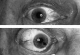 |
Figure-15 | Aberrant Regeneration of the Right Pupil | Aberrant regeneration of the right pupil in a man with a large intracavernous sinus meningioma causing a pupil-involving, incomplete third cranial nerve palsy. His pupil is round when he gazes straight ahead (top). When he tries to rotate the eye medially, the pupil constricts, but a segment of the ... | Pupil Disorders; Aberrant Regeneration; Third Nerve Palsy |
| 39 |
 |
1-19 | Aberrant Regeneration of the Seventh Nerve | Examples of patients with aberrant regeneration of the seventh nerve. First example is a patient with contractions around the mouth and dimpling, demonstrated with slow and rapid eye blinking. Second example shows contraction around nose with eye blink. | Aberrant Regeneration of the Seventh Nerve; Aberrant Regeneration |
| 40 |
 |
NOVEL_Moran_2-28 | Aberrant Regeneration of the Third | Patient with a right third nerve palsy demonstrates ptosis, anisocoria and ophthalmoplegia. During attempted downgaze, the right upper lid flutters back up (aberrant movement) and remains retracted. | Third Nerve Palsy; Aberrant Regeneration |
| 41 |
 |
1-7 | Aberrant Regeneration of the Third and Sixth Nerves | Aberrant Regeneration; Third and Sixth Nerves; Third Nerve Palsy | |
| 42 |
 |
Amsler_Grid | Amsler Grid Testing | Demonstration of Amsler Grid examination. | Examination, Ocular; Amsler Grid |
| 43 |
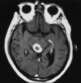 |
Figure-12 | An Enhancing Bladder Metastasis Involving the Tectum of the Midbrain | Magnetic resonance image of an enhancing bladder metastasis involving the tectum of the midbrain of a 56-year-old man who developed double vision resulting from skew deviation and divergence insufficiency. He also had a left-sided relative afferent pupillary defect measuring 1.4 log units but showed... | Physiology, Pupil; Reflex, Pupillary |
| 44 |
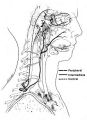 |
Figure-04 | Anatomy of the Oculosympathetic Pathway | Anatomy of the oculosympathetic pathway. (Maloney WF, Younge BR, Moyer NJ: Evaluation of the causes and accuracy of pharmacologic localization in Horner's syndrome. Am J Ophthalmol 1980;90:394-402, Ophthalmic Publishing Company with permission.) | Anatomy of the Oculosympathetic Pathway; Horner's Syndrome |
| 45 |
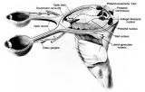 |
Figure-02 | Anatomy of the Pupillary Light Reflex Pathway | Anatomy of the pupillary light reflex pathway. (Miller NR: Walsh And Hoyt's Clinical Neuro-Ophthalmology, p 421. Vol 2, 4th ed. Baltimore: Williams & Wilkins, 1985, with permission.) | Reflex, Pupillary; Parasympathetic Pupil |
| 46 |
 |
Digre_AION | Anterior Ischemic Optic Neuropathy | PPT describing Anterior Ischemic Optic Neuropathy (AION). Covers clinical signs, such as monocular vision loss, swollen nerve, and visual field defects, as well as risk factors. | Anterior Ischemic Optic Neuropathy |
| 47 |
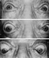 |
Figure-14 | Argyll Robertson Pupils | Argyll Robertson pupils in an elderly man treated for tabes dorsalis in 1952. His pupils are small and slightly irregular, constrict poorly in response to light stimulation (top), dilate poorly in darkness (middle), but constrict promptly in response to near stimulation (bottom). | Argyll-Robertson Pupil; Diagnosis, Pupil Disorders; Etiology, Pupil Disorders; History, Pupil Disorders; Pathology, Pupil Disorders |
| 48 |
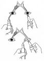 |
Figure-10 | Assessment of an Afferent Pupillary Defect When Only One Iris is Functional | Assessment of an afferent pupillary defect when only one iris is functional. In this example, a right-sided parasellar tumor is compressing both the optic and oculomotor nerves, causing an optic neuropathy and a pupil-involving third crainial nerve palsy. The pupil on the affected side has both an a... | Pupil Disorders; RAPD; Afferent Pupillary Defect |
| 49 |
 |
B-scan_technique | B-scan Technique | This video describes and demonstrates the B-scan examination technique for examination of the eye using ultrasonography. | Ultrasonography Eye Examination Techniques |
| 50 |
 |
Basal encephalocele | Basal Encephaloceles | Basal Encephaloceles |