The Emory Eye Center Neuro-Ophthalmology Collection contains a variety of lectures, videos and images relating to the discipline of neuro-ophthalmology created by faculty at Emory University in Atlanta, GA.
NOVEL: https://novel.utah.edu/
TO
Filters: Type: "Text" Collection: "ehsl_novel_eec"
| Title | Description | Creator | ||
|---|---|---|---|---|
| 26 |
 |
Optical Coherence Tomography of the Retinal Nerve Fiber Layer | A normal optical coherence tomography (OCT) of the macula is shown highlighting the position of a single retinal ganglion cell and its axon in the retinal nerve fiber layer (Figure 1). The topographical relationship of retinal ganglion cells in the retina to the visual field and position in the ante... | Jonathan A. Micieli, MD; Valérie Biousse, MD |
| 27 |
 |
Vitreopapillary Traction | A 64-year-old woman was referred for bilateral optic disc edema. Examination of her optic nerves showed indistinct margins at the nasal aspect of both eyes (Figure 1). Humphrey 24-2 SITA-Fast visual fields showed non-specific depressed points in both eyes (Figure 2). Optical coherence tomography (... | Jonathan A. Micieli, MD; Valérie Biousse, MD |
| 28 |
 |
Direct Carotid-Cavernous Sinus Fistula | A 40-year-old man presented with decreased vision and redness in his left eye. He had a significant trauma to the left side of his face about one year ago, but did not seek medical attention. External examination showed significant proptosis of the left eye (Figure 1) and conjunctival injection and ... | Jonathan A. Micieli, MD; Valérie Biousse, MD |
| 29 |
 |
Incipient Non-Arteritic Anterior Ischemic Optic Neuropathy (NAION) Evolving to Symptomatic NAION | A 54-year old woman with hypertension was seen in neuro-ophthalmology consultation for asymptomatic left optic disc edema. She had a small, crowded optic disc in the right eye known as a "disc-at-risk" (Figure 1). Her visual function including 24-2 SITA-Fast Humphrey visual fields were normal in bot... | Jonathan A. Micieli, MD; Valérie Biousse, MD |
| 30 |
 |
Typical Idiopathic Intracranial Hypertension: Optic Nerve Appearance and Brain MRI Findings | A 24-year old African American woman was referred for bilateral optic disc edema that was incidentally noted on a routine eye examination. She had excellent visual function and dilated examination showed bilateral optic disc edema with peripapillary wrinkles in the right eye and pseudodrusen in the ... | Jonathan A. Micieli, MD; Valérie Biousse, MD |
| 31 |
 |
Incipient Non-Arteritic Anterior Ischemic Optic Neuropathy (NAION) | A 61-year old white man with hypertension, diabetes, and dyslipidema was seen in neuro-ophthalmology consultation for asymptomatic right optic disc edema. He had a small, crowded optic disc in the left eye known as a "disc-at-risk" (Figure 1). He had normal visual function including normal 24-2 SITA... | Jonathan A. Micieli, MD; Valérie Biousse, MD |
| 32 |
 |
Non-Arteritic Anterior Ischemic Optic Neuropathy (NAION) With Segmental Optic Disc Edema | A 75-year old white woman with hypertension and diabetes presented with a 1 week history of vision loss in the right eye. Dilated fundus examination revealed superior segmental optic disc edema in the right eye and a small, crowded optic disc in the left eye known as a "disc-at-risk" (Figure 1). Int... | Jonathan A. Micieli, MD; Valérie Biousse, MD |
| 33 |
 |
Sequential Non-Arteritic Anterior Ischemic Optic Neuropathy (NAION) | A 68-year old woman with hypertension, obstructive sleep apnea and obesity was seen in neuro-ophthalmology consultation for vision loss in the right eye. She had right optic disc edema with a small optic disc hemorrhage a small, crowded optic disc in the left eye known as a "disc-at-risk" (Figure 1)... | Jonathan A. Micieli, MD; Valérie Biousse, MD |
| 34 |
 |
Junctional Scotoma from a Sellar Mass | This is a case of a 55-year-old woman presenting with gradual painless vision loss in both eyes. Although visual acuity was 20/20 in both eyes, there was a left relative afferent pupillary defect and diffuse pallor of both optic nerves (Figure 1). Visual fields (24-2 SITA-Fast) showed a temporal def... | Jonathan A. Micieli, MD; Valérie Biousse, MD |
| 35 |
 |
Ganglion Cell Layer Analysis by Optical Coherence Tomography (OCT) | A normal optical coherence tomography (OCT) of the macula is shown (Figure 1) and the various layers of the retina are labelled (Figure 2). The cell bodies of retinal ganglion cells (RGC) are located in the ganglion cell layer (GCL) of the retina and mostly synapse in the lateral geniculate nucleus ... | Jonathan A. Micieli, MD; Valérie Biousse, MD |
| 36 |
 |
Optic Nerve Sheath Meningioma | This is a case of an optic nerve sheath meningioma (ONSM) in a 56-year-old woman who presented with gradual, painless vision loss in her left eye. Optic disc photos at presentation showed temporal pallor of the left optic nerve (Figure 1) and Cirrus optical coherence tomography (OCT) of the retinal ... | Jonathan A. Micieli, MD; Valérie Biousse, MD |
| 37 |
 |
Typical Idiopathic Optic Neuritis | This is a case of a typical optic neuritis in a 41-year-old woman presenting with vision loss and pain with eye movements in the right eye. Optic disc photos at presentation showed subtle hyperemia in the right eye (Figure 1) and optical coherence tomography (OCT) of the retinal nerve fiber layer (R... | Jonathan A. Micieli, MD; Valérie Biousse, MD |
| 38 |
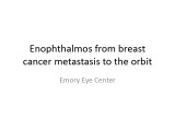 |
Enophthalmos from Breast Cancer Metastasis to the Orbit | Right painful ophthalmoplegia with right enophthalmos secondary to breast cancer metastasis to the right orbit. | Valérie Biousse, MD |
| 39 |
 |
Toxic Retinopathy: Deferoxamine Toxicity | Number of Figures and legend for each: 6 figures Figure 1: Goldmann perimetry showing large cecocentral scotomas in both eyes Figure 2: Fundus photograph of the right eye demonstrating hypopigmentation of the peripapillary and perifoveal retinal pigment epithelium (RPE) with subfoveal yellow lesions... | Will Pearce, MD; Valérie Biousse, MD |
| 40 |
 |
Normal Retinal Anatomy | Normal posterior vitreous, retinal and chroroidal anatomy (pictures, fluorescein angiography and optical coherence tomography). Figure 1: Normal fundus photograph of the left eye o a : Optic disc and fovea o b : Foveal reflex in young patients o c : Macular and foveal areas share the same center o d... | Rabih Hage, MD; Valérie Biousse, MD |
| 41 |
 |
Ocular Fundus Examination | Review of various techniques of ocular fundus examination, including direct ophthalmoscopy, binocular indirect ophthalmoscopy, slit lamp binocular indirect ophthalmoscopy, and fundus photography. Advantages and disadvantages of each technique are discussed. | Devin D. Mackay, MD; Valérie Biousse, MD |
| 42 |
 |
Rathke's Cleft Cyst Apoplexy with Junctional Scotoma | MRI features of Rathke's cleft cyst apoplexy. - Figure 1 : Humphrey visual fields at initial presentation - Figure 2 : Brain MRI without contrast at initial presentation - Figure 3 : Brain MRI with contrast at initial presentation - Figure 4 : Postoperative Humphrey visual fields | Samuel Bidot, MD; Amit M. Saindane, MD; Valérie Biousse, MD |
| 43 |
 |
Occipital Pyogenic Abscess with Homonymous Hemianopia | This is a case of right occipital abscess with a left homonymous hemianopia. Number of Figures and legend for each: 8 figures Figure 1: Humphrey visual fields: Dense left homonymous hemianopia Figure 2: T2-weighted axial MRI : Round, hyperintense lesion (yellow arrow) in the right occipital lobe sur... | Rabih Hage, MD; Valérie Biousse, MD |
| 44 |
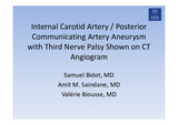 |
Internal Carotid Artery / Posterior Communicating Artery Aneurysm with Third Nerve Palsy Shown on CT Angiogram | Internal Carotid Artery / Posterior Communicating Artery Aneurysm with Third Nerve Palsy Shown on CT Angiogram ; anatomic description of vascular and bony findings on the CTA. - Figure 1 : 51 year-old man complaining of painful binocular diplopia. Orange arrows indicate the direction of gaze. In p... | Samuel Bidot, MD; Amit M. Saindane, MD; Valérie Biousse, MD |
| 45 |
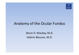 |
Anatomy of the Ocular Fundus | A review of normal features of the ocular fundus. Fundus photography using various techniques illustrate anatomic features of the ocular fundus. - Figure 1 : A) Color fundus photograph of the left optic disc and peripapillary retina showing a normal optic disc, retinal arteries, retinal veins, and... | Devin D. Mackay, MD; Valérie Biousse, MD |
| 46 |
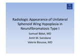 |
Radiologic Appearance of Unilateral Sphenoid Wing Hypoplasia in Neurofibromatosis Type I | MRI features of greater wing sphenoid hypoplasia in the setting of neurofibromatosis type 1. - Figure 1 : Orbital MRI with contrast showing right greater sphenoid wing hypoplasia. The lack of bone tissue leads to herniation of the right temporal lobe into the orbit, pushing forward the orbital conte... | Samuel Bidot, MD; Amit M. Saindane, MD; Valérie Biousse, MD |
| 47 |
 |
Posterior Circulation Infarctions Secondary to a Post Traumatic Vertebral Artery Dissection | A case of a young man with a vertebral artery dissection that caused multiple posterior circulation brain infarcts. Images from an MRI of the brain, digital subtraction angiography, and Humphrey visual fields are included. Figure 1 : Humphrey visual fields showed a right homonymous hemianopia with ... | Devin D. Mackay, MD; Valérie Biousse, MD |
| 48 |
 |
Sturge-Weber Syndrome | A case of Sturge-Weber syndrome (Encephalotrigeminal angiomatosis) with angiomas that involve the leptomeninges, and the skin of the ipsilateral hemiface, associated with congenital glaucoma in the same eye. Various illustrations are included to demonstrate the port wine stain, enlarged optic nerve ... | Supharat Jariyakosol, MD; Valérie Biousse, MD |
| 49 |
 |
Terson Syndrome and Subarachnoid Hemorrhage | A case of Terson syndrome resulting with subarachnoid hemorrhage and right vitreous hemorrhage resulting from a left pericallosal artery aneurysm. Figure 1 : External photograph of right eye demonstrates blunted red reflex secondary to vitreous hemorrhage Figure 2 : External photograph of left eye d... | Joshua Levinson, MD; Valérie Biousse, MD |
| 50 |
 |
Sellar Aneurysm with Chiasmal Compression | This is a case of aneurysm of the internal carotid artery, invading the sella and complicated by chiasmal compression and bitemporal hemianopia. Figure 1 : Humphrey visual fields (gray scale and pattern deviations) Figure 2a : T1-weighted axial brain MRI (1): well defined circular intracerebral mass... | Rabih Hage, MD; Valérie Biousse, MD |