Best known for his world-renowned neuro-ophthalmology unit based at the University of California, San Francisco, William Hoyt, MD collected here more than 850 of his best images covering a wide range of disorders.
William F. Hoyt, MD, Professor Emeritus of Ophthalmology, Neurology and Neurosurgery, Department of Ophthalmology, University of California, San Francisco.
NOVEL: https://novel.utah.edu/
TO
| Title | Description | Type | ||
|---|---|---|---|---|
| 26 |
 |
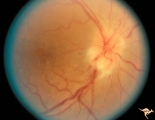
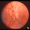
B107 Disc Swelling, Ischemic Papillopathies, AION | Pallid ischemic swelling. 41 year old man. Anatomy: Optic disc. Pathology: Axoplasmic stasis due to ischemia. Disease/ Diagnosis: AION. Clinical: Viusal loss. | Image |
| 27 |
 |
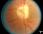
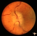
B108 Disc Swelling, Ischemic Papillopathies, AION | Pallid ischemic swelling. Woman with vasculitis. Anatomy: Optic disc. Pathology: Axoplasmic stasis due to ischemia. Disease/ Diagnosis: AION. Clinical: Visual loss. | Image |
| 28 |
 |
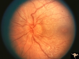
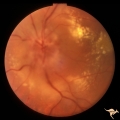
B109 Disc Swelling, Ischemic Papillopathies, AION | Ischemic swelling. Patient was diabetic. April 18, 1978. Same patient as B1-2. Anatomy: Optic disc. Pathology: Axoplasmic stasis due to ischemia. Disease/ Diagnosis: AION. Clinical: Diabetic with disc swelling and visual loss. | Image |
| 29 |
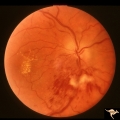 |
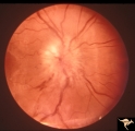
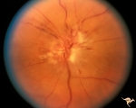
B110 Disc Swelling, Ischemic Papillopathies, AION | Pallid ischemic swelling with intraretinal exudates near the macula and a ""cotton wool"" infarct below the disc. 38 year old man. Diabetic. 20/60 vision. Altitudinal visual field defect. Anatomy: Optic disc. Pathology: Axoplasmic stasis due to ischemia. Disease/ Diagnosis: AION. Clinical: Diabetic ... | Image |
| 30 |
 |
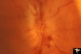
B111 Disc Swelling, Ischemic Papillopathies, AION | Acute AION. Anatomy: Optic disc. Pathology: Axoplasmic stasis due to ischemia. Disease/ Diagnosis: AION. Clinical: Visual loss. | Image |
| 31 |
 |
B112 Disc Swelling, Ischemic Papillopathies, AION | Arterioles are narrowing in resolution phase from AION. Patient had a superior altitudinal visual field defect. 20 year old man. Anatomy: Optic disc. Pathology: Axoplasmic stasis due to ischemia. Disease/ Diagnosis: AION. Clinical: Visual loss. | Image |
| 32 |
 |
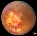
B113 Disc Swelling, Ischemic Papillopathies, AION | 57 year old woman with AION. Anatomy: Optic disc. Pathology: Axoplasmic stasis due to ischemia. Disease/ Diagnosis: AION. Clinical: Visual loss. | Image |
| 33 |
 |
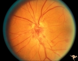
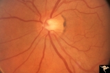
B114 Disc Swelling, Ischemic Papillopathies, AION | AION in a disc with an optic cup. Extraordinary exception with AION. Note ischemic vascular changes in disc surface. Anatomy: Optic disc. Pathology: Axoplasmic stasis due to ischemia. Disease/ Diagnosis: AION. Clinical: Visual loss. | Image |
| 34 |
 |
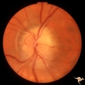
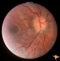
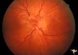
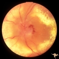
B115 Disc Swelling, Ischemic Papillopathies, AION | Normal eye in patient who later developed AION. Note generous optic cup. June 2, 1991. Same patient as B1_16b. Anatomy: Optic disc. Pathology: Normal. Clinical: Asymptomatic. | Image |
| 35 |
 |
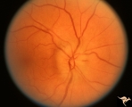
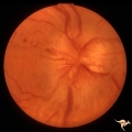
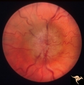
B116 Disc Swelling, Ischemic Papillopathies, AION | Typical AION in disc with optic cup. December 23, 2996. 5 years later in same patient as B1_15a. Anatomy: Optic disc. Pathology: Axoplasmic stasis due to ischemia. Disease/ Diagnosis: AION. Clinical: Visual loss. | Image |
| 36 |
 |
B201 Disc Swelling, Diabetic Papillopathy | Bilateral simultaneous diabetic papillopathy with marked exudation and remarkable recovery of vision. Right eye. Pair with B2_2b. Anatomy: Optic disc. Pathology: Axoplasmic stasis due to ischemia. Disease/ Diagnosis: Diabetic papillopathy. Clinical: Visual loss. | Image |
| 37 |
 |
B202 Disc Swelling, Diabetic Papillopathy | Bilateral diabetic papillopathy with marked exudation and remarkable recovery of vision. Left eye. Pair with B2_1a. Anatomy: Optic disc. Pathology: Axoplasmic stasis due to ischemia. Disease/ Diagnosis: Diabetic papillopathy. Clinical: Visual loss with recovery. | Image |
| 38 |
 |
B203 Disc Swelling, Diabetic Papillopathy | Disc swelling in a diabetic woman. Recovered without visual loss. Right eye. Pair with B2_04. Anatomy: Optic disc. Pathology: Axoplasmic stasis due to ischemia. Disease/ Diagnosis: Diabetic papillopathy. Clinical: Visual loss with recovery. | Image |
| 39 |
 |
B204 Disc Swelling, Diabetic Papillopathy | Disc swelling in a diabetic. Recovered without visual loss. Left eye. Pair with B2_03. Anatomy: Optic disc. Pathology: Axoplasmic stasis due to ischemia. Disease/ Diagnosis: Diabetic papillopathy. Clinical: Visual loss with recovery. | Image |
| 40 |
 |
B205 Disc Swelling, Diabetic Papillopathy | Bilateral diabetic papillopathy. Girl. Left eye. Pair with B2_06. Anatomy: Optic disc. Pathology: Axoplasmic stasis due to ischemia. Disease/ Diagnosis: Diabetic papillopathy. Clinical: Visual loss with recovery. | Image |
| 41 |
 |
B206 Disc Swelling, Diabetic Papillopathy | Bilateral diabetic papillopathy. Girl. Right eye. Pair with B2_05. Anatomy: Optic disc. Pathology: Axoplasmic stasis due to ischemia. Disease/ Diagnosis: Diabetic papillopathy. Clinical: Visual loss with recovery. | Image |
| 42 |
 |
B301 Disc Swelling, Giant Cell Arteritis | Disc swelling. Giant Cell Arteritis. Temporal. Ischemic swelling. Blind eye with pallid swelling and marked dilation of central retinal vein. | Image |
| 43 |
 |
B401 Disc Swelling, Radiation Papillopathy | Male with blind eye. Marked peripapillary intraretinal exudate. April 1985. Same patient as B402, B407. Anatomy: Optic disc. Pathology: Axoplasmic stasis due to ischemia. Disease/ Diagnosis: Radiation papillopathy; radiation optic neuropathy. Clinical: Visual loss after radiation therapy. | Image |
| 44 |
 |
B402 Disc Swelling, Radiation Papillopathy | Radiation papillopathy with arterial narrowing, exudation and venous dilation in man with blind eye. May 1985. Same patient as B401, B407. Anatomy: Optic disc. Pathology: Axoplasmic stasis due to ischemia. Disease/ Diagnosis: Radiation papillopahty; optic neuropathy. Clinical: Visual loss after radi... | Image |
| 45 |
 |
B403 Disc Swelling, Radiation Papillopathy | Man with blind eye. Ischemic hemorrhages. Vitreous haze. Anatomy: Optic disc. Pathology: Axoplasmic stasis due to ischemia. Disease/ Diagnosis: Radiation papillopathy; optic neuropathy. Clinical: Visual loss after radiation therapy. | Image |
| 46 |
 |
B404 Disc Swelling, Radiation Papillopathy | Marked vascular changes in the swollen optic disc. Probably not blind. Male. Right eye. Anatomy: Optic disc. Pathology: Axoplasmic stasis due to ischemia. Disease/ Diagnosis: Radiation Papillopathy; Optic neuropathy. Clinical: Visual loss after radiation therapy. | Image |
| 47 |
 |
B405 Disc Swelling, Radiation Papillopathy | Bilateral blindness 6 months post radiation for malignant glioma of left hemisphere. Left eye. Marked white exudation probably represents necrosis of swollen disc tissue. Japanese patient. Anatomy: Optic disc. Pathology: Axoplasmic stasis due to ischemia. Disease/ Diagnosis:Radiation papillopathy; O... | Image |
| 48 |
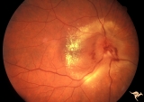 |
B406 Disc Swelling, Radiation Papillopathy | Note the marked vascular changes on the disc surface and the interesting distribution of intraretinal exudate. Patient had vision with large blind spot. Woman. Right eye. Visual field showed only an enlarged blind spot. Anatomy: Optic disc. Pathology: Axoplasmic stasis due to ischemia. Disease/ Diag... | Image |
| 49 |
 |
B407 Disc Swelling, Radiation Papillopathy | Man with blind eye. June 1985. Same patient as B401 and B402. Note the striking peripapillary intraretinal exudatation occurring at a slight distance from the disc. Anatomy: Optic disc. Pathology: Axoplasmic stasis due to ischemia; Ischemic infarction. Disease/ Diagnosis: Radiation papillopathy; Opt... | Image |
| 50 |
 |
Bilateral Chronic Papilledema | Left eye. Frisen's stage 5. Patient with long standing aqueductal stenosis. Bilateral Chronic Papilledema. Man. Anatomy: Optic disc. Pathology: Papilledema. Disease/Diagnosis: Papilledema from aqueductal stenosis. | Image |