Best known for his world-renowned neuro-ophthalmology unit based at the University of California, San Francisco, William Hoyt, MD collected here more than 850 of his best images covering a wide range of disorders.
William F. Hoyt, MD, Professor Emeritus of Ophthalmology, Neurology and Neurosurgery, Department of Ophthalmology, University of California, San Francisco.
NOVEL: https://novel.utah.edu/
TO
| Title | Description | Type | ||
|---|---|---|---|---|
| 26 |
 |
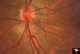
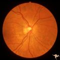
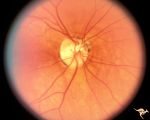
Retinal Signs of Atheromatous Embolization | Retinal signs of atheromatous embolization. Central retinal artery occlusion by soft atheromatous debris (mostly fibrin) causing blindness. Anatomy: Retina. Pathology: Carotid atheromatous disease. Disease/Diagnosis: Carotid atheromatous vascular disease. Clinical: Blindness. | Image |
| 27 |
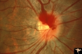 |
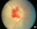
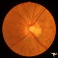
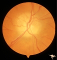
A406 Disc Swelling, Vitreous Effects | Prepapillary hemorrhage. Partial posterior vitreous detachment in myopic Asian patient. Reference: Katz B, Hoyt WF. Intrapapillary and peripapillary hemorrhage in young patients with incomplete posterior vitreous detachment. Signs of vitreopapillary traction. Ophthalmology. 1995 Feb;102(2):349-54. A... | Image |
| 28 |
 |
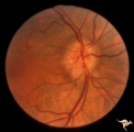
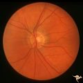
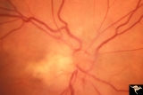
A408 Disc Swelling, Vitreous Effects | Prepapillary hemorrhage. Partial posterior vitreous detachment in myopic Asian patient. Reference: Katz B, Hoyt WF. Intrapapillary and peripapillary hemorrhage in young patients with incomplete posterior vitreous detachment. Signs of vitreopapillary traction. Ophthalmology. 1995 Feb;102(2):349-54. A... | Image |
| 29 |
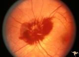 |
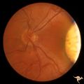
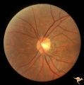
A407 Disc Swelling, Vitreous Effects | Prepapillary hemorrhage. Partial posterior vitreous detachment in myopic patient. Reference: Katz B, Hoyt WF. Intrapapillary and peripapillary hemorrhage in young patients with incomplete posterior vitreous detachment. Signs of vitreopapillary traction. Ophthalmology. 1995 Feb;102(2):349-54. Anatomy... | Image |
| 30 |
 |
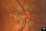
A405 Disc Swelling, Vitreous Effects | Prepapillary hemorrhage. Partial posterior vitreous detachment in myopic Asian patient. Reference: Katz B, Hoyt WF. Intrapapillary and peripapillary hemorrhage in young patients with incomplete posterior vitreous detachment. Signs of vitreopapillary traction. Ophthalmology. 1995 Feb;102(2):349-54. ... | Image |
| 31 |
 |
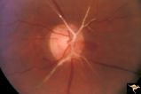
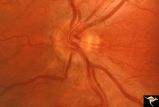
A501 Disc Swelling, Pre-Ischemic Swelling | Pre AION swelling. Asymptomatic on October 8, 1985. Same patient as A5_2b. Anatomy: Optic disc. Pathology: Axoplasmic stasis due to ischemia. Disease/ Diagnosis: Pre AION, Pre ischemic swelling. Clinical: Asymptomatic. | Image |
| 32 |
 |
H35 Segmental Hypoplasia, Retinal-Congenital Toxo | Left eye. Moving out temporally to see large chorioretinal scar. Temporal sector hypoplasia from congenital retinal toxoplasmosis. Same patient as H_34. Anatomy: Optic disc; Retina. Pathology: Hypoplasia secondary to retinal lesion. Disease/ Diagnosis: Segmental optic disc hypoplasia | Image |
| 33 |
 |
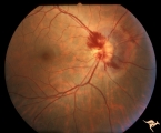
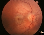
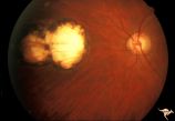
C209 Papillitis with Macular Star, Cat Scratch Disease | Proven Bartonella neuroretinitis. Woman. Anatomy: Optic disc; Retina. Pathology: Axoplasmic stasis due to inflammation. Disease/ Diagnosis: Bartonella Henslae (Cat Scratch). Clinical: Visual blurring without visual field defect; Ocular disc edema with macular star (ODEMS). | Image |
| 34 |
 |
H48 Segmental Hypoplasia, Retinal-Nasal Hypoplasia | Bilateral nasal hypoplasia with absence of nasal nerve fiber layer and corresponding flag-like temporal field defect. Right eye. Same patient as H_49. Anatomy: Optic disc. Pathology: Nasal segmental disc hypoplasia. Disease/ Diagnosis: Congential anomaly. | Image |
| 35 |
 |
H49 Segmental Hypoplasia, Retinal-Nasal Hypoplasia | Bilateral nasal hypoplasia with absence of nasal nerve fiber layer and corresponding flag-like temporal field defect. Left eye. Same patient as H_48. Anatomy: Optic disc. Pathology: Nasal segmental disc hypoplasia. Disease/ Diagnosis: Congential anomaly. | Image |
| 36 |
 |
C208 Papillitis with Macular Star, Cat Scratch Disease | Proven Bartonella neuroretinitis. Man. Anatomy: Optic disc; Retinitis. Pathology: Axoplasmic stasis due to inflammation. Disease/ Diagnosis: Bartonella Henslae (Cat Scratch). Clinical: Visual blurring; Ocular disc edema with macular star (ODEMS). | Image |
| 37 |
 |
H46 Segmental Hypoplasia, Retinal-Nasal Hypoplasia | Nasal hypoplasia with suspected nasal pit about 3:00. Right eye. Man with temporal sector field defect. Same patient as H_47. Anatomy: Optic disc. Pathology: Nasal segmental disc hypoplasia. Disease/ Diagnosis: Congenital anomaly | Image |
| 38 |
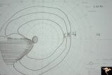 |
H42 Segmental Hypoplasia, Retinal, Nasal Hypoplasia | Visual field of patient in H_43. Flag-like temporal field defect from patient with nasal segmental disc hypoplasia. Anatomy: Optic disc. Pathology: Nasal segmental disc hypoplasia. Disease/ Diagnosis: Congenital anomaly. | Image |
| 39 |
 |
H44 Segmental Hypoplasia, Retinal, Nasal Hypoplasia | Bilateral nasal hypoplasia with bilateral flag-like temporal field defect. Right eye. Same patient as H_45. Anatomy: Optic disc. Pathology: Nasal segmental disc hypoplasia. Disease/ Diagnosis: Congenital anomaly. | Image |
| 40 |
 |
H45 Segmental Hypoplasia, Retinal-Nasal Hypoplasia | Bilateral nasal hypoplasia with bilateral flag-like temporal field defect. Left eye. Same patient as H_44. Anatomy: Optic disc. Pathology: Nasal segmental disc hypoplasia. Disease/ Diagnosis: Congenital anomaly. | Image |
| 41 |
 |
H47 Segmental Hypoplasia, Retinal-Nasal Hypoplasia | Normal left eye. Same patient as H_46. Anatomy: Optic disc. Pathology: Nasal segmental disc hypoplasia. Disease/ Diagnosis: Congenital anomaly. | Image |
| 42 |
 |
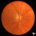
H43 Segmental Hypoplasia, Retinal, Nasal Hypoplasia | Nasal hypoplasia barely perceptible on disc. Nasal retinal nerve fibers are completely absent from 7:00 - 12:00. Anaotmy: Optic disc. Pathology: Nasal segmental disc hypoplasia. Disease/ Diagnosis: Congenital anomaly. | Image |
| 43 |
 |
H34 Segmental Hypoplasia, Retinal-Congenital Toxo | Left eye. Temporal sector hypoplasia from congenital retinal toxoplasmosis. Note the sector shaped nerve fiber loss between 2:00 and 4:00. Same patient as H_35. Anatomy: Optic disc; retina. Pathology: Hypoplasia secondary to retinal lesion. Disease/ Diagnosis: Segmental optic disc hypoplasia. | Image |
| 44 |
 |
H36 Segmental Hypoplasia, Retinal, Congenital Toxo | Left eye. Optic disc hypoplasia from congenital nasal retinal toxoplasma lesion. Chorioretinal scar. Anatomy: Optic disc, retina. Pathology: Hypoplasia secondary to retinal lesion. Disease/ Diagnosis: Segmental optic disc hypoplasia. | Image |
| 45 |
 |
P55b Chronic Papilledema, Late Stage | Left eye. Optic atrophy secondary to chronic papilledema, late stage with retinal choroidal bypass vein. Opticocilliary shunt. Glioblastoma, chemotherapy patient. Anatomy: Optic disc. Pathology: Papilledema. Disease/ Diagnosis: Chronic papilledema with atrophy. | Image |
| 46 |
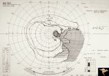 |
H51 Segmental Hypoplasia, Retinal-Nasal Hypoplasia | Visual field showing flag-like temporal field defect in patient shown in H_50. Anatomy: Optic disc. Pathology: Nasal segmental disc hypoplasia. Disease/ Diagnosis: Cogenital anomaly. | Image |
| 47 |
 |
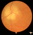
H50 Segmental Hypoplasia, Retinal-Nasal Hypoplasia | Right eye. Nasal hypoplasia with nasal pit. Same patient as H_51. Anatomy: Optic disc. Pathology: Nasal segmental disc hypoplasia. Disease/ Diagnosis: Congential anomaly. | Image |
| 48 |
 |
P55a Chronic Papilledema, Late Stage | Right eye. Optic atrophy secondary to chronic papilledema. Glioblastoma. Chemotherapy patient. Anatomy: Optic disc. Pathology: Papilledema. Disease/ Diagnosis: Chronic papilledema with atrophy. | Image |
| 49 |
 |
F101 Optic Disc Lymphosarcoma | Optic disc lymphosarcoma. This disc has been infiltrated by neoplastic cells. Anatomy: Optic disc. Pathology: Lymphosarcoma. Disease/ Diagnosis: Lymphosarcoma. | Image |
| 50 |
 |
Retinal Signs of Atheromatous Embolization | Retinal signs of atheromatous embolization. Series shows event in progress. R3_A19c shows embolus has reached the bifurcation and a second embolus (below) is beginning its transit along the same path. Anatomy: Retina. Pathology: Carotid atheromatous disease. Disease/Diagnosis: Carotid atheromatous v... | Image |