Collection of materials relating to neuro-ophthalmology as part of the Neuro-Ophthalmology Virtual Education Library.
NOVEL: https://novel.utah.edu/
TO
- NOVEL230
| Title | Creator | Description | Subject | ||
|---|---|---|---|---|---|
| 26 |
 |
Cerebral Aneurysm | Justin Gibson, MD; Charles Prestigiacomo, MD | Cerebral angiogram of a patient with an arteriovenous malformation, or AVM. | Angiogram; Cerebral Aneurysm |
| 27 |
 |
Cerebral Circulation: Neuroanatomy Video Lab - Brain Dissections | Suzanne S. Stensaas, PhD | The major vessels of the anterior and posterior circulation are demonstrated along with the Circle of Willis on both a model and in an animation. The distribution of the three major cerebral arteries is demonstrated along with the concept of a watershed zone. A gross specimen with good vessels is al... | Cerebral Circulation; Brain; Dissections |
| 28 |
 |
Cerebral Palsy | Mozammil Alam, Medical Student; Sean Gratton, MD | This video covers an overview of cerebral palsy. | Cerebral Palsy |
| 29 |
 |
Classification of Uveitis | Joanne Thomas, BS; Jessica Shantha, MD | This is an overview of the classification of uveitis. Topics discussed include: SUN anatomic classification, characterization of uveitis descriptors, AC cell and AC flare classification, and terminology of activity. | Uveitis; Classification; SUN |
| 30 |
 |
Clinical Characteristics of Ocular Lateropulsion | Jorge C Kattah, MD | A discussion of the normal mechanism that maintain the eyes in normal horizontal position. | Ocular Lateropulsion; Unilateral Gaze Palsy; Radiographic h-CGD |
| 31 |
 |
Cogan Lid Twitch | Hari Anandarajah, BA | A 50-year-old woman presented with ptosis of her left eyelid for 6 months. Several exam findings including variable and fatigable ptosis, and Cogan lid twitch, raised suspicion for Myasthenia Gravis. Acetylcholine receptor binding, blocking, and modulating antibodies were negative, and single fiber ... | Lid Twitch; Myasthenia Gravis |
| 32 |
 |
Cogan's Lid Twitch Sign | Raed Behbehani, MD | Cogan's lid twitch sign is a twitch sign of he upper lid upon looking straight from a sustained downgaze position. It is associated with Ocular Myasthenia Gavis. | Myasthenia; Ptosis; Lid Twitch |
| 33 |
 |
Common Patterns of Visual Field Defects | Sean Gratton, MD; Sarah Lam, 6th year BA/MD | Lecture covering common visual field defects, including those of the retina, optic nerve, chiasm, and retrochiasmal. | Visual Field Defects |
| 34 |
 |
Common Vitreo-Retinal Procedures and Surgeries | Luke B. Lindsell, OD, MD | Brief presentation on Common vitreo-retinal procedures and surgeries. | Vitreo-Retinal Procedures; Surgeries |
| 35 |
 |
Comparison of the Motor Systems: Neuroanatomy Video Lab - Brain Dissections | Suzanne S. Stensaas, PhD | A comparison of the three major motor systems focuses on categorizing motor problems as corticospinal tract, cerebellar, or basal ganglia. It begins with 12 minutes of gross anatomical structures and pathway review followed by clinical video clips demonstrating clinical features of disease of each o... | Corticospinal Tract; Cerebellar; Basal Ganglia; Motor Systems; Brain; Dissection |
| 36 |
 |
Complications of Strabismus Surgery and Botox | W. Walker Motley, MD | A narrated video slideshow outlining complications associated with strabismus surgery. | Strabismus; Surgery; Surgical Complications; Botox |
| 37 |
 |
Congenital Oculomotor Apraxia | Raed Behbehani, MD | Congenital Ocular Motor Apraxia is an uncommon condition that causes children to have difficulty moving their eyes horizontally or from side to side. They are usually unable to quickly move their eyes from side to side and often have to turn their head (head jerking) and not just their eyes to track... | Oculomotor Apraxia |
| 38 |
 |
Conjunctiva Muller's Muscle Resection | Patrick Burchell, MD; Jeffrey Nerad, MD | Demonstration of conjunctiva Muller's muscle resection (CMMR). | Conjunctiva Muller's Muscle Resection; CMMR |
| 39 |
 |
Control of the Eye Movements: Neuroanatomy Video Lab - Brain Dissections | Suzanne S. Stensaas, PhD | Disturbances in eye movements can provide important clues for localization of neurological damage. The role of the frontal eye fields in horizontal gaze is stressed. The need to coordinate cranial nerves on both sides of the brain stem introduces the medial longitudinal fasciculus and its role in co... | Medial Longitudinal Fasciculus; Third Cranial Nerve; Sixth Cranial Nerve; Internuclear Ophthalmoplegia; Nystagmus; Brain; Dissection |
| 40 |
 |
Control of the Pupil: Neuroanatomy Video Lab - Brain Dissections | Suzanne S. Stensaas, PhD | Through diagrams, animations and gross specimens the constriction and dilation of the pupil by the autonomic nervous system are described. Both the parasympathetic and sympathetic control are traced and the importance of a constricted pupil, Horner's Syndrome, and temporal lobe (uncal) herniation (d... | Brain; Dissections |
| 41 |
 |
Convergence-Retraction Nystagmus in Dorsal-Midbrain Syndrome | Paul Freund, MD, FRCSC; Edward Margolin, MD, FRCSC | A man in his early twenties was referred by optometrist for abnormal eye motility findings. He had a remote history of an excised pinealoma. On exam he had almost complete upgaze palsy, convergence-retraction nystagmus on attempted upgaze, and light-near dissociation of pupillary reaction, the class... | Dorsal Midbrain Syndrome; Parinaud Syndrome; Convergence-Retraction Nystagmus; Light-Near Dissociation |
| 42 |
 |
Coordination Exam: Abnormal Examples: Finger-to-nose (x2) (includes Spanish audio & captions) | Paul D. Larsen, MD | The patient places her heel on the opposite knee then runs the heel down the shin to the ankle and back to the knee in a smooth coordinated fashion. NeuroLogic Exam has been supported by a grant from the Slice of Life Development Fund at the University of Utah, the Department of Pediatrics and the O... | Coordination Examination; Finger-to-nose Test |
| 43 |
 |
Coordination Exam: Abnormal Examples: Heel-to-shin (x2) (includes Spanish audio & captions) | Paul D. Larsen, MD | The patient with ataxia of the lower extremity will have difficulty placing the heel on the knee with a side-to-side irregular over- and undershooting as the heel is advanced down the shin. Dysmetria on heel-to-shin can be seen in midline ataxia syndromes as well as cerebellar hemisphere disease so ... | Coordination Examination; Heel-shin Test |
| 44 |
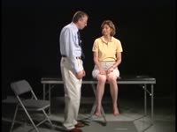 |
Coordination Exam: Normal Exam: Finger-to-nose (includes Spanish audio & captions) | Paul D. Larsen, MD | The patient moves her pointer finger from her nose to the examiner's finger as the examiner moves his finger to new positions and tests accuracy at the furthest outreach of the arm. NeuroLogic Exam has been supported by a grant from the Slice of Life Development Fund at the University of Utah, the D... | Coordination Examination; Finger-to-nose Test |
| 45 |
 |
Coordination Exam: Normal Exam: Heel-to-shin (includes Spanish audio & captions) | Paul D. Larsen, MD | The patient places her heel on the opposite knee then runs the heel down the shin to the ankle and back to the knee in a smooth coordinated fashion. NeuroLogic Exam has been supported by a grant from the Slice of Life Development Fund at the University of Utah, the Department of Pediatrics and the O... | Coordination Examination; Heel-shin Test |
| 46 |
 |
Cortical Localization: Neuroanatomy Video Lab - Brain Dissections | Suzanne S. Stensaas, PhD | The lobes of the brain are defined together with their major functions. The visual field representation in the occipital lobe is explained with a diagram. Speech areas and the major types of aphasia are discussed in the dominant hemisphere and parietal lesions of neglect and spatial orientation are ... | Cortical Localization; Brain; Dissections |
| 47 |
 |
Crafting a Grant Proposal for Research | Silvia Sörensen, PhD | Lecture describing the process of writing a grant for research. | Grant Writing |
| 48 |
 |
Cranial Nerves: Neuroanatomy Video Lab - Brain Dissections | Suzanne S. Stensaas, PhD | The approach is to learn to associate the cranial nerves with their brainstem level and blood supply. Emphasis is given to the midbrain (3, 4), pons (5, 6, 7, 8), medulla (9, 10, 11, 12) and their most important functions. | Cranial Nerves; Brain; Dissection |
| 49 |
 |
Curtain Sign (Enhanced Ptosis) | Bashaer Aldhahwani, MD; Hong Jiang, MD, PhD | This is a 78-year-old male patient who presented with diplopia, right eyelid ptosis, and ophthalmoplegia. He had severe ptosis OD and pseudo-proptosis (lid retraction) OS at baseline, but when the right eyelid was manually elevated, there was marked enhanced ptosis of the left eyelid (Video). He was... | Myasthenia GravIs; Clinical Signs |
| 50 |
 |
Diagnosis and Evaluation of Stroke: Cerebral Hypoxia | Danny Alevy; James Brian Davis; Amanda Dean Henderson | Neurons are particularly vulnerable to oxygen deprivation. Cerebral hypoxia can be caused by arterial thrombosis, embolism, hypoperfusion, cervical artery dissection, or cryptogenic causes. About 1/3 of ischemic strokes are from cryptogenic causes. Embolic strokes are the next most common (20-30%), ... | Atherosclerosis; Cerebral Hypoxia; Cervical Artery Dissection; Cryptogenic Ischemia; Embolism; Hypoperfusion; Ischemia; Stroke; Thrombosis |
