Collection of materials relating to neuro-ophthalmology as part of the Neuro-Ophthalmology Virtual Education Library.
NOVEL: https://novel.utah.edu/
TO
- NOVEL717
| Title | Creator | Description | Subject | ||
|---|---|---|---|---|---|
| 26 |
 |
Corectopia | Meagan Seay, DO | These are photos of a patient with unilateral corectopia. This patient's corectopia is of unclear etiology and possibly related to birth trauma. | Corectopia; Unilateral; Photos |
| 27 |
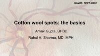 |
Cotton Wool Spots: The Basics | Arnav Gupta, BHSc; Rahul Sharma, MD, MPH | A presentation describing cotton wool spots, an abnormal finding on funduscopic exam of the retina of the eye. | Cotton Wool Spots; Retina |
| 28 |
 |
Corneal Staining | Sovik De Sirkar, MSIII; Ore-ofe Adesina, MD | Description of the corneal staining technique. | Corneal Staining |
| 29 |
 |
Craniopharyngioma and Optic Atrophy | Kathleen B. Digre, MD; James J. Corbett, MD | Slideshow describing condition. | Craniopharayngioma; Otpic Atrophy |
| 30 |
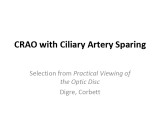 |
CRAO with Ciliary Artery Sparing | Kathleen B. Digre, MD; James J. Corbett, MD | Slideshow describing condition. | CRAO |
| 31 |
 |
Decompensated Phoria | Alex Christoff, MD | An overview of decompensated phoria and its treatment. | Decompensated Phoria |
| 32 |
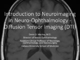 |
Diffusion Tensor Imaging (DTI) | Devin D. Mackay, MD | Explanation of using diffusion tensor imaging (DTI) in examinations. | Diffusion Tensor Imaging (DTI) |
| 33 |
 |
CT Angiography (CTA) | Devin D. Mackay, MD | Explanation of using computed tomography angiography (CTA) in examinations. | CT Angiography (CTA) |
| 34 |
 |
CT Venography (CTV) | Devin D. Mackay, MD | Explanation of using computed tomography venography (CTV). | CT Venography (CTV) |
| 35 |
 |
Diffusion Weighted Imaging (DWI) | John Pula, MD | Diffusion weighted imaging sequences are often included as part of a routine brain MRI protocol. Imaging provides examples of DWI. | Diffusion Weighted Imaging; DWI |
| 36 |
 |
Disc Cupping: The Basics | Arnav Gupta, BHSc; Rahul Sharma, MD, MPH | A presentation describing optic disc cupping, due to damage of optic nerve fibres. | Disc Cupping; Optic Disc |
| 37 |
 |
Digital Subtraction Angiography | Devin D. Mackay, MD | Explanation of using digital subtraction angiography in examinations. | Digital Subtraction Angiography |
| 38 |
 |
Doppler Ultrasonography | Devin D. Mackay, MD | Explanation of using doppler ultrasonography in examinations. | Doppler Ultrasonography |
| 39 |
 |
Dementia with Lewy Bodies: Overview and Neuro-ophthalmologic features | Pavan Vaswani, MD, PhD; Ali G. Hamedani, MD, MHS | Objectives: Recognize the difference between Dementia with Lewy Bodies and Parkinson disease dementia; Recognize the clinical presentation of DLB and differentiating features from Alzheimer disease dementia; Understand the symptomatic therapies and prognosis | Dementia; Lewy Bodies |
| 40 |
 |
Dry Eye Syndrome (Danish) | NANOS | People with abnormalities of the tear film are diagnosed with "dry eyes", but some patients with "dry eyes" may not feel that their eyes are "dry". Itching, burning, a scratchy sensation, a sensation that there is sand or grit in the eye, or intermittent blurring of the vision can all be symptoms of... | Dry Eye Syndrome; Patient Brochure |
| 41 |
 |
Paraneoplastic Disease (PowerPoint) | AAO/NANOS - American Academy of Ophthalmology / North American Neuro-Ophthalmology Society | This 46-year-old woman noted progressive bilateral visual loss over a 10-month period. She had a malignant melanoma removed from her right thigh 2 years ago and excisional biopsy of an inguinal node metastasis 8 months ago. She also complained of poor night vision and rare intermittent ""sparkles"" ... | Paraneoplastic Disease |
| 42 |
 |
Leber's Optic Neuropathy (PowerPoint) | AAO/NANOS - American Academy of Ophthalmology / North American Neuro-Ophthalmology Society | Several Primary Mutations result in Leber's hereditary optic neuropathy, including mitochondrial deletions at positions 11778, 3460, and 14484. Although the 11778 is the most common mutation, the 11484 has the best prognosis for spontaneous recovery. This case exhibits the 3460 mutation. | Leber's Optic Neuropathy |
| 43 |
 |
Central Cone Dystrophy Occult Macular Dystrophy | Gregory Van Stavern, MD | Slideshow describing condition of Central Cone Dystrophy Occult Macular Dystrophy | Central Cone Distrophy; Macular Dystrophy; Occult Macular Dystrophy |
| 44 |
 |
Optic Atrophy (PowerPoint) | William F. Hoyt, PhD | a) Evolution of optic disc pallor after optic nerve transection. Normal Right eye. Photo taken December 9, 1978. b) Injury on December 8, 1978. Evolution of optic disc pallor after optic nerve transection. Woman having rhinoplasty suffered optic nerve transection. One day after nerve transection. N... | Optic Disc Atrophy from Retrobulbar Causes (Retrograde Optic Nerve Degeneration); Severe Atrophy; Optic Atrophic |
| 45 |
 |
Temporal Atrophy (PowerPoint) | William F. Hoyt, PhD | Segmental Atrophy - Temporal pallor - Nutritional amblyopia (alcoholic). 1985. | Temporal Atrophy; Optic Disc Atrophy from Retrobulbar Causes (Retrograde Optic Nerve Degeneration); Severe Atrophy; Alcohol |
| 46 |
 |
Evolution of Optociliary Veins with Perioptic Nerve Sheath | William F. Hoyt, PhD | Series of images showing progression of disc swelling and macular degeneration. Pathology: Optociliary Vein. Disease/Diagnosis: Perioptic nerve sheath meningioma evolution. Clinical notes: Visual Loss. | Optic Disc Atrophy with Special Features; Optociliary Veins; Shunt Vessels (Meningioma) |
| 47 |
 |
Exposed Drusen (PowerPoint) | William F. Hoyt, PhD | PP25a: Left eye: Severe visual field defect. PP25b: right eye with exposed drusen and field loss: visual field defects; PP25c: right eye visual field PP25d: left eye visual field. | Pseudopapilledema; Exposed Drusen |
| 48 |
 |
Aneurysms | AAO/NANOS - American Academy of Ophthalmology / North American Neuro-Ophthalmology Society | Aneurisms may result in neuro-ophthalmologic sign and symptoms by direct compression of the afferent or efferent systems or by the secondary effects of hemorrhage. Basilar aneurisms may result in ocular motor deficits such as a unilateral or bilateral third nerve palsy. | Aneurysm |
| 49 |
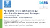 |
Paediatric Neuro-ophthalmology: Visual Acuity Assessment Strategies | Anat Bachar Zipori, MD; Nasrin Najm-Tehrani, FRCS Ed (Ophth), FRCSC | Assessing the visual function of a child can be challenging at times. When approaching a child one must understand visual development and accommodate to the child's capabilities, level of development and communication skills. The examining physician may need to apply more than one method to assess t... | Visual Acuity Assessment; Pediatric Visual Acuity Tests |
| 50 |
 |
Posterior Cortical Atrophy | Natali V. Baner, MD; Ali G. Hamedani, MD, MHS | PowerPoint providing an overview of the definition, clinical presentation and treatment of posterior cortical atrophy | Posterior Cortical Atrophy |