Collection of materials relating to neuro-ophthalmology as part of the Neuro-Ophthalmology Virtual Education Library.
NOVEL: https://novel.utah.edu/
TO
- NOVEL966
Filters: Collection: "ehsl_novel_novel"
| Title | Creator | Description | Subject | ||
|---|---|---|---|---|---|
| 26 |
 |
Conjunctival Examination | Sovik De Sirkar, MSIII; Ore-ofe Adesina, MD, | Description of the conjunctival examination. | Conjunctival Examination |
| 27 |
 |
Congenital Hydrocephalus | Mays El-Dairi, MD | Presentation covering an overview of congenital hydrocephalus. | Congenital Hydrocephalus |
| 28 |
 |
Contrast Sensitivity | Sean Gratton, MD | Explanation of contrast sensitivity. | Contrast Sensitivity |
| 29 |
 |
The Cornea and Scleral Examination | Sovik De Sirkar, MSIII; Ore-ofe Adesina, MD | Description of the cornea and scleral examination. | Cornea; Scleral Examination |
| 30 |
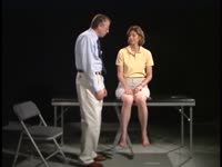 |
Coordination Exam: Normal Exam: Finger-to-nose (includes Spanish audio & captions) | Paul D. Larsen, MD | The patient moves her pointer finger from her nose to the examiner's finger as the examiner moves his finger to new positions and tests accuracy at the furthest outreach of the arm. NeuroLogic Exam has been supported by a grant from the Slice of Life Development Fund at the University of Utah, the D... | Coordination Examination; Finger-to-nose Test |
| 31 |
 |
Coordination Exam: Normal Exam: Heel-to-shin (includes Spanish audio & captions) | Paul D. Larsen, MD | The patient places her heel on the opposite knee then runs the heel down the shin to the ankle and back to the knee in a smooth coordinated fashion. NeuroLogic Exam has been supported by a grant from the Slice of Life Development Fund at the University of Utah, the Department of Pediatrics and the O... | Coordination Examination; Heel-shin Test |
| 32 |
 |
Corectopia | Meagan Seay, DO | These are photos of a patient with unilateral corectopia. This patient's corectopia is of unclear etiology and possibly related to birth trauma. | Corectopia; Unilateral; Photos |
| 33 |
 |
Cotton Wool Spots: The Basics | Arnav Gupta, BHSc; Rahul Sharma, MD, MPH | A presentation describing cotton wool spots, an abnormal finding on funduscopic exam of the retina of the eye. | Cotton Wool Spots; Retina |
| 34 |
 |
Corneal Staining | Sovik De Sirkar, MSIII; Ore-ofe Adesina, MD | Description of the corneal staining technique. | Corneal Staining |
| 35 |
 |
Cranial Nerves: Neuroanatomy Video Lab - Brain Dissections | Suzanne S. Stensaas, PhD | The approach is to learn to associate the cranial nerves with their brainstem level and blood supply. Emphasis is given to the midbrain (3, 4), pons (5, 6, 7, 8), medulla (9, 10, 11, 12) and their most important functions. | Cranial Nerves; Brain; Dissection |
| 36 |
 |
Craniopharyngioma and Optic Atrophy | Kathleen B. Digre, MD; James J. Corbett, MD | Slideshow describing condition. | Craniopharayngioma; Otpic Atrophy |
| 37 |
 |
CRAO with Ciliary Artery Sparing | Kathleen B. Digre, MD; James J. Corbett, MD | Slideshow describing condition. | CRAO |
| 38 |
 |
Decompensated Phoria | Alex Christoff, MD | An overview of decompensated phoria and its treatment. | Decompensated Phoria |
| 39 |
 |
Diffusion Tensor Imaging (DTI) | Devin D. Mackay, MD | Explanation of using diffusion tensor imaging (DTI) in examinations. | Diffusion Tensor Imaging (DTI) |
| 40 |
 |
CT Angiography (CTA) | Devin D. Mackay, MD | Explanation of using computed tomography angiography (CTA) in examinations. | CT Angiography (CTA) |
| 41 |
 |
CT Venography (CTV) | Devin D. Mackay, MD | Explanation of using computed tomography venography (CTV). | CT Venography (CTV) |
| 42 |
 |
Diffusion Weighted Imaging (DWI) | John Pula, MD | Diffusion weighted imaging sequences are often included as part of a routine brain MRI protocol. Imaging provides examples of DWI. | Diffusion Weighted Imaging; DWI |
| 43 |
 |
Disc Cupping: The Basics | Arnav Gupta, BHSc; Rahul Sharma, MD, MPH | A presentation describing optic disc cupping, due to damage of optic nerve fibres. | Disc Cupping; Optic Disc |
| 44 |
 |
Digital Subtraction Angiography | Devin D. Mackay, MD | Explanation of using digital subtraction angiography in examinations. | Digital Subtraction Angiography |
| 45 |
 |
Doppler Ultrasonography | Devin D. Mackay, MD | Explanation of using doppler ultrasonography in examinations. | Doppler Ultrasonography |
| 46 |
 |
Direct and Consensual Response | Karl C. Golnik, MD | Explanation of direct and consensual response testing. | Direct and Consensual Response |
| 47 |
 |
Distance Visual Acuity Testing | Sean Gratton, MD | Demonstration of measuring distance visual accuity. | Visual Acuity Testing |
| 48 |
 |
Dementia with Lewy Bodies: Overview and Neuro-ophthalmologic features | Pavan Vaswani, MD, PhD; Ali G. Hamedani, MD, MHS | Objectives: Recognize the difference between Dementia with Lewy Bodies and Parkinson disease dementia; Recognize the clinical presentation of DLB and differentiating features from Alzheimer disease dementia; Understand the symptomatic therapies and prognosis | Dementia; Lewy Bodies |
| 49 |
 |
Dry Eye Syndrome (Danish) | NANOS | People with abnormalities of the tear film are diagnosed with "dry eyes", but some patients with "dry eyes" may not feel that their eyes are "dry". Itching, burning, a scratchy sensation, a sensation that there is sand or grit in the eye, or intermittent blurring of the vision can all be symptoms of... | Dry Eye Syndrome; Patient Brochure |
| 50 |
 |
Paraneoplastic Disease (PowerPoint) | AAO/NANOS - American Academy of Ophthalmology / North American Neuro-Ophthalmology Society | This 46-year-old woman noted progressive bilateral visual loss over a 10-month period. She had a malignant melanoma removed from her right thigh 2 years ago and excisional biopsy of an inguinal node metastasis 8 months ago. She also complained of poor night vision and rare intermittent ""sparkles"" ... | Paraneoplastic Disease |