A collection of videos relating to the diagnosis and treatment of eye movement disorders. This collection includes many demonstrations of examination techniques.
Dan Gold, D.O., Associate Professor of Neurology, Ophthalmology, Neurosurgery, Otolaryngology - Head & Neck Surgery, Emergency Medicine, and Medicine, The Johns Hopkins School of Medicine.
A collection of videos relating to the diagnosis and treatment of eye movement disorders.
NOVEL: https://novel.utah.edu/
TO
Filters: Collection: "ehsl_novel_gold"
| Title | Description | Subject | ||
|---|---|---|---|---|
| 26 |
 |
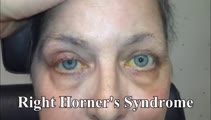
Apraclonidine Testing in Horner's syndrome | This patient experienced relatively abrupt ptosis and was seen and diagnosed with a Horner's syndrome within a few days of the onset. There were no other exam findings and history did not offer clues as to the etiology. Neuroimaging of the oculosympathetic tract was unrevealing. Apraclonidine testin... | Horner's Syndrome |
| 27 |
 |
Assessing Utricle Pathway Function and the Effects of Convergence on Nystagmus in Acute Vestibular Neuritis | A 35-year-old woman presented a few days after the onset of room-spinning vertigo. She denied diplopia, dysarthria, dysphagia, dysphonia, incoordination, numbness, and weakness. On examination, she had subtle spontaneous right-beat nystagmus (RBN). This nystagmus increased in amplitude and frequency... | Vestibular Nystagmus; Jerk Nystagmus; Acute Vestibular Syndrome; Eighth Nerve; Abnormal VHIT |
| 28 |
 |
Assessing for Hyperventilation-induced Nystagmus | 𝗢𝗿𝗶𝗴𝗶𝗻𝗮𝗹 𝗗𝗲𝘀𝗰𝗿𝗶𝗽𝘁𝗶𝗼𝗻: Hyperventilation induced nystagmus is tested by asking the patient to take quick deep breaths (~1/s) for 40-60 seconds. This decreases ICP and increases CSF pH. This can be helpful in diagnosing irritative conditions of ... | Hyperventilation |
| 29 |
 |
Atypical Ocular Motor Features (Gaze-evoked Nystagmus) in PSP | This is a 70-yo-woman who met clinical and radiologic diagnostic criteria for progressive supranuclear palsy (PSP). Typical ocular motor features of PSP include square wave jerks, hypometric saccades, choppy pursuit/VORS, impaired down>upgaze (supranuclear in origin) and impaired down>upward saccade... | Abnormal Saccades; OMS Cerebellar; Jerk Nystagmus; Upbeat Nystagmus; Gaze Evoked Nystagmus |
| 30 |
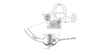 |
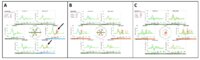
Atypical PC BPPV Variant Figures | Figure 1: Atypical posterior canal BPPV variants The labyrinth consists of the cochlea (C), two otolithic organs including utricle (U) and saccule (S), and three semicircular canals including anterior canal (AC), horizontal canal (HC), and posterior canal (PC). A. If otoconia are located within the ... | BPPV |
| 31 |
 |
Bilateral 6th Nerve Palsies Due to Idiopathic Intracranial Hypertension | This is a 25-year-old woman who presented with diplopia and blurry vision. On exam, she was found to have papilledema and bilateral 6th nerve palsies. Her opening pressure was >40 cm of water with a normal CSF analysis, and neuroimaging was unremarkable aside from subtle findings that have been asso... | Sixth Nerve Palsy; Abnormal Range |
| 32 |
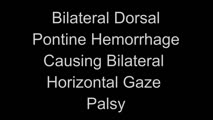 |
Bilateral Horizontal Gaze Palsy and Oculopalatal Tremor Due to Pontine Hemorrhage | This 70-yo-woman experienced headache and diplopia and was found to have a hemorrhage centrally within the dorsal pons. Months after the onset, the patient was seen in clinic and had no horizontal eye movements (pursuit, saccades, VOR) in either eye, suggestive of bilateral nuclear 6th nerve palsies... | VOR HIT Abnormal; Horizontal Gaze Palsy; OMS PONS; Pendular Nystagmus; Oculopalatal |
| 33 |
 |
Bilateral INOs Due to Stroke | This is a 65-year-old man with multiple vascular risk factors who experienced the abrupt onset of diplopia 6 months prior to this video. MRI done within 24 hours of onset was unremarkable. Examination demonstrated subtle bilateral adduction lag with horizontal saccades. There was very mild abducting... | Abnormal Saccades; Abnormal Range |
| 34 |
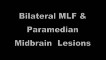 |
Bilateral INOs and Partial 3rd Nerve Palsies | This is a 45-year-old man with progressive ptosis and ophthalmoparesis. 10 years prior to presentation, he experienced diplopia and had a hyperintense lesion involving the medial longitudinal fasciculus (MLF) per report. Over time, he developed bilateral adduction paresis, ptosis and upgaze paresis ... | Abnormal Range; Third Subnuclear; INO; Mesencephalon |
| 35 |
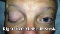 |
Bilateral Pseudo-abducens Palsies Due to Midbrain Stroke | 𝗢𝗿𝗶𝗴𝗶𝗻𝗮𝗹 𝗗𝗲𝘀𝗰𝗿𝗶𝗽𝘁𝗶𝗼𝗻: This is a man who suffered right>left midbrain strokes due to endocarditis complaining of ptosis and inability to move his eyes as well as hallucinations (peduncular hallucinosis). There was a presumed nuclear 3rd nerve p... | Bilateral Pseudo-abducens Palsies; Midbrain Stroke |
| 36 |
|
Bilateral Vestibular Loss With Gaze-Evoked Nystagmus and Saccadic Visually Enhanced VOR | This is 55-year-old man with the subacute onset of head movement-induced oscillopsia and dizziness. He had a history of psoriatic arthritis. He had not used medications known to be vestibulo-toxic such as gentamicin. ; Salient findings on his examination included 1) bilateral vestibular loss (BVL) d... | Jerk Nystagmus; Gaze-Evoked Nystagmus; Abnormal VOR-HIT; Eighth Nerve |
| 37 |
|
Bilateral riMLF Syndrome Causing Vertical Saccadic Palsy and Loss of Ipsitorsional Fast Phases | This is a 60-year-old man who developed fatigue and diabetes insipidus about 12 months prior to this video, and MRI demonstrated hypothalamic enhancement at that time. Nine months prior to this video, he gradually noticed that he was unable to look down. Work-up for ischemic, infectious, inflammator... | Midbrain OMS; Downgaze Palsy; Upgaze Palsy; Vertical Gaze Palsy; Abnormal Saccades; Normal Pursuit |
| 38 |
 |
Bilaterally Abnormal Head Impulse Test | 𝗢𝗿𝗶𝗴𝗶𝗻𝗮𝗹 𝗗𝗲𝘀𝗰𝗿𝗶𝗽𝘁𝗶𝗼𝗻: This video is an example of bilaterally abnormal head impulse test (HIT) due to bilateral vestibular loss (BVL). Typical symptoms in BVL: head movement-induced dizziness and jumping vision for years with visual jumping/b... | Abnormal VOR-HIT; Eighth Nerve |
| 39 |
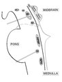 |
Brainstem Ocular Motor Machinery | Seen here is a sagittal view of the brainstem. The medulla has a significant role in gaze-holding, and the nucleus prepositus hypoglossi (NPH, along with the medial vestibular nucleus ) is the horizontal neural integrator. The abducens (6th) nucleus is located in the dorsal pons, and sends off the 6... | Medulla OMS; Pons OMS; Mesencephalon OMS; Dorsal Midbrain OMS |
| 40 |
 |
Bruns Nystagmus (During Video-Oculography) Due to Vestibular Schwannoma | A 25-year-old man with a history of right-sided hearing loss, headaches and imbalance was found to have a right vestibular schwannoma on MRI, and underwent a partial resection and radiotherapy. He denied symptoms of head movement dependent oscillopsia (i.e., suggestive of significant unilateral or b... | Jerk Nystagmus; Gaze-Evoked Nystagmus; Vestbular Nystagmus; Eighth Cranial Nerve; Vestibulocochlear Nerve; Cerebellar OMS |
| 41 |
 |
Bruns Nystagmus Due to a Cerebellopontine Angle Tumor | 𝗢𝗿𝗶𝗴𝗶𝗻𝗮𝗹 𝗗𝗲𝘀𝗰𝗿𝗶𝗽𝘁𝗶𝗼𝗻: This is a 15-yo-girl who experienced headache and imbalance leading to an MRI which showed a left sided cerebellopontine angle (CPA) tumor. Because of involvement of the left brainstem/cerebellum (e.g., dysfunction of the... | Jerk Nystagmus; Gaze Evoked Nystagmus; Vestibular Nystagmus |
| 42 |
 |
CANVAS (Cerebellar Ataxia, Neuropathy, and Vestibular Areflexia Syndrome) Video Head Impulse Test (vHIT) Figure | CANVAS (Cerebellar Ataxia, Neuropathy, and Vestibular Areflexia Syndrome) is a genetic condition consisting of slowly progressive late-onset ataxia, bilateral vestibulopathy, sensory neuropathy, chronic cough, and autonomic dysfunction. While the term vestibular areflexia typically implies bilateral... | Vestibulo-ocular Reflex and Head Impulse Testing - Abnormal; Video Head Impulse Test |
| 43 |
 |
Caloric Testing | 𝗢𝗿𝗶𝗴𝗶𝗻𝗮𝗹 𝗗𝗲𝘀𝗰𝗿𝗶𝗽𝘁𝗶𝗼𝗻: Caloric testing is a peripheral vestibular test which takes advantage of the fact that the labyrinth is sensitive to temperature changes. Warm stimulation causes excitation of the semicircular canals while cold stimulatio... | Normal VOR; Abnormal VOR; Vestibular Lab Testing; Calorics |
| 44 |
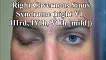 |
Cavernous Sinus Mass Causing Right 3rd and 4th Nerve Palsies | 𝗢𝗿𝗶𝗴𝗶𝗻𝗮𝗹 𝗗𝗲𝘀𝗰𝗿𝗶𝗽𝘁𝗶𝗼𝗻: 25-yo-man who complained of diplopia and was initially found to have right 4th and 6th nerve palsies in the setting of a right cavernous sinus mass (subsequently diagnosed as Ewing's sarcoma). When seen in follow-up (this... | Third (Oculomotor) Nerve Palsy; ThirdSsubnuclear; Fourth (Trochlear) Nerve Palsy |
| 45 |
 |
Central (Nuclear) 3rd Nerve Palsies | Shown here are two patients with left sided midbrain pathology (hemorrhage and ischemia) which caused damage to the 3rd nucleus. Both of the patients have ipsilateral mydriasis, adduction, supra- and infraduction paresis. Ipsilateral>contralateral ptosis is also present, and localizes to the central... | Range of Eye Movements/Motility Abnormal; Third Nuclear; Upgaze Palsy; Downgaze Palsy; Mesencephalon; Jerk Nystagmus; Rotary Nystagmus |
| 46 |
 |
Central 4th Nerve Palsy with Contralateral Horner's Syndrome | This is a 60-yo-woman who presented with a complaint of diplopia. Examination demonstrated a left hypertropia that worsened in right and down gaze as well as in left head tilt, and a left 4th nerve palsy was diagnosed. There was also evidence of a mild motility deficit in down/medial gaze OS, consis... | Horner's; Fourth Nerve Palsy; Mesencephalon |
| 47 |
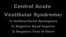 |
Central Acute Vestibular Syndrome Due to Posterior Fossa Hemorrhage | This is a patient presenting with the acute vestibular syndrome (AVS, e.g., acute prolonged vertigo, spontaneous nystagmus) whose HINTS (Head Impulse, Nystagmus, Test of Skew) testing indicated a central etiology based on negative (normal) head impulse testing (HIT). Nystagmus was unidirectional and... | VOR HIT; Alignment; Jerk Nystagmus; Vestibular Nystagmus; Acute Vestibular |
| 48 |
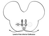 |
Central Anatomy of the Fourth Nerve | The IVth or trochlear nucleus is located ventral to the central periaqueductal grey matter, dorsal to the medial longitudinal fasciculus (MLF) and medial to the oculosympathetic tract at the level of the inferior colliculus. The fascicles of the IVth nerve travel dorsally and caudally around the cen... | Mesencephalon OMS |
| 49 |
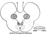 |
Central Anatomy of the Third Nerve | Seen here is an axial section of the midbrain at the level of the superior colliculus. The paired nuclei are located ventral to the periaqueductal grey, and the midline central caudal nucleus (CCN) is located between the right and left nuclei. The CCN sends projections to bilateral levator palpebrae... | Mesencephalon; Third Nerve Palsy |
| 50 |
 |
Central HINTS (With an Abnormal Head Impulse Sign) in the Acute Vestibular Syndrome Due to Lateral Pontine/Middle Cerebellar Peduncle Demyelination | 𝗢𝗿𝗶𝗴𝗶𝗻𝗮𝗹 𝗗𝗲𝘀𝗰𝗿𝗶𝗽𝘁𝗶𝗼𝗻: This is a 30-year-old man presenting with vertigo, diplopia and mild left facial weakness (not seen in the video). On exam, there was right-beating nystagmus (RBN) in primary gaze that increased in right gaze (in accordan... | Abnormal Alignment; Vestibulocochlear; OMS Pons; Rotary Nystagmus; Vestibular Nystagmus; Acute Vestibular Nystagmus; Jerk Nystagmus; VOR HIT Abnormal |