The Health Education Assets Library (HEAL) is a collection of over 22,000 freely available digital materials for health sciences education. The collection is now housed at the University of Utah J. Willard Marriott Digital Library.
TO
| Title | Description | Subject | Collection | ||
|---|---|---|---|---|---|
| 26 |
 |
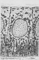
Enamel in longitudinal section of tooth - human, adult. Thin ground section. | From left to right: incremental lines (striae) of Retzius are distinctly shown as oblique broad zones (at the left) to the enamel surface; during formation of the crown successive apposition of layers of enamel is deposited and results in these so-called incremental grow lines; surface of enamel in ... | oral cavity; incremental lines; Retzius | Poja Histology Collection - Oral Cavity Subset |
| 27 |
 |
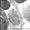
Early cap stage in tooth development - human, embryo; low magnification | Stain: Azan. From top to bottom: top left vestibular groove with gland formation; stratified ectoderm with ingrowth of the dental lamina (in the middle); bulbous growing end of dental lamina; bottom left alveolar bone formation (dark blue); and connective tissue/mesenchym stains light blue. | oral cavity; tooth development; dental lamina | Poja Histology Collection - Oral Cavity Subset |
| 28 |
 |
Enamel (odontogenic) organ in tooth development - bell stage, human, embryo | Stain: Azan. Outer surface of bell; from left to right: (avascular) Stellate reticulum; capillaries in this stage proliferate and invaginate between the outer dental epithelial cells; Part of fibrous tooth follicle. | oral cavity | Poja Histology Collection - Oral Cavity Subset |
| 29 |
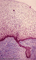 |
Early cap stage in tooth development - human, embryo | Stain: Azan. From top to bottom: stratified ectoderm with ingrowth of the dental lamina; knob-like end of the dental lamina; and collagen fibers of lamina propria are blue. | oral cavity; tooth development; dental lamina | Poja Histology Collection - Oral Cavity Subset |
| 30 |
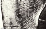 |
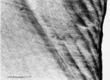
Enamel in longitudinal section of tooth - human, adult. Thin ground section. | At the left side surface of enamel in cuspal region; the parallel horizontal arrangement of stacks of rods (prisms) is evident. At the right side area of dentin (dark area). Incremental lines (striae) of Retzius run as curved lines (from bottom to middle top) and presented successive apposition of l... | oral cavity; incremental lines; Retzius; Hunter-Schreger bands | Poja Histology Collection - Oral Cavity Subset |
| 31 |
 |
Enamel in longitudinal section of tooth - human, adult. Thin ground section. | Enamel is compact and acellular, and consists of vertical stacks of rods (prisms) as well as interrod (interprismatic) regions with less calcifying substance parallel to each other. Each prism is surrounded by an enamel sheath (a non-mineralized organic substance). From left to right: surface of ... | oral cavity; enamel rods; enamel prisms | Poja Histology Collection - Oral Cavity Subset |
| 32 |
 |
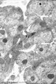
Enamel rods (prisms) in tooth development - gerbil, postnatal | Electronmicroscopy. Cross-section of rods (prisms) demonstrate round aggregates with hydroxyapatite crystals. Between the round rods the interprismatic substance with crystals orientated in a different course. Note that twisting of the crystallites can be seen in the longitudinal bundles where grey ... | oral cavity; enamel prisms; hydroxyapatite crystals | Poja Histology Collection - Oral Cavity Subset |
| 33 |
 |
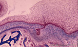
Epithelial tooth bud in tooth development - tooth germ, human, embryo | Stain: hematoxylin. At the top stratified ectoderm. The proliferating basal cell layers are palisade-arranged with a light cytoplasm close to the basement membrane (right side). Below the basement membrane subepithelially an accumulation of inductive neural crest-derived mesenchymal cells is locally... | oral cavity; tooth bud; tooth development | Poja Histology Collection - Oral Cavity Subset |
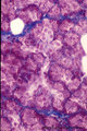
| 34 |
 |
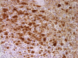
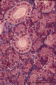
Eosinophilic granuloma mastoid | High power view (40X) of a neoplastic proliferation of Langerhan's cells (Histiocytosis) in mastoid bone stained with CD1a an immunohistochemical stain for Langerhans cells | HEAL Reviewed Collection | |
| 35 |
 |
Eosinophilic granuloma mastoid | High power view (40X) of a neoplastic proliferation of Langerhan's cells (Histiocytosis) in mastoid bone stained for S100 an immunohistochemical stain dendritic cells | HEAL Reviewed Collection | |
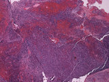
| 36 |
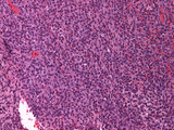 |
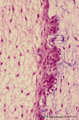
Eosinophilic granuloma mastoid | Medium power view (20X) of a neoplastic proliferation of Langerhan's cells (Histiocytosis) in mastoid bone | HEAL Reviewed Collection | |
| 37 |
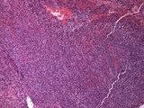 |
Eosinophilic granuloma mastoid | Low power view (10X) of a neoplastic proliferation of Langerhan's cells (Histiocytosis) in mastoid bone | HEAL Reviewed Collection | |
| 38 |
 |
Eosinophilic granuloma mastoid | Low power view (5X) of a neoplastic proliferation of Langerhan's cells (Histiocytosis) in mastoid bone | HEAL Reviewed Collection | |
| 39 |
 |
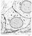
Exocrine gland (salivary gland) | Scheme electronmicroscopy. Part of an acinus of mucous cells with different amount of mucous secretion. Supranuclearly the Golgi areas with maturing mucous secretion granules, basolateral the endoplasmic reticulum. At the top the lumen. The left cell shows an accumulation of the secretory droplets, ... | oral cavity; mucous gland | Poja Histology Collection - Oral Cavity Subset |
| 40 |
 |
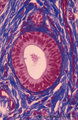
Exocrine gland (salivary gland) | Scheme electronmicroscopy. Part of an acinus of serous cells, basolaterally a well developed rough endoplasmic reticulum and apically secretion granules with different maturity towards the small lumen. On top stages of early formed secretion granule (left) and more matured ones (right). Note small j... | oral cavity; serous gland | Poja Histology Collection - Oral Cavity Subset |
| 41 |
 |
Exocrine gland - intercalated duct - salivary glands, pancreas | Scheme electronmicroscopy. Part of an intercalated duct, the low cuboidal epithelial cells contain sparsely organelles and some desmosomal structures. Between the lining cells and the basal lamina is squeezed part of a filament-rich myoepithelial cell. Lumen side top left quadrant. | oral cavity; intercalated duct | Poja Histology Collection - Oral Cavity Subset |
| 42 |
 |
Exocrine gland - intercalated duct - submandibular gland, human | Stain: Azan. Branching intercalated duct draining several serous acini, the lining duct cells are lighter stained due to the content of organelles. Serous cells are darker stained (many organelles) with round nuclei. Note few fat cells, the septa are blue stained. | oral cavity; intercalated duct; serous gland | Poja Histology Collection - Oral Cavity Subset |
| 43 |
 |
Exocrine gland - striated (intralobular) duct - salivary glands, pancreas | Scheme electronmicroscopy. Part of a striated duct with basally membrane infoldings and mitochondria accumulations in the tall columnar cells. Small junctional complexes as well as apically mitochondria and numerous vesicles are present. | oral cavity; striated duct | Poja Histology Collection - Oral Cavity Subset |
| 44 |
 |
Exocrine gland - parotid gland, rat | Electronmicroscopy. Part of a striated duct with basally membrane infoldings and numerous mitochondria perpendicularly orientated to the basal membrane. Upper side is luminal side. Apically junctional complexes as well as mitochondria and many vesicles are present. | oral cavity; serous gland | Poja Histology Collection - Oral Cavity Subset |
| 45 |
 |
Exocrine gland - striated (intralobular) ducts - parotid gland, human | Stain: Azan. Cross-sections of striated ducts in upper part between serous acini. Note at the right bottom part of (lighter stained) lining cells of an intercalated duct. Connective tissues between the structures are blue stained. | oral cavity; striated duct | Poja Histology Collection - Oral Cavity Subset |
| 46 |
 |
Exocrine gland - interlobular duct - submandibular gland, human | Stain: Azan. An interlobular duct with partly columnar as well as pseudostratified columnar epithelium. Note the dense connective tissue of the interlobular septum with small blood vessels. | oral cavity; seromucous glands | Poja Histology Collection - Oral Cavity Subset |
| 47 |
 |
Exocrine gland - intercalated duct - submandibular gland, rat | Electronmicroscopy. The dark cells form an intercalated duct from the left lower corner to the right upper corner, and end in the acinus with lightly stained cells (serous). | oral cavity; intercalated duct; serous gland | Poja Histology Collection - Oral Cavity Subset |
| 48 |
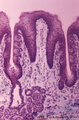 |
Foliate papillae of the tongue (dorsal side, rabbit) | Stain: Goldner trichrome. Foliate papil with primary and secondary connective tissue projections. Taste buds are localized in the lining, non-keratinized epithelium of the grooves. The serous gland of von Ebner (minor salivary gland) drains via an enlarged duct into the left groove of the papil. | oral cavity; foliate papillae; von Ebner | Poja Histology Collection - Oral Cavity Subset |
| 49 |
 |
Free denticulus (pulp stone) in the tooth - longitudinal section of root pulp; human, adult | Stain: Hematoxylin and eosin. In the center of pulp connective tissue a free pulp stone (false denticulus). Dispersed through the pulp several small stones. Left of the central stone a longitudinal sectioned thin-walled blood vessel; right of the stone bundles of nerve fibers. | oral cavity; denticulus; pulp stone | Poja Histology Collection - Oral Cavity Subset |
| 50 |
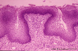 |
Fungiform papillae of the tongue (dorsal side, human) | Stain: Hematoxylin and eosin. A broad prominent papil of connective tissue (without secondary papils) covered by non-keratinized stratified epithelium. This specimen does not show any taste bud. Well vascularized lamina propria. | oral cavity; fungiform papillae | Poja Histology Collection - Oral Cavity Subset |