Collection of materials relating to neuro-ophthalmology as part of the Neuro-Ophthalmology Virtual Education Library.
NOVEL: https://novel.utah.edu/
TO
- NOVEL224
| Title | Creator | Description | Subject | ||
|---|---|---|---|---|---|
| 26 |
 |
Patient Portal: Homonymous Hemianopsia | James C. O'Brien, MD | Homonymous hemianopia refers to an absence of vision towards one side of the visual world in each eye. The damage that caused this problem is in the brain and not in the eyes. | Homonymous hemianopia; Visual pathway |
| 27 |
 |
Patient Portal: Optic Neuritis | Anthony Brune, DO | Optic neuritis is inflammation of the optic nerve. In optic neuritis, the covering around the fibers of the optic nerve (myelin) is damaged by inflammation (demyelination), which typically results in blurred or dark vision. | Optic neuritis; Myelin; Demyelination |
| 28 |
 |
Patient Portal: Transient Vision Loss | Anthony Brune, DO | Transient visual loss is the term used to describe loss of part or all of the vision in one or both eyes temporarily. Some people do not experience a complete loss of the affected vision and instead describe the abnormality as "blurring" or like "looking through a veil." The vision typically returns... | Transient visual loss |
| 29 |
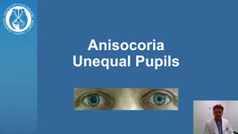 |
Patient Portal: Anisocoria | Nagham Al-Zubidi, MD | Anisocoria is a medical term for unequal pupil size. Normally our pupils are relatively the same size. While small differences in pupil size are normal and can even come and go (physiologic anisocoria), constant and significant differences in pupil sizes may be a sign of damage to the brain or the n... | Anisocoria; Horner Syndrome; 3rd Cranial Nerve Palsy; Adie Tonic Pupil |
| 30 |
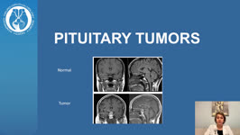 |
Patient Portal: Pituitary Adenoma | Nagham Al-Zubidi, MD | The pituitary gland is a pea-sized gland that sits underneath the base of the brain. It produces and releases many hormones. These hormones control your metabolism, stress level, growth, ovulation and menstruation in women, sperm and testosterone production in men, milk production, and urine product... | Pituitary Adenoma; Pituitary Tumor |
| 31 |
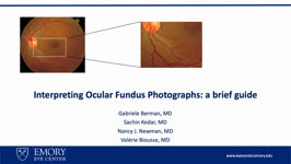 |
Interpreting Ocular Fundus Photographs: A Brief Guide | Gabriele Berman; Sachin Kedar; Nancy J. Newman; Valerie Biousse | This is a brief guide to the interpretation of the ocular fundus photograph. In this presentation we will describe the structures that comprise the normal ocular fundus followed the abnormalities that can be detected on fundus photographs. By the end of the presentation, learners should be able to d... | Fundus Photograph; Glaucoma; Papilledema; Retinal Detachment |
| 32 |
 |
Raymond Cestan Syndrome | Srujay Pandiri; Sean Gratton | This is a brief narrated powerpoint that explains the clinical presentation of Raymond Cestan Syndrome. This is a rare brainstem stroke syndrome that presents with ipsilateral internuclear ophthalmoplegia and contralateral hemiparesis as well as other features. It is sometimes referred to as upper d... | Brainstem Stroke Syndromes; Internuclear Ophthalmoplegia; Pons |
| 33 |
 |
Vergence Eye Movements | Yu Hsin Chen; Amanda Dean Henderson, MD | Vergence (e.g. convergence and divergence), a class of eye movements that rotates the eyes in opposite directions (disjunctive), serves to hold image on the fovea of both eyes in order to obtain a single, clear image. This presentation overviews the neurology and examinations of vergence response, w... | Convergence; Convergence Insufficiency; Convergence Spasm; Divergence; Divergence Insufficiency |
| 34 |
 |
Retinal Venous Occlusive Disease | Ali Alkhabbaz; James Brian Davis; Amanda Dean Henderson | Retinal venous occlusion (RVO) includes central retinal vein occlusion (CRVO), hemi-central retinal vein occlusion (HCRVO), and branch retinal vein occlusion (BRVO). The most important risk factor for RVO is hypertension, but other risk systemic factors include advanced age and cardiovascular diseas... | Branch Retinal Vein; BRVO; Central Retinal Vein; CRVO; HCRVO; Retinal Venous Occlusion |
| 35 |
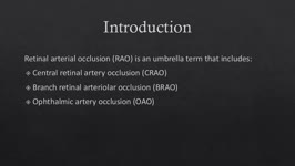 |
Retinal Artery Occlusive Disease | Ali Alkhabbaz, MD; James Brian Davis; Amanda Dean Henderson, MD | Retinal arterial occlusion includes ophthalmic artery occlusion (OAO), central retinal artery occlusion (CRAO), branch retinal arteriolar occlusion (BRAO), from proximal to distal. These can occur with or without retinal ischemia and may be permanent or transient. There are 4 subtypes of CRAO: non-a... | Branch Retinal Arteriolar Occlusion; Central Retinal Artery Occlusion; Giant Cell Arteritis; Ophthalmic Artery Occlusion |
| 36 |
 |
Methanol Toxicity | Daniel Lovasz; James Brian Davis; Amanda Dean Henderson, MD | Methanol toxicity can be very dangerous with high morbidity and mortality rates, and outcomes typically worsen with increasing acidemia, hyperglycemia, or acute kidney injury. Delayed treatment can result in irreversible injury, such as vision loss, or even death. The harmful effects of methanol com... | Acidosis; Fomepizole; Formic Acid; Methanol Toxicity; Toxic Optic Neuropathy |
| 37 |
 |
Therapeutics for Tuberous Sclerosis | Daniel Lovasz; Amanda Henderson, MD | Tuberous Sclerosis Complex (TSC) is a disease characterized by abnormal hamartomas and benign neoplasms in multiple organs. The diagnosis is made by the presence of 2 major features, or 1 major feature accompanied by 2 minor features. These features include various growths in different parts of the ... | Tuberous Sclerosis; Therapeutics; Everolimus; Sirolimus; Hamartomas; mTOR |
| 38 |
 |
Natural Language Processing in Neuro-Ophthalmology | Areeba Abid, BS; Sachin Kedar, MD | This video provides an overview of natural language processing (NLP) techniques, applications, and limitations in neuro-ophthalmology. NLP, a branch of artificial intelligence, enables computers to understand and analyze human language. This video discusses three types of NLP techniques: sentiment a... | Artificial Intelligence; Machine Learning; Natural Language Processing; Word Prediction; Word Embeddings; Sentiment Analysis |
| 39 |
 |
Thiamine (Vitamin B1) Deficiency (Video) | Nirupama Devanathan; Devin D. Mackay, MD | 33-year-old female who initially presented with heat stroke that was thought to provoke cyclical vomiting over the course of 6 weeks, perpetuated by anxiety, resulting in a 15- pound weight loss. She presented to the ED with bilateral vision loss, bilateral lower extremity weakness and vertical nyst... | Thiamine; Upbeat Nystagmus; Nutritional Optic Neuropathy; Wernicke Encephalopathy |
| 40 |
 |
Vascular Headache Disorders | Salman Ali Mahmood, MD; Sean Gratton, MD | This is a PowerPoint with embedded audio presenting a detailed summary of the evaluation and management of an important cause of secondary headaches: vascular headache disorders. Topics covered include intracranial hemorrhage, arterial dissection, giant cell arteritis, cerebral venous sinus thrombos... | Vascular Headache Disorders; Epidural Hematoma; Subdural Hematoma; Subarachnoid Hemorrhage; Intracranial Hemorrhage; Arterial Dissection; Giant Cell Arteritis; Cerebral Venous Sinus Thrombosis; Pituitary Apoplexy |
| 41 |
 |
Treatment Outcomes of Ocular Manifestations in Wernicke's Encephalopathy: Video | Whitney Stuard Sambhariya, PhD, Medical Student; Melanie Truong-Le, DO, OD | The case of a 28- year-old woman with a past medical history of gastric sleeve who was reported to have blurry vision and presented to neuro-ophthalmology with double vision. On examination the patient had bilateral abducens palsy, alternating upbeat and downbeat nystagmus with a torsional component... | Wernicke's Encephalopathy; Ocular Manifestations; Neuro-ophthalmology; Sbducens Palsy; Nystagmus; Double Vision; Blurry Vision |
| 42 |
 |
A Brief Introduction to Post-Ocular Surgery Strabismus | Srujay Pandiri, Medical Student | This is a short narrated Powerpoint that introduces concepts important to post-ocular surgery strabismus. It highlights the connection between common surgeries include cataract surgery, scleral buckle surgery, refractive surgery, among others. | Diplopia; Strabismus; Post-ocular Surgery Strabismus |
| 43 |
 |
My DEI Journey: Lessons Learned | Ore-ofe Adesina, MD | In this lecture, Dr. Adesina reviews his DEI journey in ophthalmology and the lessons of diversity, equity and inclusion he has learned along the way. He discusses why diversity, equity, and inclusion are all needed for a state of inclusive well-being, why representation matters for patient care and... | Ophthalmology; Diversity, Equity, Inclusion; Representation; Social Determinants of Health |
| 44 |
 |
Neuro-Ophthalmology of Multiple Sclerosis | Emily Sun, Medical Student; Amanda Henderson, MD | Multiple Sclerosis (MS) is the most common neurological disease in young people with an average age of onset between 15 and 35 years old. MS is an autoimmune inflammatory condition that causes demyelinating lesions in the CNS. The diagnosis is clinical, but MRI is typically used to support the diagn... | Multiple Sclerosis; Optic Neuritis; Uveitis; Internuclear Ophthalmoplegia; Nystagmus; Steroids; Demyelinating; Autoimmune |
| 45 |
 |
Diagnosis and Evaluation of Stroke: Mechanism - Vasculitis | Danny Alevy; Amanda D. Henderson, MD | In this video, we discuss the diagnosis and evaluation of several vasculitides linked to an increase in risk for cerebral ischemic stroke. We focus on ANCA-associated vasculitides, polyarteritis nodosa, Takayasu arteritis, lupus vasculitis, and Susac syndrome, while highlighting their key symptoms a... | Ischemic Stroke; Vasculitis; ANCA-associated Vasculitis; Polyarteritis Nodosa; Takayasu Arteritis; Systemic Lupus Erythematosus; Susac Syndrome |
| 46 |
 |
Postconcussion Syndrome and Postconcussion Headache | Jessica Darusz, MD; Sean Gratton, MD | This brief presentation describes the pathophysiology, evaluation, and management of concussion, with an emphasis on postconcussion headache. | Concussion; Postconcussion Syndrome; Postconcussion Headache |
| 47 |
 |
Sports Related Head Injuries | Jessica Darusz, MD; Sean Gratton, MD | This narrated PowerPoint reviews the basics of sports related head injuries. It emphasizes assessment tools and treatment decisions in assessing athletes with concussion and other head injuries. | Concussion; Sports-related Head Injuries; Post-concussion Syndrome |
| 48 |
 |
Curtain Sign (Enhanced Ptosis) | Bashaer Aldhahwani, MD; Hong Jiang, MD, PhD | This is a 78-year-old male patient who presented with diplopia, right eyelid ptosis, and ophthalmoplegia. He had severe ptosis OD and pseudo-proptosis (lid retraction) OS at baseline, but when the right eyelid was manually elevated, there was marked enhanced ptosis of the left eyelid (Video). He was... | Myasthenia GravIs; Clinical Signs |
| 49 |
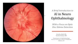 |
A Brief Introduction to AI in Neuro-ophthalmology | Areeba Abid, BS; Sachin Kedar, MD | In this video, we describe the basics of artificial intelligence and machine learning as applicable to clinical neuro-ophthalmologists. We use the example from a recent publication, where AI software was used to detect optic disc edema in fundus images. | Artificial Intelligence; Machine Learning |
| 50 |
 |
Introduction to NANOS NOTE | Karl C. Golnik, MD | Introduction to NANOS NOTE, a resource for non-neuro-ophthalmologists describing common examination techniques. | Neuro-Ophthalmology Examination Techniques |