AAO-NANOS Neuro-Ophthalmology Clinical Collection: Derived from the AAO-NANOS Clinical Neuro-Ophthalmology collection produced on CD. The images are of selected cases from the NANOS teaching slide exchange, and the CD was produced under the direction of Larry Frohman, MD and Andrew Lee, MD.
The American Academy of Ophthalmology (AAO); The North American Neuro-Ophthalmology Association (NANOS).
NOVEL: https://novel.utah.edu/
TO
| Title | Creator | Description | ||
|---|---|---|---|---|
| 326 |
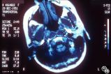 |
Neuro-Ophthalmic Imaging-MRI | Mitchell J. Wolin, MD | Axial view of Arnold-Chiari malformation on a patient with downbeat nystagmus. Note the presence of the cerebellar tonsils posterior to the caudal medulla. In addition to downbeat nystagmus, Arnold-Chiari malformations can sometimes lead to increased intracranial pressure and papilledema. |
| 327 |
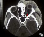 |
Neuro-Ophthalmic Imaging-CT Scan | Larry P. Frohman, MD | This patient was assaulted with a blunt object and suffered acute blindness due to traumatic optic neuropathy. Note how the lateral orbital wall has been fractured and displaced posteromedially into the region of the anterior optic canal. |
| 328 |
 |
Optic Tract Syndrome Due to Carotid Artery Dolichoectasia | Larry P. Frohman, MD | This 43-year-old man was referred for evaluation of 6 months of visual loss OU. In retrospect, he had noticed increasing difficulty with his tennis game dating back over 3 years, as balls would pass him unexpectedly when hit to his backhand (left) side. The patient did not think this was progressive... |
| 329 |
 |
Optic Tract Syndrome Due to Carotid Artery Dolichoectasia | Larry P. Frohman, MD | This 43-year-old man was referred for evaluation of 6 months of visual loss OU. In retrospect, he had noticed increasing difficulty with his tennis game dating back over 3 years, as balls would pass him unexpectedly when hit to his backhand (left) side. The patient did not think this was progressive... |
| 330 |
 |
Optic Tract Syndrome Due to Carotid Artery Dolichoectasia | Larry P. Frohman, MD | This 43-year-old man was referred for evaluation of 6 months of visual loss OU. In retrospect, he had noticed increasing difficulty with his tennis game dating back over 3 years, as balls would pass him unexpectedly when hit to his backhand (left) side. The patient did not think this was progressive... |
| 331 |
 |
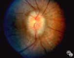
Systemic Disorders With Optic Nerve and Retinal Findings | Larry P. Frohman, MD | This 8-year-old girl had a history of acute lymphoblastic leukemia with neurologic involvement, with remission induced with radiation and chemotherapy. Her relapse showed as an isolated optic neuropathy with this fundus appearance. The other eye was normal. The MRI and spinal tap at this point were ... |
| 332 |
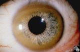 |
Ocular Manifestations of Systemic Disorders | Robert F. Saul, MD | Wilson's disease (hepatolenticular degeneration) is a progressive autosomal recessive multisystem disease that may result in cirrhosis of the liver, renal dysfunction, and motor neurologic disease. A Kayser-Fleischer ring may occur as a green-brown band at the level of Descemet's membrane in the cor... |
| 333 |
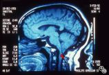 |
Neuro-Ophthalmic Imaging-MRI | Mitchell J. Wolin, MD | Sagittal view of Arnold-Chiari malformation on a patient with downbeat nystagmus. The compression of the cervicomedullary junction is clearly depicted in the sagittal view. |
| 334 |
 |
Systemic Disorders With Optic Nerve and Retinal Findings | Larry P. Frohman, MD | A 29-year-old African American woman presented with headaches, bilateral transient visual obscurations, blurred vision, numbness, and weakness of the lower extremities with myalgia and joint pains. She had an unplanned 12-pound weight loss over 2 months. A neurologist and internist diagnosed her wit... |
| 335 |
 |
Neuro-Ophthalmic Case With Notable Field Changes | Marilyn C. Kay, MD | This 28-year-old woman presented with a 4-week history of bilateral visual loss. She had a known history of multiple sclerosis. Her vision was 20/60 OD and 20/40 OS, with an RAPD OS and optic pallor OU. Her fields and MRI are shown. Optic tract lesions usually result in an incongruous homonymous hem... |
| 336 |
 |
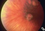
Ocular Disease With Retinal Findings | William Fletcher Hoyt, MD | Choroidal folds may result from choroidal tumors, compression on the eye wall from thyroid ophthalmopathy, orbital pseudotumor, orbital tumor, posterior scleritis, hypotony, scleral laceration, retinal detachment, marked hyperopia, or secondary to papilledema. Intraocular pressure measurements, refr... |
| 337 |
 |
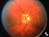
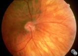
Isolated Congenital Optic Disc Anomalies | William Fletcher Hoyt, MD | This patient demonstrates bilateral tilted optic discs. Patients with this congenital optic disc anomaly may be asymptomatic or have bitemporal visual field defects that do not respect the vertical midline. Disease/Diagnosis: Tilted Discs. |
| 338 |
 |
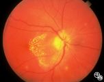
Isolated Congenital Optic Disc Anomalies | Anthony C. Arnold, MD | This image shows drusen that are especially prominent superotemporally. Pair with 92_64 and 92_67. |
| 339 |
 |
Systemic Disorders With Optic Nerve and Retinal Findings | Larry P. Frohman, MD | A 42-year old woman presented with a history of severe brow pain and 4 days of progressive visual loss OD. There was no increased pain on ocular rotation. Aside from heavy menses, she denied any significant past medical history. Her examination revealed acuity NLP OD, 20/25 OS; color vision 9/10 OS;... |
| 340 |
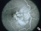 |
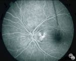
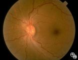
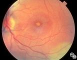
Pseudopapilledema With Subretinal Neovascularization on Fluorescein Angiogram | Joel M. Weinstein, MD | This 8-year-old boy presented with a 2-week history of decreased vision in the right eye. He had undergone a normal MRI and CSF examination, including intracranial pressure, before neuro-ophthalmologic assessment. The fundus photographs and fluorescein angiograms show subretinal neovascularization a... |
| 341 |
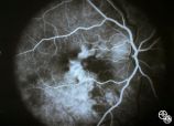 |
Pseudopapilledema With Retinal Neovascularization, Fluorescein Angiogram | Joel M. Weinstein, MD | This 8-year-old boy presented with a 2-week history of decreased vision in the right eye. He had undergone a normal MRI and CSF examination, including intracranial pressure, before neuro-ophthalmologic assessment. The fundus photographs and fluorescein angiograms show subretinal neovascularization a... |
| 342 |
 |
Isolated Optic Neuritis/Neuropathy | Anthony C. Arnold, MD | This 42-year-old male with pseudotumor cerebri and chronic papilledema demonstrated refractile bodies, which can be seen with chronic optic disc edema. This image shows the chronic papilledema at presentation, with associated refractile hyaline bodies at the disc periphery in both eyes. Pair with 96... |
| 343 |
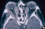 |
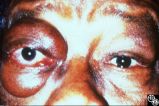
Ocular Manifestations of Systemic Disorders | Rosa A. Tang, MD | Thyroid eye disease may result in proptosis and restrictive external ophthalmoplegia. The extracoular muscles are often diffusely enlarged with sparing of the tendons. |
| 344 |
 |
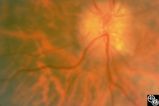
Isolated Congenital Optic Disc Anomalies | Joel M. Weinstein, MD | This 8-year-old boy presented with a 2-week history of decreased vision in the right eye. He had undergone a normal MRI and CSF examination, including intracranial pressure, before neuro-ophthalmologic assessment. The fundus photographs and fluorescein angiograms show subretinal neovascularization a... |
| 345 |
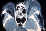 |
Ocular Manifestations of Systemic Disorders | Mitchell J. Wolin, MD | Thyroid eye disease is the most common cause of unilateral or bilateral proptosis in the adult patient. Other signs of thyroid eye disease should be sought, including lid retraction, inferior scleral show, and lid lag. Patients with markedly asymmetric or strictly unilateral proptosis should probabl... |
| 346 |
 |
Ocular Manifestations of Systemic Disorders | Mitchell J. Wolin, MD | Thyroid eye disease is the most common cause of unilateral or bilateral proptosis in the adult patient. Other signs of thyroid eye disease should be sought, including lid retraction, inferior scleral show, and lid lag. Patients with markedly asymmetric or strictly unilateral proptosis should probabl... |
| 347 |
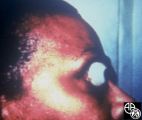 |
Ocular Manifestations of Systemic Disorders | Rosa A. Tang, MD | Thyroid eye disease can result in significant upper eyelid retraction and axial proptosis resulting in exposure keratopathy. |
| 348 |
 |
Isolated Congenital Optic Disc Anomalies | William Fletcher Hoyt, MD | This patient demonstrates bilateral tilted optic discs. Patients with this congenital optic disc anomaly may be asymptomatic or have bitemporal visual field defects that do not respect the vertical midline. Disease/Diagnosis: Tilted Discs. |
| 349 |
 |
Optic Neuropathies | Michael Wall, MD | Optic disc edema with a macular star figure may occur in infectious diseases (eg, cat-scratch disease, syphilis, tuberculosis, Lyme disease), inflammatory diseases (eg, sarcoid), ischemic diseases (anterior ischemic optic neuropathy), and in papilledema. Infectious causes should be sought in patient... |
| 350 |
 |
Optic Neuropathies | Ralph A. Sawyer, MD | Optic disc edema with a macular star figure has been referred to as neuroretinitis. |