The Health Education Assets Library (HEAL) is a collection of over 22,000 freely available digital materials for health sciences education. The collection is now housed at the University of Utah J. Willard Marriott Digital Library.
TO
Filters: Collection: ehsl_heal
| Title | Description | Subject | Collection | ||
|---|---|---|---|---|---|
| 301 |
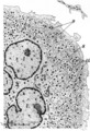 |
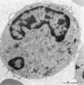
Megakaryocyte | Scheme electron microscopy. The megakaryocyte is derived from a pro-megakaryocyte which originates from splenic stem cells (CFU-S, colony forming units-spleen). The megakaryocyte is a giant polyploid cell (35-160 m) and contains a large multilobate nucleus (1). The perinuclear area shows Golgi areas... | Poja Histology Collection - Blood & Bone Marrow Subset | |
| 302 |
 |
Megakaryocyte (bone marrow, mouse) | Electron microscopy. A detail of the cytoplasm of a megakaryocyte (see inset) demonstrates distinctly a part of the intermediate zone subdivided by an interconnected tubular system (so-called demarcation membrane system or open canalicular system: OCS) that is in continuity with the cell surface. Th... | Poja Histology Collection - Blood & Bone Marrow Subset | |
| 303 |
 |
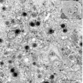
Megakaryocyte (bone marrow, rabbit) | Electron microscopy. A survey of a megakaryocyte demonstrates very well the differences in diameter between this giant polyploid cell (1) and the other young white blood cells (2-5). In the intermediate zone the (dark) granules (of the future platelets) are close associated with the electron-light i... | Poja Histology Collection - Blood & Bone Marrow Subset | |
| 304 |
 |
Megakaryocyte (peripheral blood, human) | Electron microscopy. A small part of a megakaryocyte (see also inset) shows two nuclear segments (1) and two areas of cytoplasm. Seen at this magnification and close to the perinuclear area, the granular population can be divided into homogeneous electron-dense (2) and electron-grey (3) granules. Th... | Poja Histology Collection - Blood & Bone Marrow Subset | |
| 305 |
 |
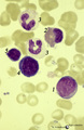
Megakaryocyte in bone marrow smear (human) | Stain: May-Grnwald-Giemsa (MGG), (black and white print). The giant polyploid cell (35-160 μm) contains a large multilobate nucleus. Note the ring-like pattern of the nuclei (1). The light-stained perinuclear zone (2) contains Golgi areas, within the encompassing greyish (intermediate) zone (3) gra... | Poja Histology Collection - Blood & Bone Marrow Subset | |
| 306 |
 |
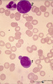
Mitosis in bone marrow smear (human) | Stain: May-Grnwald-Giemsa (MGG). Increased mitotic activity can be observed in megaloblastic anemia. (1) Mitosis figure. (2) Basophilic erythroblasts. | Poja Histology Collection - Blood & Bone Marrow Subset | |
| 307 |
 |
Monoblast | Scheme electron microscopy. The precursor of this cell is the promonoblast derived from the common stem cell CFU-GM (colony forming unit-granulocyte/monocyte). After proliferation of the promonoblasts they transform into monoblasts (up to 10 μm). The nucleus of the monoblast shows an indentation as... | Poja Histology Collection - Blood & Bone Marrow Subset | |
| 308 |
 |
Monoblast in bone marrow smear (human) | Stain: May-Grnwald-Giemsa (MGG). The monoblast (1) has a large nucleus with an irregular border, nucleoli and fine disperse chromatin, and a basophilic cytoplasm with no or a limited number of granules. (2) Two neutrophilic band forms. | Poja Histology Collection - Blood & Bone Marrow Subset | |
| 309 |
 |
Monoblast, erythroblasts and granulopoietic cells in bone marrow smear (human) | Stain: May-Grnwald-Giemsa (MGG). The monoblast (1) has a large nucleus with an irregular border, nucleoli and fine disperse chromatin, and a basophilic cytoplasm (greyish blue) with no or hardly any granules. (2) Eosinophilic metamyelocyte with large, brown granules. (3) Juvenile unsegmented (band f... | Poja Histology Collection - Blood & Bone Marrow Subset | |
| 310 |
 |
Monoblast, myeloid and erythropoietic cells in bone marrow smear (human) | Stain: May-Grnwald-Giemsa (MGG). (1) late promyelocyte or early myelocyte with nucleoli in the nucleus and ample azurophilic granules. (2) monoblast with indented nucleus and nucleoli (3) polychromatic erythroblast that will turn into an orthochromatic erythroblast (4), with progressing chromatin co... | Poja Histology Collection - Blood & Bone Marrow Subset | |
| 311 |
 |
Monocyte | Scheme electron microscopy. The diameter of monocytes ranges from 12-20 m. Characteristic is the large (kidney-shaped) nucleus with one or more nucleoli (1) and several indentations. The cytoplasm contains: (2) Golgi areas, (3) electron-dense lysosomal granules or primary or azurophilic granules wit... | Poja Histology Collection - Blood & Bone Marrow Subset | |
| 312 |
 |
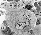
Monocyte (peripheral blood, human) | Electron microscopy. In this picture the large nucleus (1) of this cell (diameter of 12-20 μm) is twice sectioned. Golgi areas (2) and centriole, profiles of rough endoplasmic reticulum (3) and many free ribosomes are present. There are many mitochondria (4) as well as scattered homogeneous electro... | Poja Histology Collection - Blood & Bone Marrow Subset | |
| 313 |
 |
Monocyte (peripheral blood, human) | Electron microscopy. In this picture the large nucleus of this cell (diameter of 12-20 μm) is twice sectioned (1). Golgi areas (2), a few profiles of rough endoplasmic reticulum (3) and many free ribosomes are present. There are many mitochondria (4) as well as small-sized light vesicles (5). The c... | Poja Histology Collection - Blood & Bone Marrow Subset | |
| 314 |
 |
Monocyte (peripheral blood, human) | Electron microscopy. A large cell with an indented nucleus (1), few Golgi areas (2) and numerous organelles. The cytoplasm contains scattered homogeneous electron-dense lysosomal granules (3) (azurophilic granules with acid phosphatase, arylsulfatase) in variable amounts and few vacuoles. Small pseu... | Poja Histology Collection - Blood & Bone Marrow Subset | |
| 315 |
 |
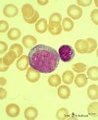
Monocyte in peripheral blood smear (human) | Stain: May-Grnwald-Giemsa (MGG). The monocyte (12-20 μm) contains a large light-stained nucleus with a characteristic indentation with a distinct nucleolus in a light blue-stained apparently transparent cytoplasm. Small primary or azurophilic granules are hardly visible. | Poja Histology Collection - Blood & Bone Marrow Subset | |
| 316 |
 |
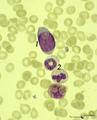
Monocyte in peripheral blood smear (human) | Stain: May-Grnwald-Giemsa (MGG). The nucleus of the mature monocyte has a kidney bean- or horseshoe-shape, is rather transparent compared to lymphocytes and granulocytes, and the cell is much larger (12-20 μm). The fine granular cytoplasm contains lysosomes, phagocytic vacuoles. Note platelets at (... | Poja Histology Collection - Blood & Bone Marrow Subset | |
| 317 |
 |
Mott cell in peripheral blood smear (human) | Stain: May-Grnwald-Giemsa (MGG). Plasma cells that contain abundant globular inclusions or vacuoles (Russell bodies) composed of immunoglobulin are called Mott cells (1) morular cells or grape cells. Russell bodies can stain blue-violet or pink but may also be dissolved during fixation and staining.... | Poja Histology Collection - Blood & Bone Marrow Subset | |
| 318 |
 |
Multilamellar bodies of type II alveolar cell in the lung (mouse) | Electron microscopy. The cytoplasm of the type II alveolar cell (pneumocyte II) contains characteristic electron-dense multilamellar bodies (*) in different maturing stages. The lamellar bodies are responsible for the vacuolated appearance of these cells, and they give rise to surfactant (phospholip... | Pneumocyte II; Multilamellar bodies; Type II alveolar cell | Poja Histology Collection - Respiratory System Subset |
| 319 |
 |
Multiple myeloma, plasma cell leukemia in bone marrow smear (human) | Stain: May-Grnwald-Giemsa (MGG). Mutiple myeloma (Kahler's disease) is characterized by proliferation of abnormal plasma cells (1, myeloma cells) in the bone marrow. In the great majority of patients secretion of a single homogenous immunoglobulin product (monoclonal component) occurs. Note the ecce... | Poja Histology Collection - Blood & Bone Marrow Subset | |
| 320 |
 |
Myeloblast | Scheme electron microscopy. A myeloblast is a large cell (10-20 μm) with a large nucleus (fine disperse chromatin) and nucleolus. In the cytoplasm the Golgi area is well developed, few large mitochondria and rough endoplasmic reticulum profiles with numerous free ribosomes and polysomes are shown a... | Poja Histology Collection - Blood & Bone Marrow Subset | |
| 321 |
 |
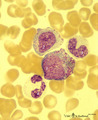
Myeloblast and promyelocyte in bone marrow smear (human) | Stain: May-Grnwald-Giemsa (MGG). The myeoloblast (1) measures 12-20 m and has a high nucleus-cytoplasm ratio and a round to oval nucleus. The nucleus has a fine disperse diffuse chromatin and one to five prominent nucleoli. The cytoplasm is pale blue (basophilic) with no or only scarce numbers of az... | Poja Histology Collection - Blood & Bone Marrow Subset | |
| 322 |
 |
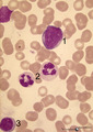
Myeloblast in bone marrow smear (human) | Stain: May-Grnwald-Giemsa (MGG). The myeloblast (1) shows a very transparent nucleus and several distinct nucleoli. The slightly basophilic cytoplasm is limited to a small rim in contrast to a promyelocyte. No granules are yet visible. (2) small lymphocyte. | Blood; Bone Marrow; Myeloblast; Lymphocyte | Poja Histology Collection - Blood & Bone Marrow Subset |
| 323 |
 |
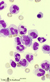
Myeloblast, neutrophilic granulocyte and lymphocyte in bone marrow smear (human) | Stain: May-Grnwald-Giemsa (MGG). The myeloblast (1) measures 12-20 m with 1-5 prominent nucleoli and fine diffuse chromatin. The slightly pale blue cytoplasm contains sometimes a few azurophilic granules (primary granules). (2) Indicates two segmented neutrophilic granulocytes with 3-5 nuclear lobes... | Poja Histology Collection - Blood & Bone Marrow Subset | |
| 324 |
 |
Myeloid cells in bone marrow smear (human) | Stain: May-Grnwald-Giemsa (MGG). (1) promyelocyte is the largest cell in the myeloid series. It has a transparent nucleus with nucleoli and ample cytoplasm with many azurophilic granules. (2) myelocyte. (3) metamyelocyte with an indented nucleus. (4) beginning of nucleus segmentation in the band neu... | Poja Histology Collection - Blood & Bone Marrow Subset | |
| 325 |
 |
Myelopoiesis in bone marrow smear (human) | Stain: May-Grnwald-Giemsa (MGG). The smear shows a group of myeloid cells in different stages of maturation, in which series the alterations in the nucleus can be noticed clearly going from (1) to (5), i.e. the nucleus gets more and more condensed, indented and subsequently segmented in lobes. (1) p... | Poja Histology Collection - Blood & Bone Marrow Subset |