AAO-NANOS Neuro-Ophthalmology Clinical Collection: Derived from the AAO-NANOS Clinical Neuro-Ophthalmology collection produced on CD. The images are of selected cases from the NANOS teaching slide exchange, and the CD was produced under the direction of Larry Frohman, MD and Andrew Lee, MD.
The American Academy of Ophthalmology (AAO); The North American Neuro-Ophthalmology Association (NANOS).
NOVEL: https://novel.utah.edu/
TO
| Title | Creator | Description | ||
|---|---|---|---|---|
| 301 |
 |
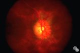
Ocular Manifestations of Congenital/Inherited Diseases | Larry P. Frohman, MD | This 14-year-old boy presented with sudden visual loss of the right eye that occurred 3 weeks before and due to a central retinal vein occlusion. His ocular history was quite complicated. He had had a resection of a lymphangioma of the left upper lid at age 7 months and underwent left orbitotomy for... |
| 302 |
 |
Ocular Manifestations of Congenital/Inherited Diseases | Mark J. Kupersmith, MD | This 9-year-old girl, who had complained of recurrent spontaneous bleeding from the palate and slight swelling and increased warmth over the left cheek, was found to have Wyburn-Mason syndrome. Image 1993_16 shows a small area of arteriovenous shunt on the left optic disc in this patient, who has no... |
| 303 |
 |
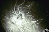
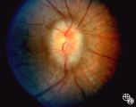
Ocular Manifestations of Congenital/Inherited Diseases | Daniel M. Jacobson MD | This 38-year-old man had unexplained poor vision in his left eye all his life and was told that he had some type of congenital vascular anomaly. He had no neurocutaneous problems. The photographs demonstrate a complex arteriovenous malformation with dilated loops of veins extending out from the nerv... |
| 304 |
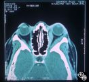 |
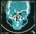
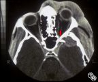
Neuro-Ophthalmic Imaging-CT Scan | Mitchell J. Wolin, MD | Idiopathic orbital pseudotumor is an inflammatory disorder that may effect any part of the ocular anatomy. The site of inflammation determines the nomenclature. For example, involvement of the sclera is referred to as scleritis. And involvement of one or more of the extraocular muscles is referred t... |
| 305 |
 |
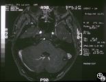
Systemic Disorders With Optic Nerve and Retinal Findings | Larry P. Frohman, MD | This 1-year-old child with familial erythrophagocytic lymphohistiocytosis was readmitted with a fever and was noted to have bilateral blindness. The spinal tap showed a protein of 148, with 178 WBC with 98% ""lymphocytes."" This MRI image demonstrates the optic nerve infiltration. He was treated wit... |
| 306 |
 |
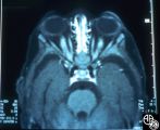
Orbital Tumors | Larry P. Frohman, MD | This 30-year-old man had a retrobulbar intraconal mass OS. The CT scans showed a heterogeneous lobulated enhancing mass, 2.2 x 1.9 x 1.8 cm. The case beautifully exhibits chorodial folds. The ultrasound showed internal reflectivity. The patient refused surgery. Pair with Images 97_60, 97_61, 97_62, ... |
| 307 |
 |
Orbital Tumors | Larry P. Frohman, MD | This 30-year-old man had a retrobulbar intraconal mass OS. The CT scans showed a heterogeneous lobulated enhancing mass, 2.2 x 1.9 x 1.8 cm. The case beautifully exhibits chorodial folds. The ultrasound showed internal reflectivity. The patient refused surgery. Pair with Images 97_60, 97_61, 97_62, ... |
| 308 |
 |
Neuro-Ophthalmic Imaging-CT Scan | Mitchell J. Wolin, MD | This is a patient with trauma leading to enucleation, with swelling years later over the implant. This is a presumed chronic abscess between orbit and dura. |
| 309 |
 |
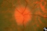
Isolated Congenital Optic Disc Anomalies | Larry P. Frohman, MD | This 63-year-old man with amblyopia OD was seen for a question of ischemic optic neuropathy with a pale, swollen disc OD. The correct diagnosis is an exophytic capillary angioma of the optic nerve head. Disease/Diagnosis: Capillary Angioma. |
| 310 |
 |
Acquired Disc Changes | Rosa A. Tang, MD | Optociliary shunt vessels are venous collaterals that form after chronic venous obstruction. The presence of optic atrophy, progressive visual loss, and optociliary shunt vessels may indicate a compressive optic nerve lesion such as meningioma. |
| 311 |
 |
Isolated Optic Neuritis/Neuropathy | Anthony C. Arnold, MD | This 42-year-old male with pseudotumor cerebri and chronic papilledema demonstrated refractile bodies, which can been seen with chronic optic disc edema. This image exhibits decreased disc edema and resolution of the refractile bodies OD after therapy. Pair with 96_01, 96_02, 96_04, 96_05, and 96_06... |
| 312 |
 |
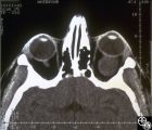
Neuro-Ophthalmic Imaging-CT Scan | Larry P. Frohman, MD | This patient was assaulted with a blunt object and suffered acute blindness due to traumatic optic neuropathy. Note how the lateral orbital wall has been fractured and displaced posteromedially into the region of the anterior optic canal. |
| 313 |
 |
Optic Tract Syndrome Due to Carotid Artery Dolichoectasia | Larry P. Frohman, MD | This 43-year-old man was referred for evaluation of 6 months of visual loss OU. In retrospect, he had noticed increasing difficulty with his tennis game dating back over 3 years, as balls would pass him unexpectedly when hit to his backhand (left) side. The patient did not think this was progressive... |
| 314 |
 |
Optic Tract Syndrome Due to Carotid Artery Dolichoectasia | Larry P. Frohman, MD | This 43-year-old man was referred for evaluation of 6 months of visual loss OU. In retrospect, he had noticed increasing difficulty with his tennis game dating back over 3 years, as balls would pass him unexpectedly when hit to his backhand (left) side. The patient did not think this was progressive... |
| 315 |
 |
Optic Tract Syndrome Due to Carotid Artery Dolichoectasia | Larry P. Frohman, MD | This 43-year-old man was referred for evaluation of 6 months of visual loss OU. In retrospect, he had noticed increasing difficulty with his tennis game dating back over 3 years, as balls would pass him unexpectedly when hit to his backhand (left) side. The patient did not think this was progressive... |
| 316 |
 |
Systemic Disorders With Optic Nerve and Retinal Findings | Larry P. Frohman, MD | This 8-year-old girl had a history of acute lymphoblastic leukemia with neurologic involvement, with remission induced with radiation and chemotherapy. Her relapse showed as an isolated optic neuropathy with this fundus appearance. The other eye was normal. The MRI and spinal tap at this point were ... |
| 317 |
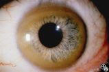 |
Ocular Manifestations of Systemic Disorders | Robert F. Saul, MD | Wilson's disease (hepatolenticular degeneration) is a progressive autosomal recessive multisystem disease that may result in cirrhosis of the liver, renal dysfunction, and motor neurologic disease. A Kayser-Fleischer ring may occur as a green-brown band at the level of Descemet's membrane in the cor... |
| 318 |
 |
Systemic Disorders With Optic Nerve and Retinal Findings | Larry P. Frohman, MD | A 29-year-old African American woman presented with headaches, bilateral transient visual obscurations, blurred vision, numbness, and weakness of the lower extremities with myalgia and joint pains. She had an unplanned 12-pound weight loss over 2 months. A neurologist and internist diagnosed her wit... |
| 319 |
 |
Neuro-Ophthalmic Case With Notable Field Changes | Marilyn C. Kay, MD | This 28-year-old woman presented with a 4-week history of bilateral visual loss. She had a known history of multiple sclerosis. Her vision was 20/60 OD and 20/40 OS, with an RAPD OS and optic pallor OU. Her fields and MRI are shown. Optic tract lesions usually result in an incongruous homonymous hem... |
| 320 |
 |
Ocular Disease With Retinal Findings | William Fletcher Hoyt, MD | Choroidal folds may result from choroidal tumors, compression on the eye wall from thyroid ophthalmopathy, orbital pseudotumor, orbital tumor, posterior scleritis, hypotony, scleral laceration, retinal detachment, marked hyperopia, or secondary to papilledema. Intraocular pressure measurements, refr... |
| 321 |
 |
Isolated Congenital Optic Disc Anomalies | William Fletcher Hoyt, MD | This patient demonstrates bilateral tilted optic discs. Patients with this congenital optic disc anomaly may be asymptomatic or have bitemporal visual field defects that do not respect the vertical midline. Disease/Diagnosis: Tilted Discs. |
| 322 |
 |
Isolated Congenital Optic Disc Anomalies | Anthony C. Arnold, MD | This image shows drusen that are especially prominent superotemporally. Pair with 92_64 and 92_67. |
| 323 |
 |
Systemic Disorders With Optic Nerve and Retinal Findings | Larry P. Frohman, MD | A 42-year old woman presented with a history of severe brow pain and 4 days of progressive visual loss OD. There was no increased pain on ocular rotation. Aside from heavy menses, she denied any significant past medical history. Her examination revealed acuity NLP OD, 20/25 OS; color vision 9/10 OS;... |
| 324 |
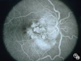 |
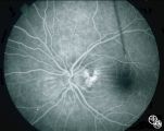
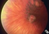
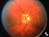
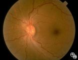
Pseudopapilledema With Subretinal Neovascularization on Fluorescein Angiogram | Joel M. Weinstein, MD | This 8-year-old boy presented with a 2-week history of decreased vision in the right eye. He had undergone a normal MRI and CSF examination, including intracranial pressure, before neuro-ophthalmologic assessment. The fundus photographs and fluorescein angiograms show subretinal neovascularization a... |
| 325 |
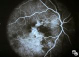 |
Pseudopapilledema With Retinal Neovascularization, Fluorescein Angiogram | Joel M. Weinstein, MD | This 8-year-old boy presented with a 2-week history of decreased vision in the right eye. He had undergone a normal MRI and CSF examination, including intracranial pressure, before neuro-ophthalmologic assessment. The fundus photographs and fluorescein angiograms show subretinal neovascularization a... |