The Health Education Assets Library (HEAL) is a collection of over 22,000 freely available digital materials for health sciences education. The collection is now housed at the University of Utah J. Willard Marriott Digital Library.
TO
Filters: Collection: "ehsl_heal"
| Title | Description | Subject | Collection | ||
|---|---|---|---|---|---|
| 301 |
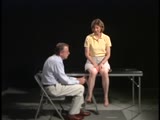 |
Coordination Exam: Normal Exam: Heel-to-shin (includes Spanish audio & captions) | The patient places her heel on the opposite knee then runs the heel down the shin to the ankle and back to the knee in a smooth coordinated fashion. NeuroLogic Exam has been supported by a grant from the Slice of Life Development Fund at the University of Utah, the Department of Pediatrics and the O... | Coordination Examination; Heel-shin Test | NeuroLogic Exam: An Anatomical Approach |
| 302 |
 |
Resection and End Colostomy | Colon resection. End colostomy. Harmann's procedure. | End Colostomy; Colon Resection; Hartmann's Procedure | Royal College of Surgeons in Ireland Illustrations |
| 303 |
|
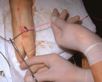
Needle stick | Picture of a needle stick in a plastic arm | Venipuncture | HEAL Reviewed Collection |
| 304 |
|
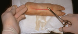
Equipment for venipuncture | This is an image of equipment for venipuncture. | Venipuncture | HEAL Reviewed Collection |
| 305 |
|
Gastrointestinal Tract (Labeled) | Gastrointestinal tract showing upper GI, ligament of Treitz, and lower GI. | Upper GI; Upper Gastrointestinal Tract; Ligament of Treitz; Lower GI; Lower Gastrointestinal Tract | Royal College of Surgeons in Ireland Illustrations |
| 306 |
|
Resection and Primary Anastomosis of Sigmoid Colon | Make private -- psd file. Colon resection. Primary anastomosis. | Primary Anastomosis; Resection | Royal College of Surgeons in Ireland Illustrations |
| 307 |
|
Male Urogenital Diaphragm | Urogenital diaphragm. Male. | Ischiopubic Ramus; Perineal Membrane; Urogenital Diaphragm; Perineal Body; Penile Veins; Prostatic Venous Plexus | Royal College of Surgeons in Ireland Illustrations |
| 308 |
|
Exposed Facial Nerve with Retracted Parotid Gland and Stylohyoid Muscle (Labeled) | Exposed facial nerve with retracted parotid gland and stylohyoid muscle. | Stylohyoid Muscle | Royal College of Surgeons in Ireland Illustrations |
| 309 |
|
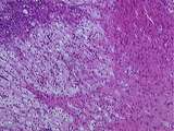
Ganglioneuroma of adrenal gland | Gross photograph showing firm white mass in adrenal medulla. Histology showed ganglioneuroma. | HEAL Reviewed Collection | |
| 310 |
 |
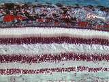
Eye - Rods and Cones | Rod and cone photoreceptors have their cell bodies in the outer nuclear layer. Between these cell bodies and the retinal pigment epithelium are the photoreceptor inner segments (where proteins are synthesized) and the outer segments (the light sensitive portion). The inner segments of cones are cone... | UCLA Histology | |
| 311 |
 |
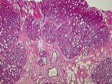
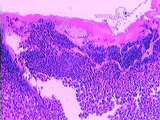
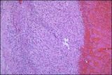
Uterus | This relatively brief phase of the endometrial cycle consists of shrinkage of the endometrium and intraendometrial hemorrhage and fragmentation. The myometrium is located to the left. A higher magnification of the ischaemic endometrium is shown in image 226-10-1. UCLA Histology Collection. | ischaemic endometrium; Uterus | UCLA Histology |
| 312 |
 |
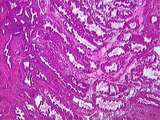
Kidney | In this section through the renal papilla, identify the terminal collecting tubules (lined by columnar epithelium). Also note a straight segment of the loop of Henle, many thin loops of Henle, and cross-sections of the vasa recta. UCLA Histology Collection. | Kidney; renal papilla; terminal collecting tubules | UCLA Histology |
| 313 |
 |
Breast | This active, lactating breast contains a large number of lobules of tightly packed alveoli. The connective tissue of the interlobular elements is highly compressed. Note the lactating glands, the excretory duct and some fat. UCLA Histology Collection. | Breast; lactating breast | UCLA Histology |
| 314 |
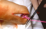 |
Suturing | After placing four throws to create a square knot, the suture is then cut just above the knot. | Knowledge Weavers Dermatology | |
| 315 |
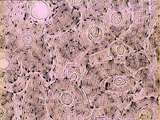 |
Bone - Haversian System | Haversian systems can be seen well in this image of dried compact bone. Haversian or central canals and concentrically arranged osteocytes are also visible. UCLA Histology Collection. | UCLA Histology | |
| 316 |
 |
Suturing | The suture is then tightened by crossing the non-dominant (left) hand over the dominant (right) hand. | Knowledge Weavers Dermatology | |
| 317 |
 |
Suturing | This demonstrates tightening of the double loop along the long axis of the wound using suture. | Knowledge Weavers Dermatology | |
| 318 |
 |
Skin | Healing skin (rat). See lymphocyte infiltration. UCLA Histology Collection. | Skin | UCLA Histology |
| 319 |
 |
Somatotropinoma | Somatotropinoma | Knowledge Weavers Pathology | |
| 320 |
 |
Eye - Retina | The retina consists of the retinal pigment epithelium and neurosensory retina. The neurosensory retina includes multiple layers of neurons and some glia. The outer nuclear layer contains the cell bodies of rod and cone photoreceptors. The inner nuclear layer contains the cell bodies of multiple neur... | UCLA Histology | |
| 321 |
 |
Ovary | This tissue looks like a dying corpus luteum, but that is a rare event. This is an example of a structure that we cannot identify unless we see a zona pellucida and/or a glassy membrane. If you had a section along the plane labelled A you would not be able to differentiate between a section through ... | Ovary | UCLA Histology |
| 322 |
 |
Corpus luteum cysts exhibit a convoluted lining with luteinized granulosa and theca cells. | Corpus luteum of the ovary, medium power. The granulosa cells have undergone proliferation and alteration to lutein cells that produce progesterone. The lutein cells are large and polyhedral and the cytoplasm is foamy and eosinophilic. | Knowledge Weavers Human Reproduction | |
| 323 |
 |
Ear | At this higher magnification, one can differentiate the hair & supporting cells, the gelatinous cupula, and the dark cells which mark the transition of the membranous labyrinth into the crista ampullaris. Bone is also evident. Movement of the endolymph against the cupula causes bending of hair cell ... | Crista Ampullaris; Ear | UCLA Histology |
| 324 |
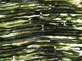 |
Peripheral Nervous System | This slide of myelinated nerves is stained with osmium to demonstrate the myelin sheaths. The region where two myelin sheaths covering the same axon contact one another is the Node of Ranvier. The region of myelin sheath between these nodes is known as the internode. UCLA Histology Collection. | myelin sheaths; osmium; Peripheral Nervous System | UCLA Histology |
| 325 |
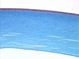 |
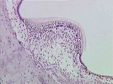
Epithelium - Cornea | The cornea of the eye is lined by simple squamous epithelium on its inner surface and stratified squamous epithelium on its outer surface. The stroma of the cornea consists of regularly arranged collagen fibers. UCLA Histology Collection. | simple squamous epithelium; stratified squamous epithelium | UCLA Histology |