The Health Education Assets Library (HEAL) is a collection of over 22,000 freely available digital materials for health sciences education. The collection is now housed at the University of Utah J. Willard Marriott Digital Library.
Filters: Collection: ehsl_heal
| Title | Description | Subject | Collection | ||
|---|---|---|---|---|---|
| 226 |
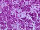 |
Gastrointestinal Tract - Submandibular Gland | Higher magnification view of the submandibular gland showing purple serous alveoli, pale mucous alveoli, and the serous demilunes (half-moons) of mixed alveoli. Note the secretory granules within many serous acinar cells. UCLA Histology Collection. | UCLA Histology | |
| 227 |
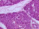 |
Gastrointestinal Tract - Submandibular Gland | This medium power view of the submandibular gland, a mixed seromucous gland, shows its subdivision into lobules by interlobular septa. Compare the location and appearance of the few intralobular ducts to the single interlobular duct within the interlobular septum. Identify the sparse, lightly staine... | UCLA Histology | |
| 228 |
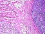 |
Gastrointestinal Tract - Tongue - Posterior | In this low view of the posterior tongue, identify the lingual tonsil composed of lymphocytic nodules, the mucous glands, adipocytes, and rows of the striated muscle of the tongue. UCLA Histology Collection. | Lingual Tonsil | UCLA Histology |
| 229 |
 |
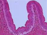
Glands of GI Tract | Higher magnification image of liver stained with PAS showing different levels of glycogen storage in hepatocytes. Some hepatocytes have numerous glycogen granules, whereas there are other hepatocytes without glycogen granules, indicating depletion of stored glycogen. UCLA Histology Collection. | glycogen; Liver; PAS stain; gastrointestinal tract | UCLA Histology |
| 230 |
 |
Glands of GI Tract | This images illustrates a section of the liver stained with PAS to visualize the glycogen storage in hepatocytes. Hepatocytes have granules in their cytoplasm due to the presence of stored glycogen. Shown here are also sinusoids and a central vein. UCLA Histology Collection. | gastrointestinal tract; glycogen; Liver; pas stain | UCLA Histology |
| 231 |
 |
Glands of GI Tract | Higher magnification image showing the portal triad with a biliary duct, a branch of hepatic artery and a branch of portal vein embedded within connective tissue, and the hepatocytes. UCLA Histology Collection. | Gastrointestinal tract; Liver; portal triad | UCLA Histology |
| 232 |
 |
Glands of GI Tract | This image shows Kupffer cells, which are macrophages that are distinguished by their phagocytation of a particulate dye. Note the hepatocyte cords and the sinusoids which separate them. UCLA Histology Collection. | Kupffer cells; Liver | UCLA Histology |
| 233 |
 |
Glands of GI Tract | In this image you can see a portal triad containing a branch of the portal vein, a small hepatic artery, and a biliary duct embedded in collagenous connective tissue (stained blue by trichrome). Also shown are dark red hepatocytes and pinkish sinusoids. UCLA Histology Collection. | gastrointestinal tract; Liver; trichrome | UCLA Histology |
| 234 |
 |
Glands of GI Tract | Low power view of the gallbladder showing the mucosa composed of simple columnar epithelium, the lamina propria, and a layer of smooth muscle. The gall bladder is encapsulated by perimuscular connective tissue containing blood vessels. Some surfaces of the gallbladder are attached to hepatic tissue.... | Gall bladder; gastrointestinal tract | UCLA Histology |
| 235 |
 |
Glands of GI Tract | Higher magnification of the gallbladder mucosa illustrating the tall cells of the simple columnar epithelium and collagen fibers within the lamina propria. UCLA Histology Collection. | Gall bladder; gastrointestinal tract | UCLA Histology |
| 236 |
 |
Glands of GI Tract | Another area of the liver showing a central vein surrounded by hepatocytes organized into cords which are separated by sinusoids. Nutrient-rich blood from the hepatic portal vein percolates through the sinusoids, where the hepatocytes regulate the nutrient content. The sinusoids drain into central v... | gastrointestinal tract; Hepatocyte; Liver | UCLA Histology |
| 237 |
 |
Glands of GI Tract | This low power view of the pancreas shows two islets of Langerhans (the endocrine portion of the gland) and the mass of serous acini with an interlobular duct surrounded by abundant connective tissue. The acini and ducts form the exocrine portion of the gland. Note also the numerous intersecting sep... | islet of Langerhans; Pancreas | UCLA Histology |
| 238 |
 |
Glands of GI Tract | Higher magnification image showing an islet of Langerhans containing endocrine cells and capillaries; also shown here are a mass of serous acini and intralobular ducts. UCLA Histology Collection. | islet of Langerhans; Pancreas | UCLA Histology |
| 239 |
 |
Glands of GI Tract | This low power view of the liver shows the mass of hepatocytes, a central vein and a portal triad composed of a biliary duct, a branch of hepatic artery and a branch of portal vein embedded within connective tissue. UCLA Histology Collection. | gastrointestinal tract; hepatic triad; Liver | UCLA Histology |
| 240 |
 |
Glands of GI Tract | High power view of the gallbladder mucosa showing the lamina propria, plasma cells, neutrophils, and the simple columnar epithelium. Note the basophilic cytoplasm and clock-face nucleus which are characteristic of plasma cells. UCLA Histology Collection. | Gall bladder; gastrointestinal tract | UCLA Histology |
| 241 |
 |
Glands of GI Tract | The staining used in this preparation reveals reticular fibers supporting the hepatocytes which line the sinusoids. Bile canaliculi also bind the dye used here, so they appear black. UCLA Histology Collection. | Gastrointestinal tract; Hepatocyte; Liver; reticular fibers | UCLA Histology |
| 242 |
 |
Glands of GI Tract | In this image you can see islets of Langerhans, (lighter staining), serous acini (darker staining), a septum and an interlobular duct. UCLA Histology Collection. | islet of Langerhans; Pancreas | UCLA Histology |
| 243 |
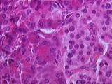 |
Glands of GI Tract | Another area of the pancreas (high magnification) showing an islet of Langerhans with its endocrine cells and capillaries. The surrounding serous acini secrete the contents of their red secretory granules into the gut via the pancreatic ducts. UCLA Histology Collection. | islet of Langerhans; Pancreas | UCLA Histology |
| 244 |
 |
healing Skin | Healing skin. The wound has disrupted the epidermis and dermis. At the base of the wound a lymphocytic infiltration has formed. Note the hair follicles and sebaceous gland. UCLA Histology Collection. | epidermis; Skin | UCLA Histology |
| 245 |
 |
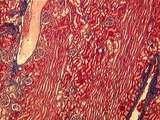
Kidney | In this section through the renal papilla, identify the terminal collecting tubules (lined by columnar epithelium). Also note a straight segment of the loop of Henle, many thin loops of Henle, and cross-sections of the vasa recta. UCLA Histology Collection. | Kidney; renal papilla; terminal collecting tubules | UCLA Histology |
| 246 |
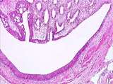 |
Kidney | The renal papilla is the site where terminal collecting ducts empty urine into the U-shaped minor calyx. The lumen of the minor calyx is lined by transitional epithelium. Strands of circular smooth muscle are found in the wall of the minor calyx. UCLA Histology Collection. | Kidney; minor calyx; renal papilla; terminal collecting ducts | UCLA Histology |
| 247 |
 |
Kidney | In this trichrome stained section of the renal cortex there are sections of an interlobular artery and interlobular vein. Also identify the long medullary rays and several renal corpuscles. UCLA Histology Collection. | Kidney; medullary rays; trichrome | UCLA Histology |
| 248 |
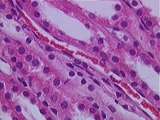 |
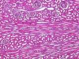
Kidney | Present here in the kidney medulla are sections of straight distal tubules (low cuboidal walls), straight proximal tubules (tall cuboidal walls), and vasa recta (lined by endothelium). UCLA Histology Collection. | Kidney; medulla; straight distal tubules; straight proximal tubules; vasa recta | UCLA Histology |
| 249 |
 |
Kidney | This section can be identified as renal cortex because of the presence of renal corpuscles and medullary rays. Sections of an interlobular artery are also present. UCLA Histology Collection. | Kidney; renal corpuscles; renal cortex | UCLA Histology |
| 250 |
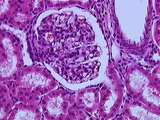 |
Kidney | A number of features of the renal corpuscle can be seen here. The parietal and visceral layers of Bowman's capsule, which consist of two types of simple squamous epithelium, line the capsular space. The filtrate leaves the glomerulus via the urinary pole, which leads to a proximal convoluted tubule ... | Bowman's capsule; Kidney; renal corpuscle; renal cortex | UCLA Histology |