Collection of materials relating to neuro-ophthalmology as part of the Neuro-Ophthalmology Virtual Education Library.
NOVEL: https://novel.utah.edu/
TO
- NOVEL966
Filters: Collection: "ehsl_novel_novel"
| Title | Creator | Description | Subject | ||
|---|---|---|---|---|---|
| 226 |
 |
Diagnostic Error of Neuro-ophthalmologic Conditions: State of the Science | Leanne Stunkel, MD; David E. Newman-Toker, MD, PhD; Nancy J. Newman, MD; Valérie Biousse, MD | Diagnostic error is prevalent and costly, occurring in up to 15% of US medical encounters and affecting up to 5% of the US population. One-third of malpractice payments are related to diagnostic error. A complex and specialized diagnostic process makes neuro-ophthalmologic conditions particularly vu... | Diagnostic Errors |
| 227 |
 |
Professionalism and Communication Skills | Karl C. Golnik, MD, MEd | Lecture covering professionalism and communication skills. | Professionalism; Communication Skills |
| 228 |
 |
Ocular Surface, Cornea, & Lens | Sari Yordi, MD | Video lecture on the anatomy of the ocular surface, cornea, and lens. | Ocular Surface; Cornea; Lens |
| 229 |
 |
Maintenance of Certification Basics: American Board of Psychiatry and Neurology | Sean Gratton, MD | Video lecture covering certification basics. | Credentialing |
| 230 |
 |
Lacrimal Pathways: Anatomy and Physiology | Sari Yordi, MD | Video lecture covering anatomy and physiology of the lacrimal pathways. | Lacrimal Pathways |
| 231 |
 |
Anaesthesia for Eye Surgery and Associated Complications | Julie Smith, MBBS, FANZCA | Lecture covering commonly performed eye surgery and anaesthetic techniques. | Eye Surgery; Anesthesia |
| 232 |
 |
Neuroablative Procedures | Benjamin Jonker, MB BS, MMed(Clin Epi), FRACS | Video lecture covering neuro-ablative procedures that are relevant to neuro-ophthalmologists. | Ablative Procedures |
| 233 |
 |
Anaesthesia for Eye Surgery and Associated Complications Slides | Julie Smith, MBBS, FANZCA | Lecture covering commonly performed eye surgery and anaesthetic techniques. | Eye Surgery; Anesthesia |
| 234 |
 |
Visual Maturation | Yesha Shah, BSA, BBA; Amanda Henderson, MD | Video lecture covering visual maturation. | Visual Maturation; Foveal Development; Vision in Infants |
| 235 |
 |
Embryology of the Eye | Yesha Shah, BSA, BBA; Amanda Henderson, MD | Video lecture covering the embryology of the eye. | Embryology; Eye |
| 236 |
 |
Brain Surface Anatomy | Arooj Ahmad, MD; Devin D. Mackay, MD | These images depict labeled structures of the surface anatomy of the different facies of the brain. | Neuroanatomy; Brain Surface Anatomy |
| 237 |
 |
The Anatomic Course of Cranial Nerve IV | Divya Chauhan, MD | Overview of the intracranial course of the trochlear nerve. | Cranial Nerve IV; Trochlear Nerve; Anatomy |
| 238 |
 |
The Anatomic Course of Cranial Nerve VI | Divya Chauhan, MD | Overview of the intracranial course of the abducens nerve. | Cranial Nerve VI; Abducens Nerve; Anatomy |
| 239 |
 |
CSF Composition | Divya Chauhan, MD | Overview of the composition of cerebrospinal fluid. | Cerebrospinal Fluid; CSF |
| 240 |
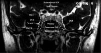 |
Cavernous Sinus | Andrew R. Carey, MD | Cavernous sinus imaging with labels. | Cavernous Sinus |
| 241 |
 |
Optic Tract Syndrome Secondary to Lacunar Infarction | Justin J. Grassmeyer, PhD; Kenan Xiao, MD; Jason T. Helvey, MD; Sachin Kedar, MD | Lesions of the optic tract produce a characteristic triad of clinical findings: contralateral homonymous hemianopia, contralateral relative afferent pupillary defect, and bilateral optic disc atrophy. This case describes clinical features, radiological findings, and optical imaging characteristics f... | Optic Tract Syndrome; Optic Tract; Lacunar Infarction; Homonymous Hemianopia |
| 242 |
 |
Lateral Orbitotomy with Bone Window | Richard C. Allen, MD, PhD, FACS | Narrated video of lateral orbitotomy with bone removal for improved orbital decompression. | Orbitotomy; Orbital Decompression |
| 243 |
 |
Lateral Orbitotomy #3 | Richard C. Allen, MD, PhD, FACS | Narrated video of lateral orbitotomy and biopsy of presumed benign cavernous hemangioma. | Orbital Biopsy; Orbitotomy |
| 244 |
 |
Lateral Orbitotomy #2 | Richard C. Allen, MD, PhD, FACS | Narrated video of lateral orbitotomy and biopsy of presumed orbital inflammatory condition. | Orbital Biopsy; Orbitotomy |
| 245 |
 |
Lateral Canthotomy Incision | Richard C. Allen, MD, PhD, FACS | Narrated video of lateral canthotomy procedure. | Lateral Canthotomy |
| 246 |
 |
The Internal Carotid Arteries and Branches | Katherine Hutchins, MD; Devin D. Mackay, MD | Illustrations, MRA, CTA, and cerebral angiography images of the internal carotid artery and its branches. | Vascular Anatomy; Internal Carotid Artery; Anterior Cerebral Artery; Middle Cerebral Artery; Anterior Circulation |
| 247 |
 |
The Vertebrobasilar System | Katherine Hutchins, MD; Devin D. Mackay, MD | Illustrations, MRA, and CTA images of the vertebrobasilar system and branches. | Vascular Anatomy; Basilar Artery; Vertebral Artery; AICA; PICA; Superior Cerebellar Artery; Posterior Cerebral Artery; Posterior Circulation |
| 248 |
 |
Ptosis | Ethan Waisberg, MB, BCh, BAO candidate | Description of ptosis including etiology, management and treatment. | Ptosis; Blepharoptosis |
| 249 |
 |
A Five-Minute, Intravenous Edrophonium -Window to Observe the CNS Response to Myasthenic Ophthalmoplegia | Jorge C Kattah, MD | A video showing how ocular motor adaptation may be observed. | Ocular Motor Adaptation |
| 250 |
 |
Hereditary Optic Neuropathy (Leber's Hereditary Optic Neuropathy) | NANOS | Hereditary Optic Neuropathy - A hereditary optic neuropathy is caused by a genetic variant (or mutation) that causes dysfunction of the neurons (nerve cells) which form the optic nerve. The optic nerve sends information from the back of the eye to the vision center in the brain.The two most common t... | Hereditary Optic Neuropathy; Patient Brochure |