Collection of materials relating to neuro-ophthalmology as part of the Neuro-Ophthalmology Virtual Education Library.
NOVEL: https://novel.utah.edu/
TO
- NOVEL716
| Title | Creator | Description | Subject | ||
|---|---|---|---|---|---|
| 226 |
 |
Blepharospasm (Telugu) | North American Neuro-Ophthalmology Society | Uncontrolled blinking, squeezing, and eyelid closure that occurs in both eyes without an apparent environmental cause. | Blepharospasm; Patient Brochure |
| 227 |
 |
Optic Disc Drusen (Telugu) | North American Neuro-Ophthalmology Society | Optic disc drusen are abnormal deposits of protein-like material in the optic disc - the front part of the optic nerve. | Drusen; Patient Brochure |
| 228 |
 |
Homonymous Hemianopia (Telugu) | North American Neuro-Ophthalmology Society | This refers to an absence of vision towards one side of the visual world in each eye. The damage that caused this problem is in the brain and not in the eyes. | Homonymous Hemianopsia; Patient Brochure |
| 229 |
 |
Myasthenia Gravis (Italian) | North American Neuro-Ophthalmology Society | This is an autoimmune condition where the body's immune system has damaged receptors on your muscles and can result in double vision or drooping lid | Myasthenia Gravis; Patient Brochure |
| 230 |
|
Myasthenia Gravis (Telugu) | North American Neuro-Ophthalmology Society | This is an autoimmune condition where the body's immune system has damaged receptors on your muscles and can result in double vision or drooping lid | Myasthenia Gravis; Patient Brochure |
| 231 |
 |
Dry Eye Syndrome (Telugu) | North American Neuro-Ophthalmology Society | People with abnormalities of the tear film are diagnosed with "dry eyes", but some patients with "dry eyes" may not feel that their eyes are "dry". Itching, burning, a scratchy sensation, a sensation that there is sand or grit in the... | Dry Eye Syndrome; Patient Brochure |
| 232 |
 |
Optochiasmal Tuberculoma | Jeanie Paik, MD; Rudrani Banik, MD | PowerPoint of case of chiasmal tuberculoma causing bitemporal defect in patient with tuberculosis on RIPE treatment; case history, differential diagnosis and treatment discussed. | Chiasmal Disorder; Chiasmal Tuberculoma; Bitemporal Visual Field Defect; Ethambutol Optic Neuropathy |
| 233 |
 |
Emianopsia Omonima (Italian) | North American Neuro-Ophthalmology Society | This refers to an absence of vision towards one side of the visual world in each eye. The damage that caused this problem is in the brain and not in the eyes. | Homonymous Hemianopsia; Patient Brochure |
| 234 |
 |
The Ice Pack Test For Myasthenia | Ryan D. Walsh, MD; Collin McClelland, MD | The ice pack test is a simple, low-tech, bedside test that can readily be applied in the outpatient clinic or hospital setting, with minimal risk or discomfort. The ice pack test is used in patients with ptosis to help support or refute a diagnosis of myasthenia. We describe how to perform and int... | Ice Pack Test; Ice Test; Myasthenia Gravis; Ocular Myasthenia; Ptosis; Diagnostic Tests |
| 235 |
|
Secchezza Degli Occhi - Dry Eye (Italian) | North American Neuro-Ophthalmology Society | People with abnormalities of the tear film are diagnosed with "dry eyes", but some patients with "dry eyes" may not feel that their eyes are "dry". Itching, burning, a scratchy sensation, a sensation that there is sand or grit in the... | Dry Eye Syndrome; Patient Brochure |
| 236 |
 |
Neurosyphilis | Timothy Sullivan, MD; Rudrani Banik, MD | Power point of case of a 66 year old male with acute vision loss OD to no light perception. Underwent extensive work-up for cardiovascular and neurologic etiologies, all negative. Subsequent serologic work-up was positive for syphilis and the diagnosis of neurosyphilis was confirmed by lumbar punc... | Neurosyphilis; Infectious/Vasculitic Optic Neuropathy |
| 237 |
 |
Straatsma Syndrome | Charles A. McBride, OD | Triad of ipsilateral retinal nerve fiber myelination, myopia and amblyopia. Includes fundus photos and OCT showing normal macular architecture OD and thickened and hyper reflective RNFL outside macular region. | Straatsma Syndrome; Myelinated Nerve Fibers; myelination; anisometropia; amblyopia |
| 238 |
 |
Non-Organic Visual Loss | Omar Ozgur, MD; Rudrani Banik, MD | Power point of case presentation of 12 year old girl with recurrent monocular visual loss. Examination is normal. Differential diagnosis discussed, including non-organic visual loss. Diagnostic testing for non-organic visual loss reviewed. Slide 4: Figure 1: Table of exam findings Slide 5: Figure 2... | Non-organic Visual Loss; Monocular Visual Loss |
| 239 |
 |
Pituitary Apoplexy and Hemifield Slide Phenomenon | Helen H. Yeung, MD; Rudrani Banik, MD | PowerPoint of case presentation of pituitary apoplexy. Patient presented with bilateral severe visual loss and bilateral ophthalmoplegia from partial third nerve palsies (pupil-sparing with no ptosis) from midbrain compression. After transsphenoidal surgery with decompression of mass and steroids, ... | Pituitary Apoplexy; Hemifield Slide; Bitemporal Defect; Partial Third Nerve Palsy |
| 240 |
 |
Acute Zonal Occult Outer Retinopathy (AZOOR) versus Multiple Evanescent White Dot Syndrome (MEWDS) | Asim V. Farooq, MD; Michael T. Andreoli, MD; Heather E. Moss, MD | PPT case report on acute zonal occult outer retinopathy (AZOOR) versus multiple evanescent white dot syndrome (MEWDS). | AZOOR; MEWDS; Paracentral Scotoma; Goldmann Visual Field; Photoreceptor Loss |
| 241 |
 |
Retinal Causes of a Neurologic-Type Visual Field Defect | Omar Ozgur, MD; Rudrani Banik, MD | Power point of case presentation of 47 year old female with history of breast cancer with new onset temporal visual field defect and photopsias. Differential diagnosis of homonymous hemianopia discussed; retinal causes of neurologic-type visual field defects reviewed including: white dot syndrome (m... | Homonymous Hemianopia; Neurologic Visual Field Defect; Temporal Visual Field Defect; White Dot Syndrome; Multiple Evanescent White Dot Syndrome (MEWDS); Cancer-Associated Retinopathy; Tamoxifen Retinopathy; Autoimmune Retinopathy |
| 242 |
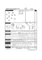 |
Montreal Cognitive Assessment (MOCA) | Ziad Nasreddine, MD | The MoCA© is a cognitive screening test designed to assist Health Professionals in detection of mild cognitive impairment. For more information, contact: info@moca.org | Cognitive Disorders; Cognitive Assessment; MOCA Test |
| 243 |
 |
Pseudotumor cerebri and Chiari Malformation | Nicole Scripsema, MD; Rudrani Banik, MD | Power point of case presentation of pseudotumor cerebri with co-existing Chiari malformation. Management of severe visual loss associated with chronic papilledema discussed, as well as possible relationship between raised intracranial pressure from pseudotumor cerebri and Chiari malformation. | Pseudotumor Cerebri; Papilledema; Chiari Malformation |
| 244 |
 |
Wallenberg Syndrome and Skew Deviation | Lauren Schneider, MD; Rudrani Banik, MD | Power point of case presentation of acute Wallenberg Syndrome associated with vertical diplopia, found by 3 step and supine testing to be consistent with skew deviation. | Wallenberg Syndrome; Skew Deviation; Vertical Diplopia |
| 245 |
 |
Prolactinoma in Pregnancy | Timothy Sullivan, MD; Rudrani Banik, MD | Power point of case of prolactinoma which became symptomatic during pregnancy with visual field loss. Discussion of prolactinomas and their management. Patient underwent observation only. Post-partum examination revealed resolution of bitemporal field defect with reduction in size of prolactinoma ... | Prolactinoma; Pregnancy; Bitemporal Defect |
| 246 |
 |
Lemierre Syndrome - A Neuroophthalmological Approach | Vinzenz A. C. Vadasz, MD; Christina Gerth-Kahlert, MD | Case report of a twenty-two year old woman with double vision after tonsillitis, caused through multiples thrombosis by an infection with fusobacterium necrophorum known as the Lemierre-Syndrome. Fig. 1: Ocular motility at ICU (lying position) Fig. 2: white arrows show thrombosis of the right opht... | Lemierre-Syndrome; Fusobacterium Necrophorum; Septic Thrombosis |
| 247 |
 |
Oculopharyngeal Muscular Dystrophy (OPMD) | Natasha Nayak, MD; Rudrani Banik, MD | Power point of case presentation of chronic, progressive ophthalmoplegia and bilateral ptosis in adult male with positive family history of similar ocular findings. Differential diagnosis with associated findings reviewed. Work up done: EMG testing consistent with myopathy. Genetic testing positiv... | Ophthalmoplegia;, Ptosis; Oculopharygneal Muscular Dystrophy; Genetic Disorder |
| 248 |
 |
Neuromyelitis Optica | Omar Ozgur, MD; Rudrani Banik, MD | Power point of case presentation of female with bilateral, sequential atypical optic neuritis. MRI Brain normal with no demyelination; MRI Spine shows enhancement at multiple levels and NMO antibody positive, confirming diagnosis of neuromyelitis optica (NMO). History of NMO discussed, diagnostic c... | Neuromyelitis Optica; Atypical Optic Neuritis; MRI; Plasmapheresis |
| 249 |
 |
Idiopathic Bilateral Neuroretinitis in a Child | Asim V. Farooq, MD; Michael T. Andreoli, MD; Molly Gilbert, MD; Heather E. Moss, MD | PPT case describing idiopathic bilateral neuroretinitis in a child. | Neuroretinitis; Pediatric; Idiopathic; Optic Atrophy |
| 250 |
 |
The History of the International Neuro-Ophthalmology Society | Klara Landau, MD, FEBO | This presentation provides an ovreview of hte hisotry of the International Neuro-ophthalmology Society (INOS), with maps and photos. | International Neuro-Ophthalmology Society: INOS |