John A. Moran Eye Center Neuro-Ophthalmology Collection: A variety of lectures, videos and images relating to topics in Neuro-Ophthalmology created by faculty at the Moran Eye Center, University of Utah, in Salt Lake City.
NOVEL: https://novel.utah.edu/
TO
Filters: Collection: "ehsl_novel_jmec"
| Title | Description | Type | ||
|---|---|---|---|---|
| 226 |
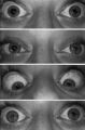 |
Pupil Signs in a 32-year-old Woman with Right-sided Adie's Pupil | Pupil signs in a 32-year-old woman with right-sided Adie's pupil. The right pupil is larger than the left pupil (top), reacts poorly to direct light stimulation (second panel), and better in response to near stimulation (third panel). The right pupil also shows a supersensitive response 30 minutes a... | Image |
| 227 |
 |
Sector Palsies and Light-Near Dissociation | Example of patient with bilateral Adie's pupils. Exam is performed with a slit-lamp. Shows iris stroma and focal segments of iris sphincter that retain their contractilty. Suggests post-ganglionic parasympathetic denervation. | Image/MovingImage |
| 228 |
 |
Benign Episodic Unilateral Mydriasis | Presentation covering benign episodic mydriasis. | Text |
| 229 |
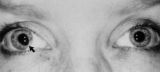 |
Tadpole-shaped Pupil | Tadpole-shaped pupil in a 20-year-old women with frequent episodes of blurred vision and achiness of the right eye lasting several minutes. The patient took a photograph of her eyes during an attack to document the peaked, segmental dilation of her right pupil (black arow). (Thompson HS, Zackon DH, ... | Image |
| 230 |
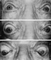 |
Argyll Robertson Pupils | Argyll Robertson pupils in an elderly man treated for tabes dorsalis in 1952. His pupils are small and slightly irregular, constrict poorly in response to light stimulation (top), dilate poorly in darkness (middle), but constrict promptly in response to near stimulation (bottom). | Image |
| 231 |
 |
Light-near Dissociation | Example of patient with Argyll Robertson pupil with neurosyphilis. Shows a lack of pupillary response to light and some pupillary response to nearness of finger. | Image/MovingImage |
| 232 |
 |
Ocular Lateropulsion (Wallenberg's Syndrome) | Example of patient with ocular lateropulsion. Patient also has central Horner syndrome and nystagmus in right gaze. When shifting gaze back to forward, eyes overshoot their mark. Eyes laterally deviate to the right upon opening. | Image/MovingImage |
| 233 |
|
Dilation Lag | Two examples of dilation lag (Horner's syndrome). In the first example, the right pupil dilates much faster than the left pupil when the light is turned out. In the second example, the left pupil dilates much faster than the right pupil when the light is turned out. Discussion of methods of document... | Image/MovingImage |
| 234 |
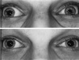 |
Left-sided Dilation Lag in a Man with Horner's Syndrome | Left-sided dilation lag in a 29-year-old man with Horner's syndrome caused by a posterior mediastinal ganglioneuroma. Note that the degree of anisocoria is greater after 5 seconds in darkness (top) compared with findings after 15 seconds in darkness (bottom). | Image |
| 235 |
 |
Fourth Nerve Palsy | Demonstration of examination of patient who experienced blurry vision and pain in the left eye. Demonstrates checking of eye movements, focusing on object while each eye is covered and uncovered, turning head both ways and repeating. Shows limitation of depression in adduction of left eye, left hype... | Image/MovingImage |
| 236 |
 |
3 Step Test | Demonstration of patient examination. | Image/MovingImage |