The Health Education Assets Library (HEAL) is a collection of over 22,000 freely available digital materials for health sciences education. The collection is now housed at the University of Utah J. Willard Marriott Digital Library.
TO
Filters: Collection: "ehsl_heal"
| Title | Description | Subject | Collection | ||
|---|---|---|---|---|---|
| 226 |
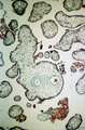 |
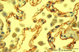
Intervillous space with villi (human placenta, midpregnancy, cross-section) | Stain: Trichrome (Goldner). In the middle a thick stem villus (cross-sections of left artery (1) and right vein (2) of the umbilical circulation in the thick embryonic connective tissue) with ramifications into terminal villi (tertiary). Most tertiary villi of varying diameters contain capillaries a... | placenta; chorionic villi; fibrinoid; cytotrophoblast; syncytiotrophoblast | Poja Histology Collection - Placenta |
| 227 |
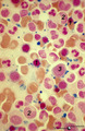 |
Iron (Perls) staining of sideroblasts in bone marrow smear (human) | Stain: Perl's stain for iron (Prussian blue). The intense blue stained hemosiderin in the erythroblasts (1) is visible as siderotic granules (pathologic siderosomes). These pathologic siderosomes are sometimes distributed in a circle around the nucleus (ringed sideroblasts). The myeloid cell types (... | Poja Histology Collection - Blood & Bone Marrow Subset | |
| 228 |
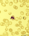 |
Karyorrhexis of polychromatic erythroblast in bone marrow smear (human) | Stain: May-Grnwald-Giemsa (MGG). The nucleus of the polychromatic erythroblast (1) shows abnormal karyorrhexis due to vitamin B12 deficiency or folic acid deficiency. Normochromic normocyte (2, normal erythrocyte), poikilocytosis and oval macrocytes are seen. | Poja Histology Collection - Blood & Bone Marrow Subset | |
| 229 |
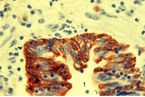 |
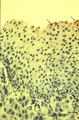
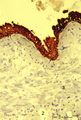
Keratin 7 in the bronchial epithelium of the lung (human, adult) | Stain: anti-keratin 7 antibody (Pan-Ck 7) immunoperoxidase staining (with aminoethylcarbazole (AEC) substrate). The epithelium of the bronchus (1) stains dark brown-red after the reaction with AEC indicating a positive reaction for cytokeratin 7. This antibody can be used in the study of development... | Bronchiolus; Immunoperoxidase; Immuno-reaction | Poja Histology Collection - Respiratory System Subset |
| 230 |
 |
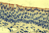
Keratin 7 in the lining alveolar epithelium of the lung (human, adult) | Stain: anti-keratin 7 antibody (Pan-Ck 7) immunoperoxidase staining (with aminoethylcarbazole (AEC) substrate). Both alveolar epithelial cell types I (1) and II (2) stain dark brown-red after the reaction with AEC indicating a positive reaction for cytokeratin 7. Note the white (non-stained) capilla... | Pneumocyte I; Pneumocyte II; Imunoperoxidase; Immuno-reaction | Poja Histology Collection - Respiratory System Subset |
| 231 |
 |
Keratin 7 staining in dysplastic epithelium of a bronchus (human, adult) | Stain: anti-keratin 7 antibody (Pan-Ck 7) immunoperoxidase staining (with aminoethylcarbazole (AEC) substrate). A red-brown staining with AEC indicates a positive reaction for cytokeratin 7. Disturbances in the growth and maturing might result in dysplasia of the epithelium with a changed reaction p... | Immunoperoxidase; Immuno-reaction | Poja Histology Collection - Respiratory System Subset |
| 232 |
 |
Keratin 7 staining in squamous metaplasia and dysplasia of the epithelium of a bronchus (human, adult) | Stain: anti-keratin 7 antibody (Pan-Ck 7) immunoperoxidase staining (with aminoethylcarbazole (AEC) substrate). A red-brown staining with AEC indicates a positive reaction for cytokeratin 7. Note that the top layer only reacts weakly positive for keratin 7 (↓). Upon squamous metaplasia the bronch... | Squamous metaplasia; Immunoperoxidase; Immuno-reaction | Poja Histology Collection - Respiratory System Subset |
| 233 |
 |
Keratin 7 staining in squamous metaplasia of the epithelium of a bronchiolus (human, adult) | Stain: anti-keratin 7 antibody (Pan-Ck 7) immunoperoxidase staining (with aminoethylcarbazole (AEC) substrate). A red-brown staining with AEC indicates a positive reaction for cytokeratin 7. Note that the top layer only is stained positively for keratin 7 (↓). Upon squamous metaplasia the bronchio... | Bronchiolus; Squamous metaplasia; Immunoperoxidase; Immuno-reaction | Poja Histology Collection - Respiratory System Subset |
| 234 |
 |
Keratin staining of bronchus epithelium (human, adult) | Stain: Anti-keratin-5-7-14-19 (OVTL 12/5 monoclonal antibody with immunoperoxidase staining on frozen sections). The pseudostratified epithelium (1) is stained keratin-positively (brown product) as well as the epithelial derivates such as the bronchial glands (2) and their excretory ducts (3). Other... | Bronchial epithelium; Keratin antibodies; Tumor diagnosis | Poja Histology Collection - Respiratory System Subset |
| 235 |
 |
Keratin staining of bronchus epithelium (human, adult) | Stain: Anti-keratin-5-7-14-19 (OVTL 12/5 monoclonal antibody with immunoperoxidase staining on frozen sedctions). The pseudostratified epithelium (1) reacts positively (brown-red product), the nuclei of the basal cells are well visible (arrows). Other structures react negatively (blue stain) such as... | Tumor diagnosis; Bronchial epithelium; Lung parenchym; Keratin antibodies | Poja Histology Collection - Respiratory System Subset |
| 236 |
 |
Lamellar body of a type II alveolar cell in the lung (dog) | Electron microscopy. Extrusion of surfactant material out of a type II alveolar cell (pneumocyte II, great alveolar cell). After fusion of a multilamellar body (1) (cytosome) with the apical cell membrane (note microvilli 2) the electron-dense lamellar content or surfactant (consisting of phosphatid... | Type II alveolar cell; Pneumocyte II; Multilamellar bodies; Cytosomes; Tubular myelin | Poja Histology Collection - Respiratory System Subset |
| 237 |
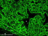 |
Laminin in the canalicular period of the lung (mouse, embryo) | Stain: Fluorescence microscopy with anti-laminin antibody. Laminin is a component of the basement membrane. Strong fluorescence visualizes the course of all basement membranes, especially around the bronchioles (B) and their branches in the developing lung tissue. In between the distal air spaces (t... | Bronchioli; Canalicular period | Poja Histology Collection - Respiratory System Subset |
| 238 |
 |
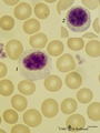
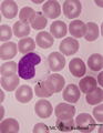
Large lymphocyte and monocyte in peripheral blood smear (human) | Stain: May-Grnwald-Giemsa (MGG). The large lymphocyte (1) has a slightly condensed nucleus and the cytoplasm contains some fine azurophilic granules, and is therefore sometimes called large granular lymphocyte (LGL) or killer cell. The monocyte (2) is generally larger than the lymphocyte with a roun... | Poja Histology Collection - Blood & Bone Marrow Subset | |
| 239 |
 |
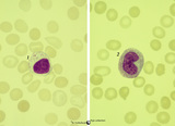
Large lymphocyte and small plasmacytoid lymphocyte in peripheral blood smear (human) | Stain: May-Grnwald-Giemsa (MGG). (1) large lymphocyte with a transparent light cytoplasm in which few fine granules are visible. (2) (inset) smaller plasmacytoid lymphocyte (activated young B cell) with light basophilic cytoplasm. | Poja Histology Collection - Blood & Bone Marrow Subset | |
| 240 |
 |
Large lymphocyte in peripheral blood smear (human) | Stain: May-Grnwald-Giemsa (MGG). The large lymphocyte has a dense chromatin structure and a large transparent cytoplasm with some granules (→). (On the contrary a monocyte exposes a more transparent nucleus and a slightly greyish cytoplasm). | Poja Histology Collection - Blood & Bone Marrow Subset | |
| 241 |
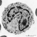 |
Late band-form of neutrophilic granulocyte (peripheral blood, human) | Electron microscopy. Band forms (9-12 μm) are the earliest stages of the motile two-lobed granulocytes. The horseshoe shaped nucleus (3) is irregularly and indicates progressing lobulation. The neutrophil contains many granules of varying sizes and densities. On base of routine electron microscopy ... | Poja Histology Collection - Blood & Bone Marrow Subset | |
| 242 |
 |
Late cap stage in tooth development - human, embryo | Stain: Azan. From top to bottom: Top side stellate reticulum (enamel pulp) consisting of a network of ectoderm-derived cells; Right side outer dental epithelium with part of the fibrous tooth follicle. This epithelium will further develop downwards as the outer layer of the Hertwig's epithelial root... | oral cavity | Poja Histology Collection - Oral Cavity Subset |
| 243 |
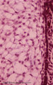 |
Late cap stage in tooth development - human, embryo | Stain: Azan. The stellate reticulum (enamel pulp) consists of a network of ectoderm-derived branched cells and fluid-filled spaces (a.o. proteoglycans). It is a specialized avascular layer as a support and protection for the inner dental epithelial cells. At the right side one layer of cuboidal oute... | oral cavity; tooth development; outer dental epithelium; stellate reticulum | Poja Histology Collection - Oral Cavity Subset |
| 244 |
 |
Late cap stage in tooth development - human, embryo | Stain: Azan. From top to bottom: At the top stellate reticulum (enamel pulp) consisting of a loose network of ectoderm-derived cells; Darker stained cell layers of the stratum intermedium; Columnar inner dental epithelium (presecreetory ameloblasts) at the distal side (secretion area) oriented towar... | oral cavity | Poja Histology Collection - Oral Cavity Subset |
| 245 |
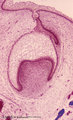 |
Late cap stage of tooth development - human, embryo; low magnification | Stain: Azan. From top to bottom: Stratified ectoderm with a distinct basal layer (red line) of cuboid cells; Dental lamina giving rise to the cap stage (center) and to the primordium of permanent tooth (right); Odontogenic organ or enamel organ (future deciduous tooth surrounded by fibrous tooth fol... | oral cavity; dental lamina | Poja Histology Collection - Oral Cavity Subset |
| 246 |
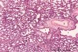 |
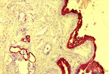
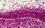
Late terminal sac period of developing lung (human, fetus, low magnification) | Stain: Hematoxylin and eosin. Cross-sections of a bronchus (1) with cartilage rings (2) closely associated with pulmonary arteries (3). The alveolar appearance is evident, and the open spaces represent future alveolar duct systems as well as alveolar sacs. The cellular septa are still thick due to n... | Lung development; Terminal sac period | Poja Histology Collection - Respiratory System Subset |
| 247 |
 |
Leukocyte Alkaline Phosphatase (LAP/NAP score) in peripheral blood smear (human) | Stain: Alkaline phosphatase staining (Ackerman). In mature neutrophils granules contain alkaline phosphatase. In eosinophils no alkaline phosphatase activity is present. A considerable number of brown stained neutrophils can be indicative for a patient with infection (leukemoid reaction, A). Image i... | Poja Histology Collection - Blood & Bone Marrow Subset | |
| 248 |
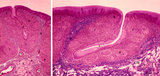 |
Lingual tonsil ('lymphoepithelial tissue', 'gut-associated lymphatic tissue' or GALT) (human) | Stain: Azan. A: Survey; B: detail of the crypt. The lingual tonsil consists of accumulations of bulging lingual lymphatic follicles in the dorsal part of the tongue behind the terminal sulcus, and belongs to the so-called Waldeyer's ring of pharyngeal lymphatic tissue. The left (A) and right (B) ... | lingual tonsil; GALT; Non-keratinized stratified squamous epithelium; crypt | Poja Histology Collection - Lymphatic Tissues and Organs Subset |
| 249 |
 |
Lingual tonsil ('lymphoepithelial tissue', 'gut-associated lymphatic tissue' or GALT) (human) | Stain: Azan. A: Left side shows a secondary lymphatic follicle (1) separated by the connective tissue of the proper lamina (2) from the lining non-keratinized stratified squamous epithelium (3). The mantle zone (4) is indicated by the peripheral dense aggregations of (memory) B lymphocytes. (5) diff... | lingual tonsil; GALT; non-keratinized stratified squamous epithelium | Poja Histology Collection - Lymphatic Tissues and Organs Subset |
| 250 |
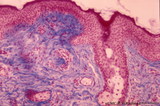 |
Lip (human), external skin surface | Stain: Azan. Keratinized squamous epithelium with one hair follicle associated with sebaceous gland. Richly vascularized connective tissue. Note the thin red cornified layer on the surface. | oral cavity | Poja Histology Collection - Oral Cavity Subset |