The Health Education Assets Library (HEAL) is a collection of over 22,000 freely available digital materials for health sciences education. The collection is now housed at the University of Utah J. Willard Marriott Digital Library.
TO
| Title | Description | Subject | Collection | ||
|---|---|---|---|---|---|
| 226 |
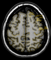 |
MRI Atlas: Brain (Axial) - Scan 8 - Labeled | Anatomical structures of the brain are identified in this scan with labeled outline from the UCLA Interactive Neurosciences MRI Atlas. | UCLA Interactive Neuroscience | |
| 227 |
 |
MRI Atlas: Brain (Axial) - Scan 8 - Labeled (Enlarged) | Anatomical structures of the brain are identified in this scan with labeled outline from the UCLA Interactive Neurosciences MRI Atlas. | UCLA Interactive Neuroscience | |
| 228 |
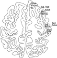 |
MRI Atlas: Brain (Axial) - Scan 8 - Labeled Outline | Anatomical structures of the brain are identified in this labeled outline from the UCLA Interactive Neurosciences MRI Atlas. | UCLA Interactive Neuroscience | |
| 229 |
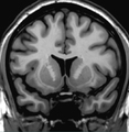 |
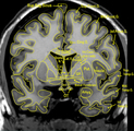
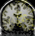
MRI Atlas: Brain (Coronal) - Scan 1 | Anatomical structures of the brain are depicted in this scan from the UCLA Interactive Neurosciences MRI Atlas. | Superior Frontal Gyrus; Mid-frontal Gyrus; Inferior Frontal Gyrus; Superior Temporal Gyrus; Mid-temporal Gyrus; Cingulate Gyrus (Body); Anterior Horn; Head of Caudate; Internal Capsule (Anterior Limb); Claustrum; Insular Cortex; Insula of Reil | UCLA Interactive Neuroscience |
| 230 |
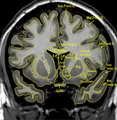 |
MRI Atlas: Brain (Coronal) - Scan 1 - Labeled | Anatomical structures of the brain are identified in this scan with labeled outline from the UCLA Interactive Neurosciences MRI Atlas. | UCLA Interactive Neuroscience | |
| 231 |
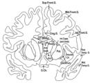 |
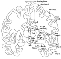
MRI Atlas: Brain (Coronal) - Scan 1 - Labeled Outline | Anatomical structures of the brain are identified in this labeled outline from the UCLA Interactive Neurosciences MRI Atlas. | UCLA Interactive Neuroscience | |
| 232 |
 |
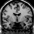
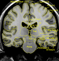
MRI Atlas: Brain (Coronal) - Scan 2 | Anatomical structures of the brain are depicted in this scan from the UCLA Interactive Neurosciences MRI Atlas. | Superior Sagittal Sinus; Superior Frontal Gyrus; Mid-frontal Gyrus; Inferior Frontal Gyrus; Superior Temporal Gyrus; Mid-temporal Gyrus; Inferior Temporal Gyrus; Cingulate Gyrus (Body); Anterior Commissure; Head of Caudate; Internal Capsule (Anterior Limb); Optic Tract; Basal Forebrain | UCLA Interactive Neuroscience |
| 233 |
 |
MRI Atlas: Brain (Coronal) - Scan 2 - Labeled | Anatomical structures of the brain are identified in this scan with labeled outline from the UCLA Interactive Neurosciences MRI Atlas. | UCLA Interactive Neuroscience | |
| 234 |
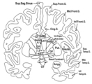 |
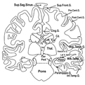
MRI Atlas: Brain (Coronal) - Scan 2 - Labeled Outline | Anatomical structures of the brain are identified in this labeled outline from the UCLA Interactive Neurosciences MRI Atlas. | UCLA Interactive Neuroscience | |
| 235 |
 |
MRI Atlas: Brain (Coronal) - Scan 3 | Anatomical structures of the brain are depicted in this scan from the UCLA Interactive Neurosciences MRI Atlas. | Superior Sagittal Sinus; Superior Frontal Gyrus; Pre-central Gyrus; Inferior Frontal Gyrus; Post-central Gyrus; Superior Temporal Gyrus; Mid-temporal Gyrus; Inferior Temporal Gyrus; Cingulate Gyrus; Head of Caudate; Internal Capsule (Anterior Limb); Optic Tract; Globus Pallidus Internus; Globus Pal... | UCLA Interactive Neuroscience |
| 236 |
 |
MRI Atlas: Brain (Coronal) - Scan 3 - Labeled | Anatomical structures of the brain are identified in this scan with labeled outline from the UCLA Interactive Neurosciences MRI Atlas. | UCLA Interactive Neuroscience | |
| 237 |
 |
MRI Atlas: Brain (Coronal) - Scan 3 - Labeled Outline | Anatomical structures of the brain are identified in this labeled outline from the UCLA Interactive Neurosciences MRI Atlas. | UCLA Interactive Neuroscience | |
| 238 |
 |
MRI Atlas: Brain (Coronal) - Scan 4 | Anatomical structures of the brain are depicted in this scan from the UCLA Interactive Neurosciences MRI Atlas.. | Superior Sagittal Sinus; Superior Frontal Gyrus; Pre-central Gyrus; Inferior Frontal Gyrus; Post-central Gyrus; Superior Temporal Gyrus; Mid-temporal Gyrus; Inferior Temporal Gyrus; Cingulate Gyrus; Heschl's Gyrus; Cerebral Peduncle | UCLA Interactive Neuroscience |
| 239 |
 |
MRI Atlas: Brain (Coronal) - Scan 4 - Labeled | Anatomical structures of the brain are identified in this scan with labeled outline from the UCLA Interactive Neurosciences MRI Atlas. | UCLA Interactive Neuroscience | |
| 240 |
 |
MRI Atlas: Brain (Coronal) - Scan 4 - Labeled Outline | Anatomical structures of the brain are identified in this labeled outline from the UCLA Interactive Neurosciences MRI Atlas. | UCLA Interactive Neuroscience | |
| 241 |
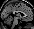 |
MRI Atlas: Brain (Sagittal) - Scan 1 | Anatomical structures of the brain are depicted in this scan from the UCLA Interactive Neurosciences MRI Atlas. | Central Sulcus; Post-central Gyrus; Pre-central Gyrus; Superior Frontal Gyrus; Cingulate Gyrus; Splenium of Body of Rostrum of Subcallosal Gyrus; Gyrus Rectus; Anterior Commissure; Red Nucleus; Midbrain; Corpus Quadrigemina; Pri Cerebellar Fissure; Cerebellar Fissure, Horizontal; Tentorium Cerebellu... | UCLA Interactive Neuroscience |
| 242 |
 |
MRI Atlas: Brain (Sagittal) - Scan 1 - Labeled | Anatomical structures of the brain are identified in this scan with labeled outline from the UCLA Interactive Neurosciences MRI Atlas. | UCLA Interactive Neuroscience | |
| 243 |
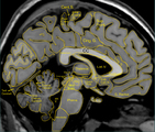 |
MRI Atlas: Brain (Sagittal) - Scan 1 - Labeled (Enlarged) | Anatomical structures of the brain are identified in this scan with labeled outline from the UCLA Interactive Neurosciences MRI Atlas. | UCLA Interactive Neuroscience | |
| 244 |
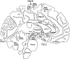 |
MRI Atlas: Brain (Sagittal) - Scan 1 - Labeled Outline | Anatomical structures of the brain are identified in this labeled outline from the UCLA Interactive Neurosciences MRI Atlas. | UCLA Interactive Neuroscience | |
| 245 |
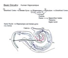 |
Supplements: Hippocampus - Basic Circuitry | Indexed diagram of the hippocampal pathways from the UCLA Interactive Neurosciences Supplements. | UCLA Interactive Neuroscience | |
| 246 |
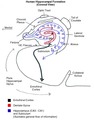 |
Supplements: Hippocampus - Circuitry - Human Hippocampal Formation (Coronal) | Indexed diagram of the hippocampal pathways from the UCLA Interactive Neurosciences Supplements. | UCLA Interactive Neuroscience | |
| 247 |
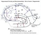 |
Supplements: Hippocampus - Hippocampal Circuitry and Neurotransmitters (Rat or Human) | Indexed diagram of the hippocampal pathways from the UCLA Interactive Neurosciences Supplements. | UCLA Interactive Neuroscience | |
| 248 |
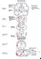 |
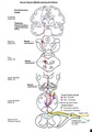
Supplements: Spinal Cord Pathways - Anterolateral System | Indexed diagram of the anterolateral system including the somatosensory cortex, thalamus, and collaterals to the mesencephalon. From the Interactive Neurosciences v3.0 Supplements. | Spinothalamic Fibers; Spinoreticular Fibers; Spinomesencephalic Fibers; Ventral Postero-Lateral Nucleus; Forebrain; Slide 85; Midbrain; Slide 23; Collaterals to Mesencephalon; Tectum; Slide 19; Upper Medulla; Slide 10; Lower Medulla; Slide 2; Cervical Level; Lamina I; Lamina IV; Lamina V; Anterior W... | UCLA Interactive Neuroscience |
| 249 |
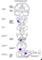 |
Supplements: Spinal Cord Pathways - Corticospinal | Indexed diagram of the anterolateral system focusing on the corticospinal pathway. From the Interactive Neurosciences v3.0 Supplements. | Corticospinal Tract; Forebrain; Slide 85; Internal Capsule; Midbrain; Slide 23; Cerebral Peduncle; Basis Pedunculi; Slide 19; Upper Medulla; Slide 10; Medullary Pyramid; Lower Medulla; Slide 2; Decussation of Pyramids; Cervical Level; Alpha Motor Neuron; Skeletal Muscle | UCLA Interactive Neuroscience |
| 250 |
 |
Supplements: Spinal Cord Pathways - Dorsal Column | Indexed diagram of the anterolateral system focusing on the dorsal column pathway. From the Interactive Neurosciences v3.0 Supplements. | Dorsal Column; Forebrain; Slide 85; Thalamus; Ventral Postero-Lateral Nucleus; Midbrain; Slide 23; Medial Lemniscus; Slide 19; Upper Medulla; Slide 10; Lower Medulla; Gracile Nucleus; Cuneate Nucleus; Internal Arcuate Fibers; Fasciculus Gracilis; Fasciculus Cuneatus; Upper Limb; Lower Limb; Cervical... | UCLA Interactive Neuroscience |