The Health Education Assets Library (HEAL) is a collection of over 22,000 freely available digital materials for health sciences education. The collection is now housed at the University of Utah J. Willard Marriott Digital Library.
TO
Filters: Collection: ehsl_heal
| Title | Description | Subject | Collection | ||
|---|---|---|---|---|---|
| 1 |
 |
An introduction and literature references to the Human Placenta as part of the POJA collection on FEMALE REPRODUCTIVE ORGANS | An introduction and literature references to the Human Placenta as part of the POJA collection on FEMALE REPRODUCTIVE ORGANS (PDF File) | placenta; references; preface | Poja Histology Collection - Placenta |
| 2 |
 |
Anchoring villi (human placenta, early midpregnancy) | Stain: Hematoxylin -azophloxine. Survey (A) and detail (B). The maternal side at the bottom shows reddish matrix-type fibrinoid accumulations (1) (Rohr's fibrinoid layer) close to an anchoring villus (2) and the cytotrophoblastic cell columns (3). The anchoring villus is lined by cytotrophoblast... | placenta; chorionic villi; Hofbauer cell; anchoring villus; cytotrophoblast | Poja Histology Collection - Placenta |
| 3 |
 |
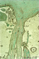
Anchoring villi (human placenta, early midpregnancy) | Stain: Hematoxylin -azophloxine. The maternal side at the bottom shows reddish matrix-type fibrinoid accumulations (1) (Rohr's fibrinoid layer) close to an anchoring villus (2) and a cytotrophoblastic cell column (3). The anchoring villus is lined by cytotrophoblasts (4) which are covered at th... | placenta; anchoring villus; fibrinoid; deciduas; cytotrophoblast | Poja Histology Collection - Placenta |
| 4 |
 |
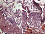
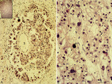
Chorioamnionitis (human) | Histologically chorioamnionitis describes the progression of the inflammatory process. Bacteria firstly colonized the chorioamniotic surface. In first two days polymorphonuclear granulocytes (PMN) migrate to the chorion (chorionitis) marginate and adhere to the bottom of the chorionic plate (stage 1... | chorioamnionitis; placenta; trophoblast | Poja Histology Collection - Placenta |
| 5 |
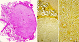 |
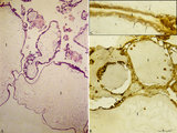
Chorioangioma (human) | Stain: (A) Hematoxylin-eosin (survey) and (B, C) van Gieson. (A) chorioangioma (1), chorionic plate (2), stem villus (3) and villi (5). (B) shows in (1) a large cellular mass with accumulated capillaries. At (2) part of the chorionic plate. Subchorionic fibrinoid stains yellow (4). (C) reveals t... | placenta; chorioangioma; villus; chorionic plate | Poja Histology Collection - Placenta |
| 6 |
 |
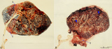
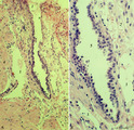
Choriocarcinoma (human) | Stain: Hematoxylin-eosin. (A) Inset: Survey tumor. Within the uterus (1) a choriocarcinoma forms solitary or multiple nodules composed of hemorrhagic necrotic areas (2) surrounded by neoplastic cells. It resembles an early implanted blastocyst with aggregations of mononuclear lighter-stained cyt... | choriocarcinoma; placenta; uterus; cytotrophoblast; GTD (gestational trophoblastic disease) | Poja Histology Collection - Placenta |
| 7 |
 |
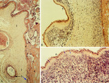
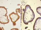
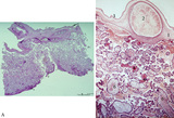
Complete hydatidiform mole (human) | Stain: (A) Hematoxylin-eosin. (B+C) Immunoperoxidase staining with diaminobenzidin (DAB) and hematoxylin counterstaining for keratin 7 (OVTL 12-30 antibody). (A)Trophoblast cells surround the dilated chorionic villi (1), with centrally large amounts of edematous mesenchymal stroma with almost no b... | trophoblast; complete hydatidiform mole; placenta; villus; immunohistochemistry; keratin-7 | Poja Histology Collection - Placenta |
| 8 |
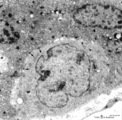 |
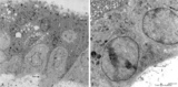
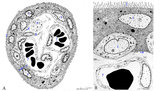
Electron microscopy of cytotrophoblast cell (human placenta, early pregnancy) | Below a single electron-light cytotrophoblast cell (CTC or Langhans cell, 1) covered by a syncytiotrophoblast cell (STC, 2) with two nuclei. A continuous basal lamina (3) separates the CTC and STC from the embryonic connective tissue. The CTC appears as a blast-like cell with a large electron-l... | placenta; tertiary villi; syncytiotrophoblast; placental barrier | Poja Histology Collection - Placenta |
| 9 |
 |
Electron microscopy of syncytiotrophoblast cells in tertiary villus (human placenta, midpregnancy) | The left photograph (A) shows the apical cytoplasm of a syncytiotrophoblast cell (STC, nucleus). The free cell surface displays small protrusions and a characteristic pattern of differently shaped microvilli; pinocytotic invaginations and a single macropinocytotic vacuole. Note the apical localized ... | placenta; tertiary villi; syncytiotrophoblast; electron microscopy | Poja Histology Collection - Placenta |
| 10 |
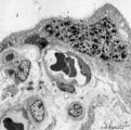 |
Electron microscopy of syncytiotrophoblast knot in tertiary villus (human placenta, almost full-term) | Electron microscopy. The mature villus, surrounded by intervillous space (1), contains capillaries (2) with erythrocytes and pericytes (3) embedded in fetal connective tissue elements (4). Two capillaries are localised close to the covering multinucleated syncytiotrophoblast cell (STC, 5) (vasculosy... | placenta; chorionic villi; syncytial knot; electron microscopy; vasculosyncytial membrane | Poja Histology Collection - Placenta |
| 11 |
 |
Electron microscopy of tertiary villus (human placenta, early pregnancy) | At the left (A), part of a tertiary (terminal) villus with the multinucleated syncytiotrophoblast cell (1) (STC) at the top. The apex shows protrusions and extensive microvilli of varying sizes (brushborder). Nuclei (1a) are sectioned at different levels and the cytoplasm contains abundant organelle... | placenta; chorionic villi; placental barrier; syncytiotrophoblast; electron microscopy | Poja Histology Collection - Placenta |
| 12 |
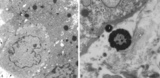 |
Electron microscopy of tertiary villus (human placenta, midpregnancy) | In the left photograph (A) is shown part of a tertiary villus with the organelle-rich cytoplasm of a syncytiotrophoblast cell (STC, 1). Below a single electron-light linked cytotrophoblast cell (CTC or Langhans cell, 2) covered by the STC. A higher magnification of another CTC (right photograph) sh... | placenta; tertiary villi; syncytiotrophoblast; electron microscopy; placental barrier | Poja Histology Collection - Placenta |
| 13 |
 |
Fetal cotyledons (normal term human placenta) | After dissection of one placental lobe several cotyledons (2) are visible in (A). Each fetal cotyledon (2) consists of a main stem villus (1) and all its branches. (B) After trypsinization of a cotyledon the tree of arborisation of the stem villus and its branches becomes visible. (By courtesy... | placenta; cotyledon; villus; trypsinization | Poja Histology Collection - Placenta |
| 14 |
 |
Immunohistochemistry of tertiary villi (human placenta, midpregnancy ) | Stain: Immunoperoxidase staining with diaminobenzidin (DAB) and hematoxylin counterstaining for keratin 7 (OVTL 12-30 antibody) at the left (A) and for human anti-chorionic gonadotrophin (hCG-DAKO 231antibody) at the right (B). Tertiary villi show a keratin-positive reaction (brown-stained) in all ... | placenta; chorionic villi; HCG; syncytiotrophoblast; keratin 7; immunohistochemistry | Poja Histology Collection - Placenta |
| 15 |
 |
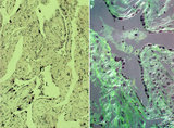
Intervillous space with villi (human placenta, midpregnancy, cross-section) | Stain: Trichrome (Goldner). In the middle a thick stem villus (cross-sections of left artery (1) and right vein (2) of the umbilical circulation in the thick embryonic connective tissue) with ramifications into terminal villi (tertiary). Most tertiary villi of varying diameters contain capillaries a... | placenta; chorionic villi; fibrinoid; cytotrophoblast; syncytiotrophoblast | Poja Histology Collection - Placenta |
| 16 |
 |
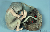
Macroscopy of fetus (human) | The fetus (1)is completely wrapped in a shiny transparent amnion (2) and closely associated with the brown-coloured placenta (3). (By courtesy of the Museum of Anatomy and Pathology, University Medical Center, St. Radboud University, Nijmegen, The Netherlands) | fetus; placenta; amnion | Poja Histology Collection - Placenta |
| 17 |
 |
Normal term placenta (human) | (A) Left (fetal surface): 30-90 cm long umbilical cord (1), remnant of ruptured transparent amnion (2→) , near the eccentric attachment of the umbilical cord to the chorion plate ramification of the umbilical vessels (3) (arteries are smaller than the veins). (B) Right (maternal surface). The su... | placenta; amnion; chorion plate; cotyledon | Poja Histology Collection - Placenta |
| 18 |
 |
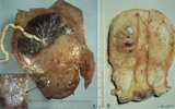
Normal term placenta with umbilical cord attached to the fetus (human) | The macroscopy shows the expelled placenta as a discoidal mass with a circular outline about 15-20 cm in the ruptured amnionitic and chorionic sacs. Amnion and smooth chorion (chorion leave) are fused and continuous with the margins of the placenta. The fetal surface (inner surface) is covered by... | placenta; amnion; chorion; umbilical cord; fetus | Poja Histology Collection - Placenta |
| 19 |
 |
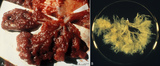
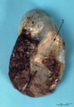
Partial hydatidiform mole and invasive mole (human) | (A) Macroscopy: The partial mole (1) occupies a large part of the placenta and is distinct from the normal chorionic plate where the umbilical cord (2) inserts eccentrically with branches of the umbilical vessels. The inset shows a circumscript area with swollen transparent grape-like vesicles (chor... | partial hydatidiform mole ; chorioadenoma | Poja Histology Collection - Placenta |
| 20 |
 |
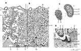
Scheme of electron microscopy of tertiary villus (human placenta, midpregnancy) | (See also POJA-L1227). (A) Survey and (B) detail of a tertiary placental villus lined by the multinucleated syncytiotrophoblast cell (1) (STC). The apex shows protrusions and extensive microvilli of varying sizes (brushborder, 2). Nuclei are sectioned at different levels and the cytoplasm contains a... | placenta; tertiary villi; placental barrier; electron microscopy | Poja Histology Collection - Placenta |
| 21 |
 |
Scheme of placental tissues (human) | See the http://www.healcentral.org/http://library.med.utah.edu/healassets/content/collections/poja_placenta/POJA-L1227-FemReprOrg-Placenta-scheme+introduction Placenta.pdf>detailed Legends for this slide (PDF file). | placenta; uterus; chorionic villi; decidua; trophoblast; cytotrophoblast | Poja Histology Collection - Placenta |
| 22 |
 |
Spiral artery in endometrium (human, midpregnancy) | Stain: Hematoxylin-eosin. (A) cross-sections of spiral arteries in endometrium. (B) semi-polarized section of part of a spiral artery. Dark-stained large trophoblast cells (3), partly replacing the cuboidal hypertrophic (B2) endothelial cells. (4) smooth muscle cells. (L) lumen of vessel with cell... | placenta; spiral artery; endometrium; trophoblast | Poja Histology Collection - Placenta |
| 23 |
 |
Spiral artery in endometrium (human, midpregnancy) | Stain: Hematoxylin-eosin. (A) Emerging into partly damaged intervillous space (1), filled with hemorrhage and fibrin (2). (3) uterine vein and (4) smooth muscle cell bundles at the endometrial-myometrial junction. (B) Lumen with hypertrophic endothelium at higher magnification (5). Note as an unu... | placenta; trophoblast; spiral artery; endometrium | Poja Histology Collection - Placenta |
| 24 |
 |
Stem villus (human placenta, midpregnancy) | Stain: Trichrome (Goldner). At the top the fetal site with part of the chorionic plate (1) with a thick stem villus (2) ramifying in two smaller villi with large blood vessels (3). The chorionic plate consists of a thick fibrous connective layer (green). Towards the intervillous space it is covere... | placenta; chorionic villi; stem villus; chorion plate | Poja Histology Collection - Placenta |
| 25 |
 |
Survey and detail of the chorionic plate and intervillous space (human placenta, full-term) | Stain: (A) Perjodic acid-Schiff reaction (PAS); (B) Hematoxylin-azophloxine. (A) At the top the chorionic plate (1) with cross-sections of umbilical vessels (2). At (3) the folded amnion covering the chorionic plate. Ramifications of thicker stem villi (6) demonstrate free-floating terminal villi ... | placenta; chorionic plate; amnion; terminal villi | Poja Histology Collection - Placenta |