Best known for his world-renowned neuro-ophthalmology unit based at the University of California, San Francisco, William Hoyt, MD collected here more than 850 of his best images covering a wide range of disorders.
William F. Hoyt, MD, Professor Emeritus of Ophthalmology, Neurology and Neurosurgery, Department of Ophthalmology, University of California, San Francisco.
NOVEL: https://novel.utah.edu/
TO
| Title | Description | Type | ||
|---|---|---|---|---|
| 1 |
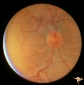 |
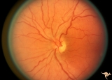
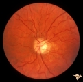
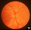
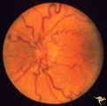
IE04 Acute Leber Optic Neuropathy | Microangiopathy without visual loss in a patient with acute Leber's optic neuropathy in the left eye. Pair with IE_05. Anatomy: Optic disc. Pathology: Optic neuropathy. Disease/ Diagnosis: Leber's optic neuropathy. Clinical: Central vision loss. | Image |
| 2 |
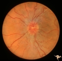 |
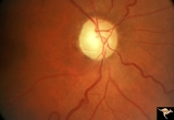
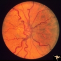
IE05 Acute Leber Optic Neuropathy | Patient has just begun to lose vision in his left eye due to Leber's optic neuropathy. Pair with IE_04. Anatomy: Optic disc. Pathology: Optic neuropathy. Disease/ Diagnosis: Leber's optic neuropathy. Clinical: Central vision loss. | Image |
| 3 |
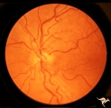 |
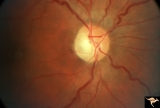
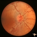
IE11 Subacute Leber Optic Neuropathy | Subacute Leber's Optic Neuropathy with distinct temporal wedge pallor and adjacent microangiopathy. 1973. Anatomy: Optic disc. Pathology: Optic neuropathy. Disease/ Diagnosis: Leber's optic neuropathy. Clinical: Blindness. | Image |
| 4 |
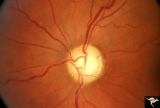 |
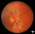
End Stage Leber Optic Neuropathy | End stage Leber's Optic Neuropathy. Severe diffuse pallor. Left eye. Pair with 15a. Anatomy: Optic disc. Pathology: Optic neuropathy. Disease/ Diagnosis: Leber's optic neuropathy. Clinical: Blindness. | Image |
| 5 |
 |
IE12a Acute Leber Optic Neuropathy | Subacute stage of Leber's Optic Neuropathy showing microangiopathy showing temporal pallor. Atrophy more advanced in left eye (12b) 1971, right eye. Anatomy: Optic disc. Pathology: Optic neuropathy. Disease/ Diagnosis: Leber's optic neuropathy. Clinical: Blindness. | Image |
| 6 |
 |
IE12b Subacute Leber Optic Neuropathy | Subacute stage of Leber's Optic Neuropathy showing microangiopathy showing temporal pallor. Atrophy more advanced in left eye. 1971, left eye. Anatomy: Optic disc. Pathology: Optic neuropathy. Disease/ Diagnosis: Leber's optic neuropathy. Clinical: Blindness. | Image |
| 7 |
 |
IE13a End Stage Leber Optic Neuropathy | End stage Leber's Optic Neuropathy. Note modest arteriolar narrowing. Note also the generalized pallor of the disc. Microangiopathy is no longer visible. Right eye. Pair with 13b. Anatomy: Optic disc. Pathology: Optic neuropathy. Disease/ Diagnosis: Leber's optic neuropathy. Clinical: Blindness. | Image |
| 8 |
 |
IE13b End Stage Leber Optic Neuropathy | End stage Leber's Optic Neuropathy. Note modest arteriolar narrowing. Note also the generalized pallor of the disc. Microangiopathy is no longer visible. Left eye. Pair with 13a. Anatomy: Optic disc. Pathology: Optic neuropathy. Disease/ Diagnosis: Leber's optic neuropathy. Clinical: Blindness. | Image |
| 9 |
 |
IE14a End Stage Leber Optic Neuropathy | End stage Leber's Optic Neuropathy. Dense temporal pallor. Microangiopathy is absent. Right eye. Pair with 14b. Anatomy: Optic disc. Pathology: Optic neuropathy. Disease/ Diagnosis: Leber's optic neuropathy. Clinical: Blindness. | Image |
| 10 |
 |
IE14b End Stage Leber Optic Neuropathy | End stage Leber's Optic Neuropathy. Dense temporal pallor. Microangiopathy is absent. Left eye. Pair with 14a. Anatomy: Optic disc. Pathology: Optic neuropathy. Disease/ Diagnosis: Leber's optic neuropathy. Clinical: Blindness. | Image |
| 11 |
 |
IE15a End Stage Leber Optic Neuropathy | End stage Leber's Optic Neuropathy. Severe diffuse pallor. Right eye. Pair with 15b. Anatomy: Optic disc. Pathology: Optic neuropathy. Disease/ Diagnosis: Leber's optic neuropathy. Clinical: Blindness. | Image |
| 12 |
 |
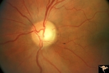
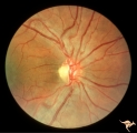
IE03 Acute Leber Optic Neuropathy | Acute stage of Leber optic neuropathy with microangiopathy and peripapillary nerve fiber layer thickening. The temporal nerve fiber layer is already showing atrophy. Central vision is grossly reduced. 1971. Anatomy: Optic disc. Pathology: Optic neuropathy. Disease/ Diagnosis: Leber's optic neuropath... | Image |
| 13 |
 |
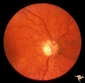
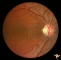
IE06 Subacute Leber Optic Neuropathy | Subacute Leber's optic neuropathy with microangiopathy with distinct temporal disc pallor. 1971. Anatomy: Optic disc. Pathology: Optic neuropathy. Disease/ Diagnosis: Leber's optic neuropathy. Clinical: Large central vision loss. | Image |
| 14 |
 |
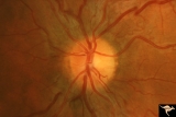
IE07 Subacute Leber Optic Neuropathy | Subacute Leber's optic neuropathy with microangiopathy. 1973. Anatomy: Optic disc. Pathology: Optic neuropathy. Disease/ Diagnosis: Leber's optic neuropathy. Clinical: Early central vision loss. | Image |
| 15 |
 |
IE08a Subacute Leber Optic Neuropathy | Subacute Leber Optic Neuropathy with temporal atrophy. August 5, 1980. Pair with IE_1, 2a&b, IE_8b, IE_9a&b. Anatomy: Optic disc. Pathology: Optic neuropathy. Disease/ Diagnosis: Leber's optic neuropathy. Clinical: Visual loss. | Image |
| 16 |
 |
IE08b Subacute Leber Optic Neuropathy | Subacute Leber Optic Neuropathy with temporal atrophy. August,1980. Pair with IE_1, 2a&b, IE_8a, IE_9a&b. Anatomy: Optic disc. Pathology: Optic neuropathy. Disease/ Diagnosis: Leber's optic neuropathy. Clinical: Visual loss. | Image |
| 17 |
 |
IE09a Chronic Leber Optic Neuropathy | Chronic Leber Optic Neuropathy with advancing temporal pallor. Notice the nerve fiber layer thickening has diminished. November 13, 1980. Pair with IE_1, 2a&b, IE_9b, IE_8a&b. Anatomy: Optic disc. Pathology: Optic neuropathy. Disease/ Diagnosis: Leber's optic neuropathy. Clinical: Blindness. | Image |
| 18 |
 |
IE09b Chronic Leber Optic Neuropathy | Chronic Leber Optic Neuropathy with advancing temporal pallor. Notice the nerve fiber layer thickening has diminished. November 13, 1980. Pair with IE_1, 2a&b, IE_9a, IE_8a&b. Anatomy: Optic disc. Pathology: Optic neuropathy. Disease/ Diagnosis: Leber's optic neuropathy. Clinical: Blindness. | Image |
| 19 |
 |
IE10a Chronic Leber Optic Neuropathy | Chronic Leber's Optic Neuropathy, August 8, 1969. Anatomy: Optic disc. Pathology: Optic neuropathy. Disease/ Diagnosis: Leber's optic neuropathy. Clinical: Blindness. | Image |
| 20 |
 |
IE10b Subacute Leber Optic Neuropathy | Subacute Leber's Optic Neuropathy, August 8, 1969, Left eye, pair with IE_10a, c. Anatomy: Optic disc. Pathology: Optic neuropathy. Disease/ Diagnosis: Leber's optic neuropathy. Clinical: Blindness. | Image |
| 21 |
 |
IE10c Chronic Leber Optic Neuropathy | February 12, 1970, Chronic Leber's Optic Neuropathy, 6 month follow up from 10b. Thickening of the nerve fiber layer is gone. Left eye, pair with IE_10a, c. Anatomy: Optic disc. Pathology: Optic neuropathy. Disease/ Diagnosis: Leber's optic neuropathy. Clinical: Blindness. | Image |
| 22 |
 |
Bilateral Papilledema from Non-tumor Etiology | Bilateral Papilledema with tortuous dilated veins from chronic lung disease with cyanosis. Note the remarkable tortuosity of retinal veins, evidence of retinal cyanosis. Anatomy: Optic disc. Pathology: Bilateral papilledema. Disease/Diagnosis: Pseudotumor due to chronic lung disease. Clinical notes:... | Image |
| 23 |
 |
Bilateral Papilledema from Non-tumor Etiology | Right eye. Bilateral Papilledema with tortuous dilated veins from chronic lung disease with cyanosis. Note the remarkable toruosity of retinal veins, evidence of retinal cyanosis. Anatomy: Optic disc. Pathology: Bilateral papiledema. Disease/Diagnosis: Pseudotumor due to chronic lung disease. Clinic... | Image |
| 24 |
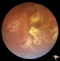 |
Bilateral Papilledema with Exudative Retinopathy | Right eye. Bilateral Papilledema with exudative retinopathy from vitamin A toxicity. Young boy. Near blind. Anatomy: Optic disc; Retina. Pathology: Bilateral papilledema; exudative retinopathy. Disease/Diagnosis: Hypervitaminosis A causing blindness. Clinical notes: Nearly blind; Headache. | Image |
| 25 |
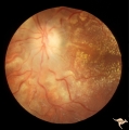 |
Bilateral Papilledema with Exudative Retinopathy | Left eye. Bilateral Papilledema with exudative retinopathy from vitamin A toxicity. Young boy. Near blind. Anatomy: Optic disc; Retina. Pathology: Bilateral papilledema; exudative retinopathy. Disease/Diagnosis: Hypervitaminosis A causing blindness. Clinical notes: Nearly blind; Headache. | Image |