Best known for his world-renowned neuro-ophthalmology unit based at the University of California, San Francisco, William Hoyt, MD collected here more than 850 of his best images covering a wide range of disorders.
William F. Hoyt, MD, Professor Emeritus of Ophthalmology, Neurology and Neurosurgery, Department of Ophthalmology, University of California, San Francisco.
NOVEL: https://novel.utah.edu/
TO
| Title | Description | Type | ||
|---|---|---|---|---|
| 1 |
 |
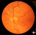
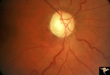
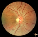
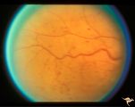
IE04 Acute Leber Optic Neuropathy | Microangiopathy without visual loss in a patient with acute Leber's optic neuropathy in the left eye. Pair with IE_05. Anatomy: Optic disc. Pathology: Optic neuropathy. Disease/ Diagnosis: Leber's optic neuropathy. Clinical: Central vision loss. | Image |
| 2 |
 |
IE05 Acute Leber Optic Neuropathy | Patient has just begun to lose vision in his left eye due to Leber's optic neuropathy. Pair with IE_04. Anatomy: Optic disc. Pathology: Optic neuropathy. Disease/ Diagnosis: Leber's optic neuropathy. Clinical: Central vision loss. | Image |
| 3 |
 |
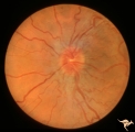
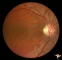
IE11 Subacute Leber Optic Neuropathy | Subacute Leber's Optic Neuropathy with distinct temporal wedge pallor and adjacent microangiopathy. 1973. Anatomy: Optic disc. Pathology: Optic neuropathy. Disease/ Diagnosis: Leber's optic neuropathy. Clinical: Blindness. | Image |
| 4 |
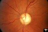 |
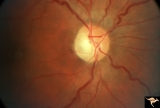
End Stage Leber Optic Neuropathy | End stage Leber's Optic Neuropathy. Severe diffuse pallor. Left eye. Pair with 15a. Anatomy: Optic disc. Pathology: Optic neuropathy. Disease/ Diagnosis: Leber's optic neuropathy. Clinical: Blindness. | Image |
| 5 |
 |
IE12a Acute Leber Optic Neuropathy | Subacute stage of Leber's Optic Neuropathy showing microangiopathy showing temporal pallor. Atrophy more advanced in left eye (12b) 1971, right eye. Anatomy: Optic disc. Pathology: Optic neuropathy. Disease/ Diagnosis: Leber's optic neuropathy. Clinical: Blindness. | Image |
| 6 |
 |
IE12b Subacute Leber Optic Neuropathy | Subacute stage of Leber's Optic Neuropathy showing microangiopathy showing temporal pallor. Atrophy more advanced in left eye. 1971, left eye. Anatomy: Optic disc. Pathology: Optic neuropathy. Disease/ Diagnosis: Leber's optic neuropathy. Clinical: Blindness. | Image |
| 7 |
 |
IE13a End Stage Leber Optic Neuropathy | End stage Leber's Optic Neuropathy. Note modest arteriolar narrowing. Note also the generalized pallor of the disc. Microangiopathy is no longer visible. Right eye. Pair with 13b. Anatomy: Optic disc. Pathology: Optic neuropathy. Disease/ Diagnosis: Leber's optic neuropathy. Clinical: Blindness. | Image |
| 8 |
 |
IE13b End Stage Leber Optic Neuropathy | End stage Leber's Optic Neuropathy. Note modest arteriolar narrowing. Note also the generalized pallor of the disc. Microangiopathy is no longer visible. Left eye. Pair with 13a. Anatomy: Optic disc. Pathology: Optic neuropathy. Disease/ Diagnosis: Leber's optic neuropathy. Clinical: Blindness. | Image |
| 9 |
 |
IE14a End Stage Leber Optic Neuropathy | End stage Leber's Optic Neuropathy. Dense temporal pallor. Microangiopathy is absent. Right eye. Pair with 14b. Anatomy: Optic disc. Pathology: Optic neuropathy. Disease/ Diagnosis: Leber's optic neuropathy. Clinical: Blindness. | Image |
| 10 |
 |
IE14b End Stage Leber Optic Neuropathy | End stage Leber's Optic Neuropathy. Dense temporal pallor. Microangiopathy is absent. Left eye. Pair with 14a. Anatomy: Optic disc. Pathology: Optic neuropathy. Disease/ Diagnosis: Leber's optic neuropathy. Clinical: Blindness. | Image |
| 11 |
 |
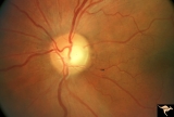
IE15a End Stage Leber Optic Neuropathy | End stage Leber's Optic Neuropathy. Severe diffuse pallor. Right eye. Pair with 15b. Anatomy: Optic disc. Pathology: Optic neuropathy. Disease/ Diagnosis: Leber's optic neuropathy. Clinical: Blindness. | Image |
| 12 |
 |
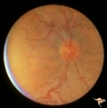
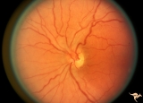
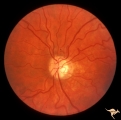
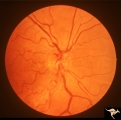
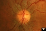
IE03 Acute Leber Optic Neuropathy | Acute stage of Leber optic neuropathy with microangiopathy and peripapillary nerve fiber layer thickening. The temporal nerve fiber layer is already showing atrophy. Central vision is grossly reduced. 1971. Anatomy: Optic disc. Pathology: Optic neuropathy. Disease/ Diagnosis: Leber's optic neuropath... | Image |
| 13 |
 |
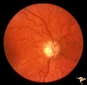
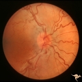
IE06 Subacute Leber Optic Neuropathy | Subacute Leber's optic neuropathy with microangiopathy with distinct temporal disc pallor. 1971. Anatomy: Optic disc. Pathology: Optic neuropathy. Disease/ Diagnosis: Leber's optic neuropathy. Clinical: Large central vision loss. | Image |
| 14 |
 |
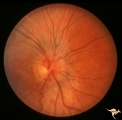
IE07 Subacute Leber Optic Neuropathy | Subacute Leber's optic neuropathy with microangiopathy. 1973. Anatomy: Optic disc. Pathology: Optic neuropathy. Disease/ Diagnosis: Leber's optic neuropathy. Clinical: Early central vision loss. | Image |
| 15 |
 |
IE08a Subacute Leber Optic Neuropathy | Subacute Leber Optic Neuropathy with temporal atrophy. August 5, 1980. Pair with IE_1, 2a&b, IE_8b, IE_9a&b. Anatomy: Optic disc. Pathology: Optic neuropathy. Disease/ Diagnosis: Leber's optic neuropathy. Clinical: Visual loss. | Image |
| 16 |
 |
IE08b Subacute Leber Optic Neuropathy | Subacute Leber Optic Neuropathy with temporal atrophy. August,1980. Pair with IE_1, 2a&b, IE_8a, IE_9a&b. Anatomy: Optic disc. Pathology: Optic neuropathy. Disease/ Diagnosis: Leber's optic neuropathy. Clinical: Visual loss. | Image |
| 17 |
 |
IE09a Chronic Leber Optic Neuropathy | Chronic Leber Optic Neuropathy with advancing temporal pallor. Notice the nerve fiber layer thickening has diminished. November 13, 1980. Pair with IE_1, 2a&b, IE_9b, IE_8a&b. Anatomy: Optic disc. Pathology: Optic neuropathy. Disease/ Diagnosis: Leber's optic neuropathy. Clinical: Blindness. | Image |
| 18 |
 |
IE09b Chronic Leber Optic Neuropathy | Chronic Leber Optic Neuropathy with advancing temporal pallor. Notice the nerve fiber layer thickening has diminished. November 13, 1980. Pair with IE_1, 2a&b, IE_9a, IE_8a&b. Anatomy: Optic disc. Pathology: Optic neuropathy. Disease/ Diagnosis: Leber's optic neuropathy. Clinical: Blindness. | Image |
| 19 |
 |
IE10a Chronic Leber Optic Neuropathy | Chronic Leber's Optic Neuropathy, August 8, 1969. Anatomy: Optic disc. Pathology: Optic neuropathy. Disease/ Diagnosis: Leber's optic neuropathy. Clinical: Blindness. | Image |
| 20 |
 |
IE10b Subacute Leber Optic Neuropathy | Subacute Leber's Optic Neuropathy, August 8, 1969, Left eye, pair with IE_10a, c. Anatomy: Optic disc. Pathology: Optic neuropathy. Disease/ Diagnosis: Leber's optic neuropathy. Clinical: Blindness. | Image |
| 21 |
 |
IE10c Chronic Leber Optic Neuropathy | February 12, 1970, Chronic Leber's Optic Neuropathy, 6 month follow up from 10b. Thickening of the nerve fiber layer is gone. Left eye, pair with IE_10a, c. Anatomy: Optic disc. Pathology: Optic neuropathy. Disease/ Diagnosis: Leber's optic neuropathy. Clinical: Blindness. | Image |
| 22 |
 |
Slow Flow (Chronic Hypoxic) Retinopathy | Examples of Slow flow (chronic hypoxic) retinopathy showing dilated and tortuous retinal veins and multiple capillary hemorrhages. This kind of retinopathy is produced by impaired arteriole circulation to the retina from various causes. Anatomy: Retina. Pathology: Ophthalmic artery venous malformati... | Image |
| 23 |
 |
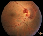
Retinal Signs of Atheromatous Embolization | Retinal signs of atheromatous embolization. Central retinal artery occlusion by soft atheromatous debris (mostly fibrin) causing blindness. Anatomy: Retina. Pathology: Carotid atheromatous disease. Disease/Diagnosis: Carotid atheromatous vascular disease. Clinical: Blindness. | Image |
| 24 |
 |
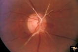
A406 Disc Swelling, Vitreous Effects | Prepapillary hemorrhage. Partial posterior vitreous detachment in myopic Asian patient. Reference: Katz B, Hoyt WF. Intrapapillary and peripapillary hemorrhage in young patients with incomplete posterior vitreous detachment. Signs of vitreopapillary traction. Ophthalmology. 1995 Feb;102(2):349-54. A... | Image |
| 25 |
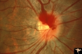
 |
A408 Disc Swelling, Vitreous Effects | Prepapillary hemorrhage. Partial posterior vitreous detachment in myopic Asian patient. Reference: Katz B, Hoyt WF. Intrapapillary and peripapillary hemorrhage in young patients with incomplete posterior vitreous detachment. Signs of vitreopapillary traction. Ophthalmology. 1995 Feb;102(2):349-54. A... | Image |