Home
Browse
Ask Us
Chat
Harmful Language Statement
Log in
Eccles Health Sciences Library
Advanced Search
Year
2018
2019
2020
2021
2022
2023
2024
TO
2018
2019
2020
2021
2022
2023
2024
Type
Image
89
Format
image/jpeg
88
application/pdf
1
Collection
Discovery and Innovation at Universit...
2
NOVEL - Neil R. Miller Collection
87
Filters:
Subject:
"Brain"
Type:
"Image"
76
-
100
of
89
<
1
2
3
4
>
Gallery view
Number of results to display per page
10
25
50
100
200
Sort by Relevance
Sort by Title A-Z
Sort by Title Z-A
Sort by Date Ascending
Sort by Date Descending
Sort by Last Modified Ascending
Sort by Last Modified Descending
Title
Date
Type
Setname
76
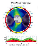
OCT Showing Band Atrophy in a Patient with an Optic Chiasmal Syndrome
2024-07
Image
ehsl_novel_nrm
77
Optic Chiasm Damage Following Excision of a Cavernous Angioma
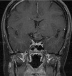
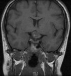
2024-07
Image
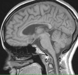
ehsl_novel_nrm
78
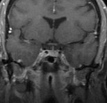
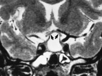
Optic Chiasm: Normal Appearance on Coronal MRI
2024-07
Image
ehsl_novel_nrm
79
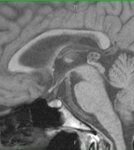
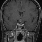
Optic Chiasm: Normal Appearance on MRI
2024-07
Image
ehsl_novel_nrm
80
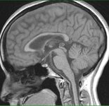
Optic Chiasm: Normal Appearance on Sagittal MRI
2024-07
Image
ehsl_novel_nrm
81
Optic Chiasm: Normal Appearance on Sagittal MRI
2024-07
Image
ehsl_novel_nrm
82
Optic Chiasm: Normal Appearance on Sagittal MRI
2024-07
Image
ehsl_novel_nrm
83
Optic Chiasmal Cavernoma
2024-07
Image
ehsl_novel_nrm
84
Optic Chiasmal Cavernoma
2024-07
Image
ehsl_novel_nrm
85
Optic Chiasmal Cavernoma
2024-07
Image
ehsl_novel_nrm
86
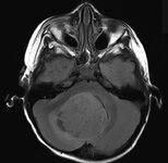
Posterior Fossa Mass Causing Hydrocephalus
2024-07
Image
ehsl_novel_nrm
87
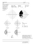
Temporal Hemianopia in the Left Eye of a Patient with Bitemporal Field Defects from a Cavernous Sngioma s/p Hemorrhage
2024-07
Image
ehsl_novel_nrm
88
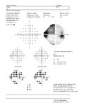
Temporal Hemianopia in the Right Eye of a Patient with Bitemporal Field Defects from a Cavernous Angioma s/p Hemorrhage
2024-07
Image
ehsl_novel_nrm
89
Upward Displacement of the Optic Chiasm by a Large Mass
2024-07
Image
ehsl_novel_nrm
76
-
100
of
89
<
1
2
3
4
>