Best known for his world-renowned neuro-ophthalmology unit based at the University of California, San Francisco, William Hoyt, MD collected here more than 850 of his best images covering a wide range of disorders.
William F. Hoyt, MD, Professor Emeritus of Ophthalmology, Neurology and Neurosurgery, Department of Ophthalmology, University of California, San Francisco.
NOVEL: https://novel.utah.edu/
TO
Filters: Collection: "ehsl_novel_wfh"
| Title | Description | Type | ||
|---|---|---|---|---|
| 126 |
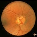 |
H29 Dysplasia with Hypoplasia (Elevated Dysplasia with Anomalous Vessels) | Right eye. Dysplasia with anomalous cilioretinal arterioles. Central retinal artery may be absent. Same patient as H_30. Anatomy: Optic disc. Pathology: Dysplasia of the optic disc. Disease/ Diagnosis: Elevated dysplasia with hypoplasia. | Image |
| 127 |
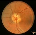 |
H30 Dysplasia with Hypoplasia (Elevated Dysplasia with Anomalous Vessels) | Left eye. Elevated dysplasia with anomalous cilioretinal vessels. Central retinal artery may be absent. Same patient as H_29. Anatomy: Optic disc. Pathology: Dysplasia of the optic disc. Disease/ Diagnosis: Elevated dysplasia with hypoplasia. | Image |
| 128 |
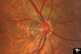 |
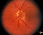
H48 Segmental Hypoplasia, Retinal-Nasal Hypoplasia | Bilateral nasal hypoplasia with absence of nasal nerve fiber layer and corresponding flag-like temporal field defect. Right eye. Same patient as H_49. Anatomy: Optic disc. Pathology: Nasal segmental disc hypoplasia. Disease/ Diagnosis: Congential anomaly. | Image |
| 129 |
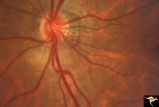 |
H49 Segmental Hypoplasia, Retinal-Nasal Hypoplasia | Bilateral nasal hypoplasia with absence of nasal nerve fiber layer and corresponding flag-like temporal field defect. Left eye. Same patient as H_48. Anatomy: Optic disc. Pathology: Nasal segmental disc hypoplasia. Disease/ Diagnosis: Congential anomaly. | Image |
| 130 |
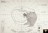 |
H51 Segmental Hypoplasia, Retinal-Nasal Hypoplasia | Visual field showing flag-like temporal field defect in patient shown in H_50. Anatomy: Optic disc. Pathology: Nasal segmental disc hypoplasia. Disease/ Diagnosis: Cogenital anomaly. | Image |
| 131 |
 |
H23 Dysplasia with Hypoplasia (Elevated Hysplasia with Anomalous Vessels) | Elevated dysplasia with anomalous vessels. Left eye. Hypoplasia with central glial tissue remnant. Japanese girl. Same patient as H_24. Anatomy: Optic disc. Pathology: Dysplasia of the optic disc. Disease/ Diagnosis: Elevated dysplasia with hypoplasia. | Image |
| 132 |
 |
H24 Dysplasia with Hypoplasia (Elevated Dysplasia with Anomalous Hessels) | Elevated dysplasia with anomalous vessels. Right eye. Hypoplastic with dysplasia. Japanese girl. Same patient as H_23. Anatomy: Optic disc. Pathology: Dysplasia of the optic disc. Disease/ Diagnosis: Elevated dysplasia with hypoplasia. | Image |
| 133 |
 |
H25 Dysplasia with Hypoplasia (Elevated Dysplasia with Anomalous Vessels) | Right eye. Elevated dysplasia with anomalous blood vessel pattern and peri-papillary choroidal malformation. Same patient as H_26. Anatomy: Optic disc. Pathology: Dysplasia of the optic disc. Disease/ Diagnosis: Elevated dysplasia with hypoplasia. | Image |
| 134 |
 |
H26 Dysplasia with Hypoplasia (Elevated Dysplasia with Anomalous Vessels) | Left eye. Dysplasia with grossly anomalous vascular pattern. Elevated dysplasia. Same patient as H_25. Anatomy: Optic disc. Pathology: Dysplasia of the optic disc. Disease/ Diagnosis: Elevated dysplasia with hypoplasia. | Image |
| 135 |
 |
H31 Dysplasia with Hypoplasia (Elevated Hysplasia with Anomalous Vessels) | Left eye. 26 year old man. Dysplasia with hypoplasia. Father of patient in H_32. Same patient as H_10. Son of patient in H_11 an H_12. Anatomy: Optic disc. Pathology: Dysplasia of the optic disc. Disease/ Diagnosis: Elevated dysplasia with hypoplasia. | Image |
| 136 |
 |
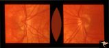
H06 Panhypoplasia | Bilateral hypoplasia. Top is Right eye - moderate. Bottom is Left eye - severe. Note venous tortuosity. Good example of double ring sign. De Morsier's syndrome.Septo-optic dysplasia. Same patient as H_7. Anatomy: Optic disc. Pathology: Hypoplasia of the optic nerve. Disease/ Diagnosis: Hypoplasia. ... | Image |
| 137 |
 |
H32 Dysplasia with Hypoplasia (Elevated Dysplasia with Anomalous Vessels) | Left eye. 6 year old boy. Severe dysplasia. Elevated dysplasia with medullated (myelinated) nerve fibers and anomalous vessels. Son of patient in H_31 and H_10. Grandson of patient in H_11 an H_12. Anatomy: Optic disc. Pathology: Dysplasia of the optic disc. Disease/ Diagnosis: Elevated dysplasia wi... | Image |
| 138 |
 |
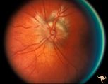
H37 Segmental Hypoplasia, Retinal, Tilted (Dysverted) Disc | Tilted (dysverted) disc in patient with high myopia. Note inferior nasal crescents with accompanying segmental hypoplasia. Man with bitemporal visual field defect. Anatomy: Optic disc, retina. Pathology: Hypoplasia secondary to retinal lesion. Disease/ Diagnosis: Segmental optic disc hypoplasia. Cli... | Image |
| 139 |
 |
H38 Segmental Hypoplasia; Retinal; Tilted (Dysverted) Disc | Right eye. Man with tilted (dysverted) disc with inferior nasal crescent and high myopia. Same patient as H_39. Anatomy: Optic disc; Retina. Pathology: Hypoplasia secondary to retinal lesion. Disease/ Diagnosis: Segmental optic disc hypoplasia. Clinical: Man with bitemporal visual field defects. | Image |
| 140 |
 |
H39 Segmental Hypoplasia, Retinal, Tilted (Dysverted) Disc | Visual field of patient in H_38 showing upper temporal field depression caused by inferior nasal hypoplasia. Anatomy: Optic disc; Retina. Pathology: Hypoplasia secondary to retinal lesion. Disease/ Diagnosis: Segmental optic disc hypoplasia. Clinical: Man with bitemporal visual field defects. | Image |
| 141 |
 |
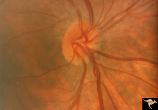
H40 Segmental Hypoplasia, Retinal, Tilted (Dysverted) Disc | 60 year old woman with incidental bitemporal visual field depression. Extreme tilting of optic disc with inferior nasal segmental hypoplasia. Nasal retinal ectasia. Same patient as H_41. Anatomy: Optic disc; retina. Pathology: Hypoplasia secondary to retinal lesion. Disease/ Diagnosis: Segmental opt... | Image |
| 142 |
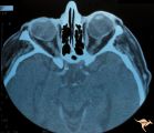 |
H41 Segmental Hypoplasia, Retinal, Tilted (Dysverted) Disc | CT scan of patient in H_40 showing marked nasal ectasia of the eyeballs. CT scan shows obliquely inserted optic nerves and marked nasal dysplasia of the eyeballs. Anatomy: Optic disc; retina. Pathology: Hypoplasia secondary to retinal lesion. Disease/ Diagnosis: Segmental optic disc hypoplasia. Imag... | Image |
| 143 |
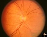 |
H53 Superior Segmental Optic Hypoplasia (SSOH) Topless Disc Syndrome | Superior segmental optic hypoplasia. High exit point of central retinal vessels. Anatomy: Optic disc. Pathology: Superior segmental optic hypoplasia (SSOH). Disease/ Diagnosis: Superior segmental optic hypoplasia (SSOH). | Image |