Best known for his world-renowned neuro-ophthalmology unit based at the University of California, San Francisco, William Hoyt, MD collected here more than 850 of his best images covering a wide range of disorders.
William F. Hoyt, MD, Professor Emeritus of Ophthalmology, Neurology and Neurosurgery, Department of Ophthalmology, University of California, San Francisco.
NOVEL: https://novel.utah.edu/
TO
| Title | Description | Type | ||
|---|---|---|---|---|
| 101 |
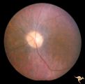
 |
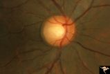
C204 Papillitis with Macular Star, Cat Scratch Disease | Proven Bartonella neuroretinitis. Left eye. October 17, 1986. Same eye as C2_03. Ocular disc edema with macular star (ODEMS). Woman. Anatomy: Optic disc; Retina. Pathology: Exudates in Henle's layer. DIsease/ Diagnosis: Neuroretinitis due to Bartonella Henslae (Cat Scratch). Clinical: Visual blurrin... | Image |
| 102 |
 |
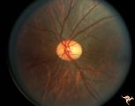
C205 Papillitis with Macular Star, Cat Scratch Disease | Proven Bartonella neuroretinitis. Macular star present, but not visible on image. 33 year old woman. Anatomy: Optic disc; Retina. Pathology: Axoplasmic stasis due to inflammation. Disease/ Diagnosis: Bartonella Henslae (Cat Scratch). Clinical: Visual blurring. | Image |
| 103 |
 |
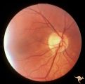
C206 Papillitis with Macular Star, Cat Scratch Disease | Proven Bartonella neuroretinitis. Resolved papillitis with residual retinal exudate. Man. Anatomy: Optic disc; Retina. Pathology: Axoplasmic stasis due to inflammation. Disease/ Diagnosis: Bartonella Henslae (Cat Scratch). Clinical: Visual blurring; Ocular edema with macular star (ODEMS). | Image |
| 104 |
 |
C208 Papillitis with Macular Star, Cat Scratch Disease | Proven Bartonella neuroretinitis. Man. Anatomy: Optic disc; Retinitis. Pathology: Axoplasmic stasis due to inflammation. Disease/ Diagnosis: Bartonella Henslae (Cat Scratch). Clinical: Visual blurring; Ocular disc edema with macular star (ODEMS). | Image |
| 105 |
 |
C24 Empty Disc | Left eye. Multiple cilioretinal arteries. Child of C_25 Anatomy: Optic disc. | Image |
| 106 |
 |
C25 Empty Disc | Right eye. Multiple cilioretinal arteries. Visual function normal. Father of C_24. Anatomy: Optic disc | Image |
| 107 |
 |
C26 Empty Disc | Right eye. Multiple cilioretinal arteries. Patient had dysplastic kidneys. Papillorenal Syndrome (PRS). Hand motion vision. 17 year old girl. Anatomy: Optic disc. | Image |
| 108 |
 |
C27 Empty Disc | Multiple cilioretinal arteries. Chronic interstitial nephritis. Renal and optic disc dysplasia. Papilorenal Syndrome (PRS). No central retinal artery. Anatomy: Optic disc. | Image |
| 109 |
 |
C28 Empty Disc | Left eye. Woman. Multiple cilioretinal vessels. Visual function normal. Anatomy: Optic disc. | Image |
| 110 |
 |
C29 Empty Disc | Left eye. Papilorenal Syndrome (PRS). Pair with C_30. Anatomy: Optic disc. | Image |
| 111 |
 |
C30 Empty Disc | Right eye. Papillorenal Syndrome (PRS). Pair with C_29. Anatomy: Optic disc. | Image |
| 112 |
 |
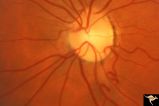
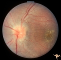
C303 Nodular Papillopathies (Sarcoid) | Lumpy infiltrative papillopathy in a patient with proven sarcoid. Anatomy: Optic disc. Pathology: Axoplasmic stasis due to sarcoid infiltration. Disease/ Diagnosis: Sarcoid papillopathy. Clinical: Unknown? | Image |
| 113 |
 |
C304 Nodular Papillopathies (Sarcoid) | Lumpy nodular disc infiltration from sarcoid. Anatomy: Optic disc. Pathology: Axoplasmic stasis due to sarcoid infiltration. Disease/ Diagnosis: Sarcoid papillopathy. Clinical: Unknown? | Image |
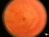
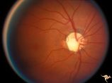
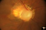
| 114 |
 |
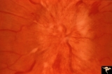
C305 Nodular Papillopathies (Sarcoid) | July 1984 shows multiple infiltrative nodules on the optic disc in addition to circumferential subretinal yellow exudates. 32 year old black woman. Same patient as C3_06 and C3_07. Anatomy: Optic disc; Retina. Pathology: Axoplasmic stasis due to sarcoid infiltration and retinal exudation. Disease/ ... | Image |
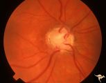
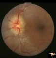
| 115 |
 |
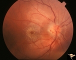
C306 Nodular Papillopathies (Sarcoid) | Lumpy disc swelling with retinal folds and a macular star in a patient with sarcoid. Presentation in October 1983. Same patient as C3_05 and C3_07. Anatomy: Optic disc; Retina. Pathology: Axoplasmic stasis due to sarcoid infiltration. Disease/ Diagnosis: Axoplasmic stasis due to sarcoid infiltration... | Image |
| 116 |
 |
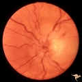
C307 Nodular Papillopathies (Sarcoid) | Fluorescein angiogram shows striking staining of the sarcoid nodules. July 1984. Same patient as C3_05 and C3_06. Corresponds with July 1984 image, C3_06. Anatomy: Optic disc. Pathology: Axoplasmic stasis due to sarcoid infiltration. Disease/ Diagnosis: Sarcoid papillopathy. Clinical: Unknown? | Image |
| 117 |
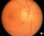 |
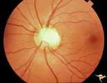
C308 Nodular Papillopathies (Sarcoid) | Nodular infiltrative papillopathy in a patient with sarcoid. Woman. Anatomy: Optic disc. Pathology: Axoplasmic stasis due to sarcoid infiltration. Disease/ Diagnosis: Sarcoid papillopahty. Clinical: Unknown? | Image |
| 118 |
 |
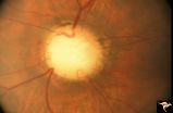
C33 Anomalous Pale Disc | Woman. Multiple cilioretinal arteries. Veins all empty into eye. Anomalous venous exit from nasal edge of optic disc. Visual function normal. Pair with C_36. Anatomy: Optic disc. | Image |
| 119 |
 |
C35 Anomalous Pale Disc | Macro disc appears pale because of large diameter. Woman. Right eye. Anatomy: Optic disc. | Image |
| 120 |
 |
C36 Anomalous Pale Disc | Multiple cilioretinal arteries. Pale appearance. Normal optic nerve function. Good example of "empty disc". Pair with C_33. Anatomy: Optic disc. | Image |
| 121 |
 |
C37 Anomalous Pale Disc | "Watermelon" disc. Woman. Normal function. Left eye. Anatomy: Optic disc. | Image |
| 122 |
 |
C38 Anomalous Pale Disc | Megalopapilla in -8 myopic eye. Right eye. Anatomy: Optic disc. Clinical: High myope. | Image |
| 123 |
 |
C401 Luetic Papillopathy (Gumma of the Optic Disc) | Diffuse optic disc swelling with tortuous capillary dilations indicating inflammatory cellular infiltration. October 2001. Same eye as C4_02. Anatomy: Optic disc. Pathology: Axoplasmic stasis due to syphillitic infection. Luetic papillopathy (Syphyllis). Clinical: Visual loss. | Image |
| 124 |
 |
C402 Luetic Papillopathy (Gumma of the Optic Disc) | November 2001. Same eye as C4_01 after treatment with penicillin. Disc swelling went away and good visual function returned. Anatomy: Optic disc. Pathology: Axoplasmic stasis due to syphillitic infection. Disease/ Diagnosis: Luetic papillopathy (Syphillis). Clinical: Improving visual loss. | Image |
| 125 |
 |
Cerebellar Macular Degeneration | Cerebellar retinal degeneration with narrowed arterioles. Disc pallor. Granular retinal degeneration. 10 year old boy with mental degenerations and seizures. Anatomy: Retina. Pathology: Optic atrophy. Disease/Diagnosis: Congenital retinal cerebellar degeneration. Clinical: Severe mental retardation ... | Image |