Best known for his world-renowned neuro-ophthalmology unit based at the University of California, San Francisco, William Hoyt, MD collected here more than 850 of his best images covering a wide range of disorders.
William F. Hoyt, MD, Professor Emeritus of Ophthalmology, Neurology and Neurosurgery, Department of Ophthalmology, University of California, San Francisco.
NOVEL: https://novel.utah.edu/
TO
| Title | Description | Type | ||
|---|---|---|---|---|
| 51 |
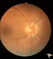 |
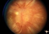
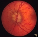
Bilateral Papilledema | Left eye. Chronic Bilateral Papilledema. Anatomy: Optic disc. Pathology: Chronic bilateral papilledema. Disease/Diagnosis: Pseudotumor long standing. Clinical notes: Chronic headache; Obesity. | Image |
| 52 |
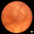 |
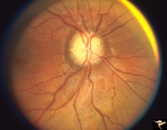
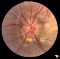
Bilateral Papilledema | Left eye. Bilateral Papilledema from vitamin A toxicity in young girl. Anatomy: Optic disc. Pathology: Bilateral papilledema. Disease/Diagnosis: Pseudotumor due to vitamin A toxicity in a young girl. Clinical notes: Headache. | Image |
| 53 |
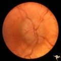 |
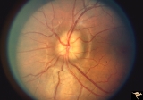
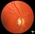
Bilateral Papilledema | Right eye. Bilateral Papilledema from vitamin A toxicity in young girl. Anatomy: Optic disc. Pathology: Bilateral papilledema. Disease/Diagnosis: Pseudotumor due to vitamin A toxicity in a young girl. Clinical notes: Headache. | Image |
| 54 |
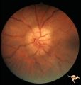 |
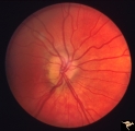
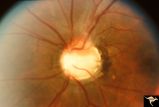
Bilateral Papilledema | Chronic Bilateral Papilledema. Anatomy: Optic disc. Pathology: Chronic bilateral papilledema. Disease/Diagnosis: Pseudotumor long standing. Clinical notes: Chronic headache; Obesity. | Image |
| 55 |
 |
Bilateral Papilledema from Pseudotumor | Right eye. Pseudotumor syndrome. Multiple endocrine adenomas. Woman. Anatomy: Optic disc. Pathology: Bilateral papilledema. Disease/Diagnosis: Pseudotumor associated with multiple endocrine adenomas. Clinical notes: Headache; Obesity. | Image |
| 56 |
 |
Bilateral Papilledema in Pseudotumor | Left eye. Pseudotumor syndrome. Multiple endocrine adenomas. Woman. Anatomy: Optic disc. Pathology: Bilateral papilledema. Disease/Diagnosis: Pseudotumor associated with multiple endocrine adenomas. Clinical notes: Headache; Obesity. | Image |
| 57 |
 |
Bilateral Papilledema with Cyanotic Heart Disease | Bilateral Papilledema with cyanotic heart disease in a young boy. Anatomy: Optic disc. Pathology: Papilledema. Disease/Diagnosis: Pseudotumor due to cyanotic heart disease. Clinical notes: Young boy with clubbing. | Image |
| 58 |
 |
Bilateral Papilledema with Exudative Retinopathy | Bilateral Papilledema with exudative retinopathy from vitamin A toxicity. Young boy. Near blind. Anatomy: Optic disc; Retina. Pathology: Bilateral papilledema; exudative retinopathy. Disease/Diagnosis: Hypervitaminosis A causing blindness. Clinical notes: Nearly blind; Headache. | Image |
| 59 |
 |
Bilateral Papilledema with Pseudotumor Cerebri | Right eye. Mild bilateral papilledema in a 7 year old boy. Cause of swelling unknown. Growth failure treated with thyroid medication. Anatomy: Optic disc. Pathology: Bilateral papilledema. Disease/Diagnosis: Intracranial hypertension due to treatment of growth failure with thyroid medicaltion. Clini... | Image |
| 60 |
 |
Bilateral Papilledema with Pseudotumor Cerebri | Left eye. Mild bilateral papilledema in a 7 year old boy. Cause of swelling unknown. Growth failure treated with thyroid medication. Anatomy: Optic disc. Pathology: Bilateral papilledema. Disease/Diagnosis: Intracranial hypertension due to treatment of growth failure with thyroid medication. Clinica... | Image |
| 61 |
 |
Bilateral Papilledema with Pseudotumor Cerebri | Chronic appearance of swelling in right eye. 29 year old woman. Bilateral papilledema. Anatomy: Optic disc. Pathology: Bilateral papilledema. Disease/Diagnosis: Intracranial hypertension due to treatment of growth failure with thyroid medication. Clinical: symptoms: headache, signs: bilateral papill... | Image |
| 62 |
 |
Bilateral Severe Hemorrhagic Papilledema | Right eye. Bilateral hyperacute papilledema with rapid blindness associated with dural sinus occlusion. Both eyes were nearly blind. Young man. Anatomy: Optic disc. Pathology: Papilledema. Disease/Diagnosis: Bilateral hyperacute papilledema | Image |
| 63 |
 |
Bilateral Severe Hemorrhagic Papilledema | Left eye. Bilateral hyperacute papilledema with rapid blindess associated with dural sinus occlusion. Both eyes were nearly blind. Boy. | Image |
| 64 |
 |
Buried and Visible Drusen | PP_19b: right eye : visible drusen in an eleven year old girl; PP_19a: left eye with buried drusen. Anatomy: Optic disc Pathology: Drusen of the optic disc Disease/Diagnosis: Drusen of the optic disc Clinical: Normally functioning eye with drusen. | Image |
| 65 |
 |
Buried and Visible Drusen | PP_19a Left eye with buried drusen. PP_19b: right eye : visible drusen. Eleven year old girl. Anatomy: Optic disc. Pathology: Drusen of the optic disc. Disease/Diagnosis: Drusen of the optic disc. Clinical notes: Normally functioning eye with drusen. | Image |
| 66 |
 |
Buried Drusen | Young woman with pseudo papilledema from buried drusen with associated visual field defects. Barely visible in the upper arcuate nerve fibers is a slit like defect. Anatomy: Optic disc. Pathology: Drusen of the optic disc. Disease/Diagnosis: Drusen of the optic disc. Clinical notes: This patient had... | Image |
| 67 |
 |
Buried Drusen | Suspected buried drusen in a girl. Anatomy: Optic disc. Pathology: Drusen of the optic disc. Disease/Diagnosis: Drusen of the optic disc. Clinical notes: Normally functioning eye with suspected drusen. | Image |
| 68 |
 |
Buried Drusen | Buried drusen; PP_13a: Right eye. Note lumpy disc margin, especially temporally. Also note absence of optic cup. Excellent example of pseudo papilledema with buried drusen. Anatomy: Optic disc. Pathology: Drusen of the optic disc. Disease/Diagnosis: Drusen of the optic disc. Clinical notes: Patient ... | Image |
| 69 |
 |
Buried Drusen | Buried drusen. Left eye. Note lumpy disc margin, especially temporally. Also note absence of optic cup. Excellent example of pseudo papilledema with buried drusen. Pair with PP_13a. Anatomy: Optic disc. Pathology: Drusen of the optic disc. Disease/Diagnosis: Drusen of the optic disc. Clinical notes... | Image |
| 70 |
 |
Buried Drusen | Excellent example of pseudo papilledema with sub surface drusen at 10:00 and 1:00. Anatomy: Optic disc. Pathology: Drusen of the optic disc. Disease/Diagnosis: Drusen of the optic disc. Clinical notes: Normally functioning eye with drusen. | Image |
| 71 |
 |
Buried Drusen with Sub-retinal Neovascular Net | Buried drusen with sub-retinal neovascular net. Both PP29a and PP29b are left eye: 17 year old girl; Visual acuity 10/400. Anatomy: Optic disc. Pathology: Drusen of the optic disc. Disease/Diagnosis: Drusen of the optic disc. Clinical notes: Loss of central vision due to subretinal neovascularizatio... | Image |
| 72 |
 |
Buried Drusen with Sub-retinal Neovascular Net | Buried drusen with sub-retinal neovascular net. This is the same left eye. Appearance of the central retina of the left eye. Both PP29a & b are left eye: 17 year old girl; Visual acuity 10/400. Anatomy: Optic disc. Pathology: Drusen of the optic disc. DIsease/Diagnosis: Drusen of the optic disc. Cl... | Image |
| 73 |
 |
C06 Pits of the Optic Disc | Right eye. Temporal pit. 6 year old with see-saw nystagmus. Anatomy: Optic disc. Clinical: Six-year old with see-saw nystagmus. | Image |
| 74 |
 |
C07 Pits of the Optic Disc | Left eye. Temporal pit. Man. Anatomy: Optic disc. | Image |
| 75 |
 |
C08 Pits of the Optic Disc | Left eye. Large cavitary anomaly (pit). Man. 20/100 visual acuity. Superior nasal visual field defect. May not have a central retinal artery. Anatomy: Optic disc. Clinical: Man. 20/100 visual acuity. Superior nasal visual field defect. | Image |