Best known for his world-renowned neuro-ophthalmology unit based at the University of California, San Francisco, William Hoyt, MD collected here more than 850 of his best images covering a wide range of disorders.
William F. Hoyt, MD, Professor Emeritus of Ophthalmology, Neurology and Neurosurgery, Department of Ophthalmology, University of California, San Francisco.
NOVEL: https://novel.utah.edu/
TO
| Title | Description | Type | ||
|---|---|---|---|---|
| 1 |
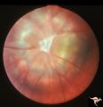
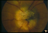 |
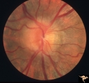
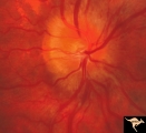
F401 Pigment Epithelial Hamartoma of Optic Disc | Optic disc tumor discovered incidentally in a 32 year old Asian woman who had no complaints about visual function in her involved left eye. Fundus slide shows granular elevation of left disc obscurring major disc vessels. Some of the granules has a shiny crystalline appearance. Near the vessel entra... | Image |
| 2 |
 |
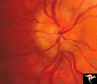
F403 Pigment Epithelial Hamartoma of Optic Disc | Optic disc tumor discovered incidentally in a 32 year old Asian woman who had no complaints about visual function in her involved left eye. Fundus slide shows granular elevation of left disc obscuring major disc vessels. Some of the granules has a shiny crystalline appearance. Near the vessel entran... | Image |
| 3 |
 |
Pigment Epithelial Hamartoma of Optic Disc | Optic disc tumor discovered incidentally in a 32 year old Asian woman who had no complaints about visual function in her involved left eye. Fundus slide shows granular elevation of left disc obscurring major disc vessels. Some of the granules has a shiny crystalline appearance. Near the vessel entra... | Image |
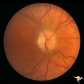
| 4 |
 |
Bilateral Papilledema | Right eye. Bilateral Papilledema in a patient with hyperthyroidism. Woman. Anatomy: Optic disc. Pathology: Papilledema. Disease/Diagnosis: Bilateral papilledema with hyperthyroidism. | Image |
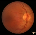
| 5 |
 |
Bilateral Papilledema | Left eye. Bilateral Papilledema in a patient with hyperthyroidism. Woman. Anatomy: Optic disc. Pathology: Papilledema. Disease/Diagnosis: Bilateral papilledema. | Image |
| 6 |
 |
Bilateral Severe Hemorrhagic Papilledema | Right eye. Bilateral hyperacute papilledema with rapid blindness associated with dural sinus occlusion. Both eyes were nearly blind. Young man. Anatomy: Optic disc. Pathology: Papilledema. Disease/Diagnosis: Bilateral hyperacute papilledema | Image |
| 7 |
 |
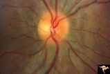
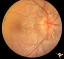
Chronic Papilledema due to Brain Tumor | Right eye. Chronic papilledema wth white centrally located exudates in a man with hemispheric glioma. Anatomy: Optic disc. Pathology: Papilledema. Disease/Diagnosis: Chronic papilledema. | Image |
| 8 |
 |
Chronic Papilledema due to Brain Tumor | Left eye. Chronic papilledema with white centrally located exudates in a man with hemispheric glioma. Anatomy: Optic disc. Pathology: Papilledema. Disease/Diagnosis: Chronic papilledema. | Image |
| 9 |
 |
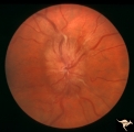
Chronic Papilledema due to Brain Tumor - Resolved | Right eye - same as P_40a - follow up after 4 months. Chronic papilledema resolved after treatment showing residual atrophy. Anatomy: Optic disc. Pathology: Papilledema. Disease/Diagnosis: Chronic papilledema. | Image |
| 10 |
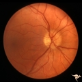 |
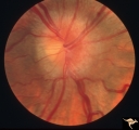
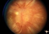
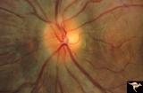
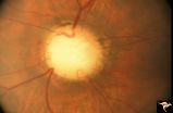
P43a Asymmetrical Papilledema due to Brain Tumor | Right eye. Early papilledema. Incipient papilledema barely recognizable. Early papilledema due to posterior fossa meningioma in a boy. Anatomy: Optic disc. Pathology: Papilledema. Disease/ Diagnosis: Asymmetrical papilledema due to posterior fossa meningioma. | Image |
| 11 |
 |
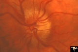
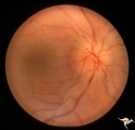
Papilledema with Choroidal Folds | Chronic papilledema with choroidal folds. Frontal astrocytoma. Man. Anatomy: Optic disc. Pathology: Papilledema. Disease/Diagnosis: Chronic papilledema. | Image |
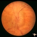
| 12 |
 |
Post Papilledema | Right eye. Post Papilledema with minimal optic disc changes after treatment for temporal lobe glioma. Minimal optic disc haze. Optic disc. Pathology: Papilledema. Disease/Diagnosis: Post Papilledema due to temporal lobe glioma. | Image |
| 13 |
 |
Post Papilledema Disc Blurring | Left eye. 8 year old boy. Post papilledema due to brain tumor. Note the entire peripapillary nerve fiber is blurred but the optic discs are barely elevated. Anatomy: Optic disc. Pathology: Brain tumor. Disease/Diagnosis: Papilledema. Clinical: Post papilledema due to brain tumor. | Image |
| 14 |
 |
Post Papilledema Disc Blurring | Right eye. 8 year old boy. Post papilledema due to brain tumor. Note the entire peripapillary nerve fiber is blurred but the optic discs are barely elevated. Anatomy: Optic disc. Pathology: Post papilledema. Disease/Diagnosis: Post papilledema due to brain tumor. | Image |
| 15 |
 |
Progression of Papilledema due to Metastatic Melanoma | Right eye. Rapid progression of papilledema due to metastatic occipital melanoma. Papilladema has increased so that it has almost filled in the optic cup. Anatomy: Optic disc. Pathology: Papilledema. Disease/Diagnosis: Papilledema due to metastatic occipital melanoma. | Image |
| 16 |
 |
Progression of Papilledema due to Metastatic Melanoma | Left eye at presentation. Early stage. Rapid progression of papilledema due to metastatic occipital melanoma. Anatomy: Optic disc. Pathology: Papilledema. Disease/Diagnosis: Papilledema due to metastatic occipital melanoma. | Image |
| 17 |
 |
Progression of Papilledema due to Metastatic Melanoma | Right eye at presentation. Early stage bilateral papilledema in a man. Note increased papilledema. Rapid progression of papilledema due to occipital metastatic melanoma. Anatomy: Optic disc. Pathology: Papilledema. Disease/Diagnosis: Early stage bilateral papilledema. | Image |
| 18 |
 |
Progression of Papilledema due to Metastatic Melanoma | Right eye at presentation. Early stage bilateral papilledema in a man. Rapid progression of bilateral papilledema due to metastatic occipital melanoma. Anatomy: Optic disc. Pathology: Papilledema. Disease/Diagnosis: Papilledema. | Image |
| 19 |
 |
C29 Empty Disc | Left eye. Papilorenal Syndrome (PRS). Pair with C_30. Anatomy: Optic disc. | Image |
| 20 |
 |
C30 Empty Disc | Right eye. Papillorenal Syndrome (PRS). Pair with C_29. Anatomy: Optic disc. | Image |
| 21 |
 |
C202 Papillitis with Macular Star Cat Scratch Disease. | Proven Bartonella neuroretinitis. 23 year old man. Ocular disc edema with macular star (ODEMS). Anatomy: Optic disc; Retina. Pathology: Axoplasmic stasis due to inflammation; Exudate in Henle's layer. Neuroretinitis due to Bartonella Henslae (or cat scratch). Clinical: Visual blurring; Optic disc sw... | Image |
| 22 |
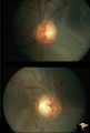 |
H83 Chiasmal Hemioptic Hypoplasia | De Morsier synrome with congenital bitemporal hemianopia. Note nasal hypoplasia of both optic discs. Left eye above, right eye below. Anatomy: Optic disc. Pathology: Chiasmal hemioptic hypoplasia. Disease/ Diagnosis: Congenital anomaly involving chiasm. | Image |
| 23 |
 |
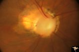
Bilateral Papilledema | Right eye. Bilateral Papilledema with hypoparathyroidism. Woman. Anatomy: Optic disc. Pathology: Papilledema. Disease/Diagnosis: Papilledema with hypoparathyroidism. | Image |
| 24 |
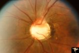 |
C08 Pits of the Optic Disc | Left eye. Large cavitary anomaly (pit). Man. 20/100 visual acuity. Superior nasal visual field defect. May not have a central retinal artery. Anatomy: Optic disc. Clinical: Man. 20/100 visual acuity. Superior nasal visual field defect. | Image |
| 25 |
 |
C17 Morning Glory Disc | "Morning Glory" disc. CT normal. Anatomy: Optic disc. Clinical: CT normal. | Image |