Best known for his world-renowned neuro-ophthalmology unit based at the University of California, San Francisco, William Hoyt, MD collected here more than 850 of his best images covering a wide range of disorders.
William F. Hoyt, MD, Professor Emeritus of Ophthalmology, Neurology and Neurosurgery, Department of Ophthalmology, University of California, San Francisco.
NOVEL: https://novel.utah.edu/
TO
Filters: Collection: "ehsl_novel_wfh" Date: "1992"
1 - 25 of 11
| Title | Description | Type | ||
|---|---|---|---|---|
| 1 |
 |
A405 Disc Swelling, Vitreous Effects | Prepapillary hemorrhage. Partial posterior vitreous detachment in myopic Asian patient. Reference: Katz B, Hoyt WF. Intrapapillary and peripapillary hemorrhage in young patients with incomplete posterior vitreous detachment. Signs of vitreopapillary traction. Ophthalmology. 1995 Feb;102(2):349-54. ... | Image |
| 2 |
 |
H01 Panhypoplasia | Extreme hypoplasia. Very small disc. Peri-papillary halo (choroidal). Right eye. Note: normal vessels. Same patient as H_2. Anatomy: Optic disc. Pathology: Hypoplasia of the optic nerve. Disease/ Diagnosis: Hypoplasia. Clinical: Blind child. | Image |
| 3 |
 |
H02 Panhypoplasia | Extreme hypoplasia. Very small disc. Peri-papillary halo (choroidal). Left eye. Note: normal vessels. Same patient as H_1. Anatomy: Optic disc. Pathology: Hypoplasia of the optic nerve. Disease/ Diagnosis: Hypoplasia. Clinical: Blind child. | Image |
| 4 |
 |
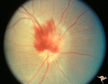
Vascular Complications of Drusen | PP34a: Right eye. Superior retinal vein drains into the choroid at 12:00. It has occluded between center of disc and 12:00. Note white ghost vessel. Note that other veins drain into the disc edge at 4:00. There is no evidence of a central retinal vein in the middle of the disc. PP34b: Visible drus... | Image |
| 5 |
 |
Vascular Complications of Drusen | PP34a: Right eye. Superior retinal vein drains into the choroid at 12:00. It has occluded between center of disc and 12:00. Note white ghost vessel. Note that other veins drain into the disc edge at 4:00. There is no evidence of a central retinal vein in the middle of the disc. PP34b: Visible drus... | Image |
| 6 |
 |
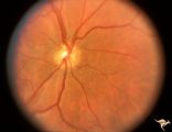
Venous Anomalies - Exit Anomalies | Intrapapillary drusen causing diversion of retinal venous blood into the disc edge at 12:00 and 4:00. Note the white ghost veins between the disc edge and the center of the disc. Anatomy: Optic disc. Pathology: Optic nerve drusen. Disease/Diagnosis: Complication of optic nerve drusen. Venous rerouti... | Image |
| 7 |
 |
Von Hippel Lindau Disease (Retinal Hemangioblastoma) | Von Hippel Lindau Disease with large retinal hemangioblastoma. Continued view of the arteriole and venous channels leading to the tumor. Group with R1_C3a, R1_C3c, R1_C3d. Anatomy: Retina. Pathology: Hemangioblastoma. Disease/Diagnosis: Von Hippel Lindau disease. Clinical: No visual symptoms. | Image |
| 8 |
 |
Von Hippel Lindau Disease (Retinal Hemangioblastoma) | Von Hippel Lindau Disease with large retinal hemangioblastoma. Huge arteriole and venous loops directed upward and nasally toward the tumor. Group with R1_C3b, R1_C3c, R1_C3d. Anatomy: Retina. Pathology: Hemangioblastoma. Disease/Diagnosis: Von Hippel Lindau disease. Clinical: No visual symptoms. | Image |
| 9 |
 |
Von Hippel Lindau Disease (Retinal Hemangioblastoma) | Von Hippel Lindau Disease with large peripheral retinal hemangioblastoma. View of the tumor. Larger artery entering and the vein leaving the tumor are evidence of rapid arteriovenous shunting within the tumor. Group with R1_C3b, R1_C3a, R1_C3d. Anatomy: Retina. Pathology: Hemangioblastoma. Disease/... | Image |
| 10 |
 |
Von Hippel Lindau Disease (Retinal Hemangioblastoma) | Von Hippel Lindau Disease with large peripheral retinal hemangioblastoma. View of the tumor. Larger artery entering and the vein leaving the tumor are evidence of rapid arteriovenous shunting within the tumor. Group with R1_C3b, R1_C3c, R1_C3c. Anatomy: Retina. Pathology: Hemangioblastoma. Disease/... | Image |
| 11 |
 |
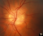
C206 Papillitis with Macular Star, Cat Scratch Disease | Proven Bartonella neuroretinitis. Resolved papillitis with residual retinal exudate. Man. Anatomy: Optic disc; Retina. Pathology: Axoplasmic stasis due to inflammation. Disease/ Diagnosis: Bartonella Henslae (Cat Scratch). Clinical: Visual blurring; Ocular edema with macular star (ODEMS). | Image |
1 - 25 of 11