Home
Browse
Ask Us
Chat
Harmful Language Statement
Log in
NOVEL - Neil R. Miller Collection
Advanced Search
About
The Neil R. Miller Collection covers a broad range of neuro-ophthalmologic conditions and syndromes in videos, images, and lecture presentations.
Year
2024
TO
2024
Type
Image
87
Format
image/jpeg
86
application/pdf
1
Collection
NOVEL - Neil R. Miller Collection
87
Filters:
Collection:
"ehsl_novel_nrm"
Subject:
"Brain"
51
-
75
of
87
<
1
2
3
4
>
Gallery view
Number of results to display per page
10
25
50
100
200
Sort by Relevance
Sort by Title A-Z
Sort by Title Z-A
Sort by Date Ascending
Sort by Date Descending
Sort by Last Modified Ascending
Sort by Last Modified Descending
Title
Date
Type
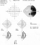
51
Chiasmal Compression - Vascular - OCSOD1
2024-07
Image
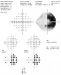
52
Chiasmal Compression - Vascular - OCSOD2
2024-07
Image
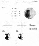
53
Chiasmal Compression - Vascular - OCSOS1
2024-07
Image
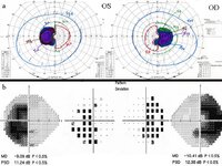
54
Chiasmal Compression - Vascular - VF
2024-07
Image
55
Chiasmal Compression by a Vascular Loop
2024-07
Image
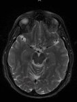
56
Chiasmal Compression from a Dilated Third Ventricle in a Patient with Severe Hydrocephalus (MRI)
2024-07
Image
57
Chiasmal Compression from a Vascular Loop
2024-07
Image
58
Complete Bitemporal Hemianopia from Chiasmal Damage
2024-07
Image
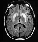
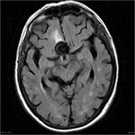
59
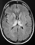
Creuzfeldt-Jakob Disease - FLAIR sCJD - Arrows
2024-07
Image
60
Creuzfeldt-Jakob Disease - FLAIR vCJD - Arrows
2024-07
Image
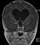
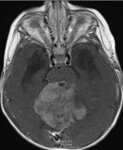
61
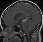
Hydrocephalus Caused by a Large Pineal Region Mass
2024-07
Image
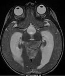
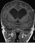
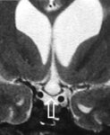
62
Hydrocephalus Causing Downward Compression and Displacement of the Optic Chiasm
2024-07
Image
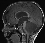
63
Hydrocephalus Causing Downward Compression of the Optic Chiasm by an Enlarged Third Ventricle
2024-07
Image
64
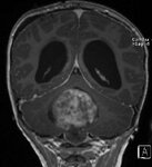
Hydrocephalus from a Large Posterior Fossa Mass
2024-07
Image
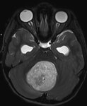
65
Hydrocephalus from Large Posterior Fossa Mass
2024-07
Image
66
Hydrocephalus from Large Posterior Fossa Mass
2024-07
Image
67
Hydrocephalus from Large Posterior Fossa Mass
2024-07
Image
68
Hyrocephalus with Downward Compression of the Optic Chiasm by an Enlarged Third Ventricle
2024-07
Image
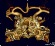
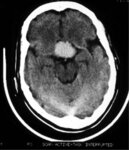
69
Large Anterior Circulation Aneurysm (CT)
2024-07
Image
70
Large Anterior Vascular System Aneurysm (MRI)
2024-07
Image
71
Large Posterior Fossa Mass with Brainstem Compression
2024-07
Image
72
Marked Compression and Downward Displacement of the Chiasm by an Enlarged Third Ventricle in a Patient with Hydrocephalus
2024-07
Image
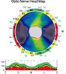
73
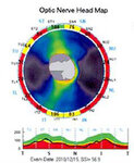
OCT Showing Band Atrophy in a Patient with an Optic Chiasmal Syndrome
2024-07
Image
74
OCT Showing Band Atrophy in a Patient with an Optic Chiasmal Syndrome
2024-07
Image
75
Optic Chiasm Damage Following Excision of a Cavernous Angioma
2024-07
Image
51
-
75
of
87
<
1
2
3
4
>