John A. Moran Eye Center Neuro-Ophthalmology Collection: A variety of lectures, videos and images relating to topics in Neuro-Ophthalmology created by faculty at the Moran Eye Center, University of Utah, in Salt Lake City.
NOVEL: https://novel.utah.edu/
TO
Filters: Collection: "ehsl_novel_jmec"
| Title | Description | Type | ||
|---|---|---|---|---|
| 1 |
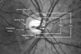 |
2-4a - Disc Anatomy | The optic disc appearance is determined by: the size of the eye, the size of the scleral canal, how the nerve is inserted into the globe, the appearance of the lamina cribrosa, where myelination stops, and what is left behind in normal development. Even though this is a disc with a very large cup, i... | Image |
| 2 |
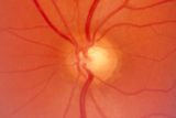 |
2-53a - Venous Pulsations | On the disc, look for spontaneous venous pulsations. Spontaneous venous pulsations can be seen in the large trunks of veins at the level of the disc margin. They are normally present and seen in 37-90% of normals -- depending on the experience of the examiner and the shape of the disc. The spontaneo... | Image |
| 3 |
 |
Aberrant Regeneration of the Third | Patient with a right third nerve palsy demonstrates ptosis, anisocoria and ophthalmoplegia. During attempted downgaze, the right upper lid flutters back up (aberrant movement) and remains retracted. | Image/MovingImage |
| 4 |
 |
Bilateral Internuclear Ophthalmoplegia | Example of patient with bilateral internuclear ophthalmoplegia. Patient is led through instructions for direction and distance of gaze. | Image/MovingImage |
| 5 |
 |
Brainstem Trauma | Image/MovingImage | |
| 6 |
 |
Cogan's Lid Twitch | Image/MovingImage | |
| 7 |
 |
Third Nerve Palsy | Patient with third nerve palsy (no audio) | Image/MovingImage |
| 8 |
 |
Vestibular Nystagmus | Example of patient with vestibular nystagmus. Patient is led through instructions for direction of gaze. Shown also with Frenzel goggles. | Image/MovingImage |
| 9 |
 |
Abducting (Dissociated) Nystagmus | Example of a patient with abducting (dissociated) nystagmus. Patient has a subtle internuclear ophthalmoplegia. Right eye has right-beating jerk nystagmus, with smaller oscillations in the left eye. Disease/Diagnosis: Abducting Nystagmus | Image/MovingImage |
| 10 |
 |
Before Tensilon | Example of patient with myasthenia gravis. Demonstration of baseline examination, followed by administration of 2mg of tensilon, which is a test dose. Procedure for administration of tensilon test is described, including variations. Patient is then shown after being given 4mg of tensilon, with very ... | Image/MovingImage |
| 11 |
 |
Congenital Ocular Motor Apraxia | Two examples of congenital ocular motor apraxia. Patients have trouble initiating saccades, and compensate with head movement. Discussion of how to distinguish this condition from simply not seeing well. | Image/MovingImage |
| 12 |
 |
Dissociated Nystagmus | Example of a patient with dissociated nystagmus. Demonstrates difference in movements between each eye. | Image/MovingImage |
| 13 |
 |
Internuclear Ophthalmoplegia (2 Examples) | Two examples of patients with internuclear ophthalmoplegia. First patient has a right internuclear ophthalmoplegia. Patient had subacute bacterial endocarditis with a bacterial abscess in the brain stem. Ductions and gaze to the right look good, but when gazing to the left, the right eye does not ad... | Image/MovingImage |
| 14 |
 |
Latent Nystagmus | Example of a patient with latent nystagmus. Demonstrates a lack of oscillations in forward gaze, followed by the occlusion of each eye, showing how this generates a jerking oscillation in the non-occluded eye away from the occluded eye. | Image/MovingImage |
| 15 |
 |
Opsoclonus | Example of patients with opsoclonus, a saccadic abnormality. | Image/MovingImage |
| 16 |
 |
Paradoxical Constriction of Pupils to Darkness (Flynn Phenomenon) | Example of patients both with and without paradoxical constriction of pupils. Observed in many congenital retinal disorders, such as achromatopsia, congenital stationary night-blindness, and Leber's congenital amaurosis. Sometimes seen in optic nerve disorders, such as dominant optic atrophy. | Image/MovingImage |
| 17 |
 |
Parinaud's Syndrome | Two examples of patients with Parinaud's syndrome, a dorsal midbrain syndrome. Discussion of hallmarks of this syndrome, including convergence retraction nystagmus, vertical gaze palsies, light-near dissociation, and Collier's Sign. Discussion of age-dependent disorders associated with this syndrome... | Image/MovingImage |
| 18 |
 |
Physiologic (End-Gaze) Nystagmus | Demonstration of physiological nystagmus, where oscillations do not represent pathology, but occur when the patient's gaze is drawn too far laterally. | Image/MovingImage |
| 19 |
|
Progressive Supranuclear Palsy | Example of patient with progressive supranuclear palsy. Discussion of difference between saccadic movement in supranuclear palsy and nystagmus. Shows saccadic intrusions in forward gaze, pursuit, saccades, and doll's head maneuver. | Image/MovingImage |
| 20 |
 |
Spasm of the Near Reflex | Example of patient with spasm of the near reflex and voluntary nystagmus. Discussion of similar-looking conditions (e.g. six nerve palsy, limitation of abduction, lateral rectus muscle problems) and how to tell them apart from spasm of the near reflex by observing the myosis evoked by the near respo... | Image/MovingImage |
| 21 |
|
Transillumination Ocular Melanoma | Video describing condition. | Image/MovingImage |
| 22 |
 |
Upbeat Nystagmus | Example of a patient with upbeat nystagmus. Shows vertical jerk nystagmus with fast phases in the up direction. Localizes to brain stem, and occurs with strokes, demyelination, and tumors. | Image/MovingImage |
| 23 |
|
Vestibular Nystagmus | Discussion of vestibular nystagmus. Seen with peripheral disorders and central disorders, and in two varieties: spontaneous and positional. Horizontal jerk with small amplitude. | Image/MovingImage |
| 24 |
 |
Aberrant Regeneration of the Third and Sixth Nerves | Image/MovingImage | |
| 25 |
 |
How to Use the Direct Ophthalmoscope in an Exam | Demonstration of using the direct ophthalmoscope to examine the optic disc. Covers hand placement , which eye to use, and distance from patient. | Image/MovingImage |