A collection of videos relating to the diagnosis and treatment of eye movement disorders. This collection includes many demonstrations of examination techniques.
Dan Gold, D.O., Associate Professor of Neurology, Ophthalmology, Neurosurgery, Otolaryngology - Head & Neck Surgery, Emergency Medicine, and Medicine, The Johns Hopkins School of Medicine.
A collection of videos relating to the diagnosis and treatment of eye movement disorders.
NOVEL: https://novel.utah.edu/
TO
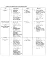
| Title | Description | Type | ||
|---|---|---|---|---|
| 1 |
 |
Video Head Impulse Testing | 𝗢𝗿𝗶𝗴𝗶𝗻𝗮𝗹 𝗗𝗲𝘀𝗰𝗿𝗶𝗽𝘁𝗶𝗼𝗻: The video head impulse test (vHIT) is a clinical assessment technique used to assess the function of the semicircular canals-the angular acceleration detectors-which initiate the vestibulo-ocular reflex (VOR). The HIT and... | Text |
| 2 |
 |
Basics of Acute Stroke Treatment | A brief overview of management of acute stroke treatment. | Text |
| 3 |
 |
Secondary Stroke Prevention | A brief overview of secondary stroke prevention. (TIA = Transient Ischemic Attack) | Text |
| 4 |
 |
ENG, VNG, & VOG | 𝗢𝗿𝗶𝗴𝗶𝗻𝗮𝗹 𝗗𝗲𝘀𝗰𝗿𝗶𝗽𝘁𝗶𝗼𝗻: Electronystagmography (ENG), and videonystagmography (VNG) or videooculography (VOG) are a collection of tests of eye movements that are performed either using surface electrodes around the eye (ENG) or with video goggles... | Text |
| 5 |
 |
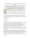
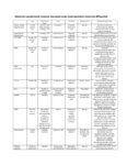
The Most Common Audiovestibular Laboratory Tests, and the Specific Conditions in Which They May Assist in Making or Supporting the Diagnosis | VN = vestibular neuritis; VM = vestibular migraine; VP = vestibular paroxysmia; vHIT = video head impulse test; VNG = video-nystagmography; ENG = electronystagmography; VOG = video-oculography; VEMPs = vestibular evoked myogenic potentials; SCDS = superior canal dehiscence syndrome; BPPV = benign pa... | Text |
| 6 |
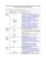 |
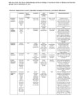
The Most Common Vestibular Conditions Categorized by Timing and Triggers, with Specific Ocular Motor and Vestibular Features that Should be Sought for Each | HINTS+ = Head Impulse, Nystagmus, Test of Skew, ‘Plus' bedside assessment of auditory function; HIT = head impulse test; NP = nerve palsy; BPPV = benign paroxysmal positional vertigo; SCDS = superior canal dehiscence syndrome; BVL = bilateral vestibular loss; PPPD = persistent postural perceptual ... | Text |
| 7 |
 |
Vertical Vergence and Fusional Amplitude | Essential information on vertical fusional vergences. | Text |
| 8 |
 |
Vestibular Evoked Myogenic Potentials (VEMPs) | 𝗢𝗿𝗶𝗴𝗶𝗻𝗮𝗹 𝗗𝗲𝘀𝗰𝗿𝗶𝗽𝘁𝗶𝗼𝗻: Vestibular-evoked myogenic potentials (VEMP) are electromyographic potential reflex tests that reflect the function of the saccule in cervical VEMP and the utricle in ocular VEMP.1 In the cervical VEMP an inhibitory refle... | Text |
| 9 |
 |
An Optokinetic Stimulation Home Exercise Program | 𝗢𝗿𝗶𝗴𝗶𝗻𝗮𝗹 𝗗𝗲𝘀𝗰𝗿𝗶𝗽𝘁𝗶𝗼𝗻: A plainly written program of optokinetic exercise intended for patient use at home. 𝗡𝗲𝘂𝗿𝗼-𝗼𝗽𝗵𝘁𝗵𝗮𝗹𝗺𝗼𝗹𝗼𝗴𝘆 𝗮𝗻𝗱 𝗡𝗲𝘂𝗿𝗼-𝗼𝘁𝗼𝗹𝗼𝗴𝘆 ... | Text |
| 10 |
 |
Common Neuro-Ophthalmic Ancillary Tests to Assist in the Diagnosis and Localization of Afferent Disorders | Chart of the common neuro-ophthalmic ancillary tests to assist in the diagnosis and localization of afferent disorders. | Text |
| 11 |
 |
Expanded Acute Onset Persistent Vision Loss Differential | Text | |
| 12 |
 |
Expanded Nystagmus & Saccadic Intrusions/Oscillations Differential | Expanded nystagmus & saccadic intrusions/ oscillations differential | Text |
| 13 |
 |
Localization of Ophthalmoplegia | 𝗢𝗿𝗶𝗴𝗶𝗻𝗮𝗹 𝗗𝗲𝘀𝗰𝗿𝗶𝗽𝘁𝗶𝗼𝗻: A table describing the localization of ophthalmoplegia. | Text |
| 14 |
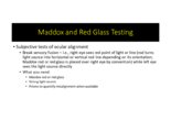 |
Maddox Rod and Red Glass Testing | 𝗢𝗿𝗶𝗴𝗶𝗻𝗮𝗹 𝗗𝗲𝘀𝗰𝗿𝗶𝗽𝘁𝗶𝗼𝗻: Describing the basics of strabismus. | Text |
| 15 |
 |
Summary of the Most Common Audio-Vestibular Testing | Chart describing common audio-vestibular testing. | Text |
| 16 |
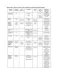 |
HINTS 'Plus' Patterns in the Acute Vestibular Syndrome Based on Location | HINTS ‘Plus' patterns in the acute vestibular syndrome based on location | Text |
| 17 |
 |
Audiometry | 𝗢𝗿𝗶𝗴𝗶𝗻𝗮𝗹 𝗗𝗲𝘀𝗰𝗿𝗶𝗽𝘁𝗶𝗼𝗻: Audiometry is the measurement of the sensitivity and range of an individual's hearing. As many etiologies of imbalance, nystagmus, vertigo and/or dizziness can have an otologic origin the audiogram is an important piece o... | Text |
| 18 |
 |
Rotary Chair Testing | 𝗢𝗿𝗶𝗴𝗶𝗻𝗮𝗹 𝗗𝗲𝘀𝗰𝗿𝗶𝗽𝘁𝗶𝗼𝗻: Rotary chair testing includes rotation around a vertical axis, and evaluates the horizontal semicircular canal vestibulo-ocular reflex (VOR). The patient sits in a mechanized chair with the head secured in a neutral posi... | Text |
| 19 |
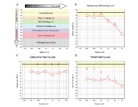 |
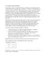
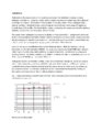
Audiometry: What Does It Look Like and How Do I Interpret It? | An audiogram measures a patient's auditory threshold responses with pure-tone stimuli across a range of sound frequencies that are important for human communication, typically 250 Hz to 8000 Hz. The threshold is the sound intensity level at which an individual detects the tone 50% of the time. Heari... | Text |
| 20 |
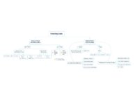 |
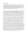
What is the Cause of My Patient's Hearing Loss? | This is a flowsheet differentiating multiple causes of hearing loss. The onset and chronicity of hearing loss is a critical starting point in understanding whether urgent action is needed, such as in the setting of suspected stroke or sudden sensorineural hearing loss. For hearing loss that has been... | Text |
| 21 |
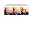 |
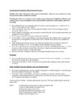
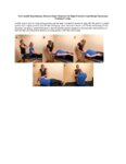
The Affected-Ear-up 90 degree Maneuver (HC-Canalithiasis) | The Affected-Ear-up 90 degree Maneuver is used to treat horizontal canal-canalithaisis. 1. The patient starts in a supine position. 2. The patient rotates their head 90 degrees towards the unaffected side. 3. The patient sits up. | Text |
| 22 |
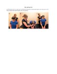 |
Bow and Lean Test | The Bow and Lean Test is used to identify the affected side and is designed to be used in conjunction with or after the Supine Roll Test. Within this test a null point may exist where the nystagmus will extinguish because the cupula is in a gravity neutral position. As this test involves the patient... | Text |
| 23 |
 |
The Canalith Repositioning Maneuver/Epley Maneuver for Right Posterior Canal Benign Paroxysmal Positional Vertigo | Posterior canal (PC) accounts for 70-90% cases of BPPV [1-3] and resolves with canalith repositioning maneuvers 90% of the time [4-20]. The Epley maneuver is considered a gold-standard treatment, with class 1 evidence for use. | Text |
| 24 |
 |
The Gans Maneuver for Right Posterior Canal Benign Paroxysmal Positional Vertigo | This maneuver is recommended for individuals with cervical restrictions or precautions, as the maneuver avoids cervical hyperextension and may reduce cervical pain associated with repositioning maneuvers. The Epley maneuver has higher subjective and objective success rates compared to the Gans maneu... | Text |
| 25 |
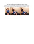 |
Gufoni Maneuver for Right Horizontal Canal-Cupulolithiasis (Apgeotropic Nystagmus) | The Gufoni Maneuver can be used to treat horizontal canal cupulolithaisis. 1. The patient starts in a seated position. 2. The patient transitions quickly to lying on their affected side. 3. The patient lies on their affected side for two minutes with the head in a neutral position. 4. The patient's ... | Text |