The Emory Eye Center Neuro-Ophthalmology Collection contains a variety of lectures, videos and images relating to the discipline of neuro-ophthalmology created by faculty at Emory University in Atlanta, GA.
NOVEL: https://novel.utah.edu/
TO
Filters: Collection: "ehsl_novel_eec"
| Title | Description | Creator | ||
|---|---|---|---|---|
| 1 |
 |
Anatomy of the Orbits on CT and MRI | MRI and CT imaging of the orbit. | Valérie Biousse, MD |
| 2 |
 |
Ophthalmic Artery Aneurysm | Slideshow describing ophthalmic artery aneurysm with MRI imaging. | Valérie Biousse, MD |
| 3 |
 |
Coexisting Thyroid Eye Disease and Myasthenia Gravis | A case of coexisting thyroid orbitopathy and myasthenia gravis. External photographs of the eyes and eyelids, as well as images from an MRI of the orbits, are included. Figure 1 : External photograph of eyes showing right lid retraction and left upper lid ptosis. Figure 2 : External photograph of ... | Devin D. Mackay, MD; Valérie Biousse, MD |
| 4 |
 |
Posterior Circulation Infarctions Secondary to a Post Traumatic Vertebral Artery Dissection | A case of a young man with a vertebral artery dissection that caused multiple posterior circulation brain infarcts. Images from an MRI of the brain, digital subtraction angiography, and Humphrey visual fields are included. Figure 1 : Humphrey visual fields showed a right homonymous hemianopia with ... | Devin D. Mackay, MD; Valérie Biousse, MD |
| 5 |
 |
Bilateral Lens Subluxation in Marfan Syndrome | This is a case of known Marfan syndrome with bilateral progressive visual loss. The ocular examination showed bilateral lens dislocation. Figure 1a: Typical superonasal lens subluxation in both eyes Figure 1b: The arrows show the inferior edges of the lenses Figure 2: Optical section of the lenses u... | Rabih Hage, MD; Valérie Biousse, MD |
| 6 |
 |
Occipital Hemorrhagic Infarction Secondary to Bacterial Endocarditis-Congruent Homonymous Scotomatous Hemianopic Defect | MRI features of occipital hemorrhage secondary to bacterial endocarditis Figure 1 : Humphrey visual fields showing a congruent homonymous scotomatous hemianopic defect Figure 2 : axial gradient echo T2*w brain MRI Figure 3 : axial T1w and T2w brain MRI Figure 4 : axial postcontrast T1w brain MRI | Samuel Bidot, MD; Amit M. Saindane, MD; Valérie Biousse, MD |
| 7 |
 |
Occipital Infarction with Incomplete Congruent Homonymous Hemianopia | CT appearance of a remote occipital infarction. Congruent homonymous hemianopia. | Samuel Bidot, MD; Amit M. Saindane, MD; Valérie Biousse, MD |
| 8 |
 |
Cavernous Sinus Meningioma Extending into the Orbital Apex | Septic left cavernous sinus and superior ophthalmic vein thrombosis, secondary to left maxillary tooth abscess. MRI characteristics Figure 1 : MRI Orbits (Coronal T2 with fat suppression) : Left periorbital edema (increased T2 signal, yellow arrows) extends inferiorly along the premalar tissues to t... | Supharat Jariyakosol, MD; Valérie Biousse, MD |
| 9 |
 |
Sturge-Weber Syndrome | A case of Sturge-Weber syndrome (Encephalotrigeminal angiomatosis) with angiomas that involve the leptomeninges, and the skin of the ipsilateral hemiface, associated with congenital glaucoma in the same eye. Various illustrations are included to demonstrate the port wine stain, enlarged optic nerve ... | Supharat Jariyakosol, MD; Valérie Biousse, MD |
| 10 |
 |
MRI Findings in Gliomatosis Cerebri | Single case of gliomatosis cerebri identified on mulltiple MRI images pre- and post-contrast administration with axial and coronal views. Figure 1 : Axial brain MRI (T2) - Diffusely thickened and T2 hyperintense area (yellow arrows) centered within the genu of the corpus callosum with extension into... | Jason Peragallo, MD; Valérie Biousse, MD |
| 11 |
 |
MRI Findings in Optic Nerve and Chiasmal Glioma in Childhood | Multiple cases of optic pathway gliomas are demonstrated using MRI imaging. The optic pathway gliomas are identified at multiple points in the optic pathways. Figure 1 : Patient 1: Axial orbital MRI (T1 postcontrast with fat suppression) showing thickening and enhancement of the right optic nerve a... | Jason Peragallo, MD; Valérie Biousse, MD |
| 12 |
 |
Orbital and Cavernous Sinus Involvement by Herpes Zoster Ophthalmicus | A single case of the effects of herpes zoster is demonstrated using external photographs and MRI imaging. The effects demonstrated include the typical dermatomal rash as well as extraocular muscle invovlement and cavernous sinus involvement. Figure 1 : External photograph of dermatomal rash and s... | Jason Peragallo, MD; Valérie Biousse, MD |
| 13 |
 |
Anterior and Posterior Scleritis | A case of anterior and posterior scleritis secondary to idiopathic orbital inflammation, also known as orbital pseudotumor. Various imaging modalities are included to demonstrate optic disc edema, macular edema, and fluid in tenon's capsule which may be seen in posterior scleritis. Figure 1 : Exter... | Joshua Levinson, MD; Valérie Biousse, MD |
| 14 |
 |
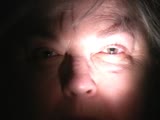
Terson Syndrome and Subarachnoid Hemorrhage | A case of Terson syndrome resulting with subarachnoid hemorrhage and right vitreous hemorrhage resulting from a left pericallosal artery aneurysm. Figure 1 : External photograph of right eye demonstrates blunted red reflex secondary to vitreous hemorrhage Figure 2 : External photograph of left eye d... | Joshua Levinson, MD; Valérie Biousse, MD |
| 15 |
 |
Left RAPD | Video clip displaying pupillary examination and RAPD measurement. | Valérie Biousse, MD |
| 16 |
 |
Nonfunctiong Pituitary Adenoma with Chiasmal Compression | This is a case of large non-functioning pituitary adenoma with mass effect on the optic chiasm inducing loss of optic nerve fibers and subsequent visual field. Figure 1: Fundus photographs demonstrating bilateral temporal optic nerve head pallor Figure 2: Humphrey visual fields demonstrating a bitem... | William Pearce, MD; Valérie Biousse, MD |
| 17 |
|
Vertical Diplopia Secondary to Skew Deviation With Ocular Tilt Reaction With Multiple Posterior Fossa Metastases | This is a case of multiple brain metastases in the posterior fossa resulting in a skew deviation. Figure 1 : Photograph of the patient demonstrating a spontaneous right head tilt. The patient's head is tilted toward his right shoulder to suppress his diplopia Figure 2 : Ocular movements : There is a... | Rabih Hage, MD; Valérie Biousse, MD; Jason Peragallo, MD |
| 18 |
 |
Iris Transillumination | Single case of iris transillumination in a patient with albinism. Figure 1 : Anterior segment photograph demonstrating reddish hue to iris in albinism Figure 2 : Slit lamp photograph with retroillumination demonstrating iris transillumination Figure 3 : Slit lamp photograph with retroillumination... | Jason Peragallo, MD; Valérie Biousse, MD |
| 19 |
 |
Large Sellar and Suprasellar Mass (Pituitary Macroadenoma) With Left Third Nerve Palsy and Left Optic Tract Compression | A case of a large sellar and suprasellar pituitary macroadenoma with an associated left third nerve palsy and left optic tract compression. Images from an MRI of the brain with contrast illustrate the imaging characteristics and extent of the tumor. Figure 1 : Humphrey Visual Fields (24-2 SITA-Fast)... | Devin D. Mackay, MD; Valérie Biousse, MD |
| 20 |
 |
Geniculate Nucleus Metastasis with Homonymous Sectoranopia | This is a case of multiple brain metastases in the setting of bladder cancer complicated with right homonymous horizontal sectoranopia. Figure 1: Pet-scan showing liver (yellow arrows) and kidneys (red arrow) metastases Figure 2: Goldmann Visual Fields: Right homonymous horizontal sectoranopia Figu... | Rabih Hage, MD; Valérie Biousse, MD |
| 21 |
 |
Colloid Cyst Hydrocephalus | This is a case of colloid cyst of the third ventricle complicated by severe hydrocephalus, raised intracranial pressure and papilledema. Figure 1: Fundus photographs demonstrating bilateral optic nerve head edema Figure 2a and 2b: T1-weighted axial brain MRI without contrast: Dilation of both later... | Rabih Hage, MD; Valérie Biousse, MD |
| 22 |
 |
Large Frontal Meningioma with Mass Effect and Increased Intracranial Pressure | This is a case of frontal meningioma presenting with raised intracranial pressure and bilateral papilledema responsible for visual loss. Figure 1: Goldmann visual field of the left eye. In the right eye, there was no response to the V4e. The visual field is severely constricted in the left eye. Fig... | Rabih Hage, MD; Valérie Biousse, MD |
| 23 |
 |
Large Right Hypophyseal Aneurysm Causing a Junctional Scotoma | Right, multi-lobulated superior hypophyseal artery aneurysm measuring 1.6 x 1.2 x 2.2 cm with 6 mm neck causing a right junctional scotoma . Images from a brain CT with contrast, a brain CT angiography with contrast, cerebral angiogram, Humphrey visual fields and ocular fundus photographs are includ... | Laurel N. Vuong, MD; Valérie Biousse, MD |
| 24 |
 |
Sellar Aneurysm with Chiasmal Compression | This is a case of aneurysm of the internal carotid artery, invading the sella and complicated by chiasmal compression and bitemporal hemianopia. Figure 1 : Humphrey visual fields (gray scale and pattern deviations) Figure 2a : T1-weighted axial brain MRI (1): well defined circular intracerebral mass... | Rabih Hage, MD; Valérie Biousse, MD |
| 25 |
 |
Metastatic Ovarian Cancer to the Left Occipital Lobe With Complete Right Homonymous Hemianopia | A case of metastatic ovarian cancer to the left occipital lobe with a complete right homonymous hemianopia. Humphrey visual fields as well as images from an MRI of the brain are included. Figure 1 : Humphrey visual fields showing a complete right homonymous hemianopia Figure 2 : MRI brain T1 axial... | Devin D. Mackay, MD; Valérie Biousse, MD |