The Health Education Assets Library (HEAL) is a collection of over 22,000 freely available digital materials for health sciences education. The collection is now housed at the University of Utah J. Willard Marriott Digital Library.
TO
Filters: Collection: "ehsl_heal"
| Title | Description | Subject | Collection | ||
|---|---|---|---|---|---|
| 1 |
 |
Base of the Brain | Anatomical structures of a dissected brain are identified in this first video from UCLA Interactive Neurosciences. Required Applications - RealNetworks RealPlayer (version 8 or later recommended), Windows Media Player (PC), or QuickTime Player (Mac). | Posterior Communicating Artery | UCLA Interactive Neuroscience |
| 2 |
|
Cerebellum and Brain Stem | Anatomical structures of a dissected brain are identified in this second video from UCLA Interactive Neurosciences. Required Applications - RealNetworks RealPlayer (version 8 or later recommended), Windows Media Player (PC), or QuickTime Player (Mac). | Posterior Inferior Cerebellar Artery; ArchiPaleoNeoVermis Cerebelli; Foramen of Monro; Bachium Pontis; Arbor Vitae Cerebelli | UCLA Interactive Neuroscience |
| 3 |
|
Hippocampus and Lateral Ventricle | Anatomical structures of a dissected brain are identified in this fifth video from UCLA Interactive Neurosciences. Required Applications - RealNetworks RealPlayer (version 8 or later recommended), Windows Media Player (PC), or QuickTime Player (Mac). | Subiculum; Uncus; Alveus Hippocampi; Hippocampal Fissure; Collateral Sulcus | UCLA Interactive Neuroscience |
| 4 |
|
Human Brain Atlas: Brain (Coronal) - Image Map | Eighteen slides of the anatomical structures of the brain are mapped in this image from the UCLA Interactive Neurosciences Human Brain Atlas. | Slide 76; Slide 77; Slide 78; Slide 79; Slide 80; Slide 81; Slide 82; Slide 83; Slide 84; Slide 85; Slide 86; Slide 87; Slide 88; Slide 89; Slide 90; Slide 91; Slide 92; Slide 93 | UCLA Interactive Neuroscience |
| 5 |
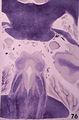 |
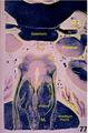
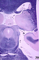
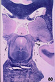
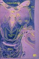
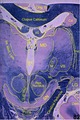
Human Brain Atlas: Brain (Coronal) - Slide 76 | Anatomical structures of the brain are depicted in this slide from the UCLA Interactive Neurosciences Human Brain Atlas. | Splenium of Fornix; Transverse Fissure; Parabrachium Pontis; Brachium Conjunctivum; Nucleus of 4; Trochlear Nucleus; Medial Longitudinal Fasciculus; Central Tegmental Tract | UCLA Interactive Neuroscience |
| 6 |
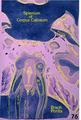 |
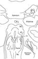
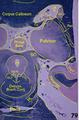
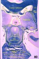
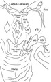
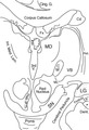
Human Brain Atlas: Brain (Coronal) - Slide 76 - Labeled | Anatomical structures of the brain are identified in this labeled slide from the UCLA Interactive Neurosciences Human Brain Atlas. | UCLA Interactive Neuroscience | |
| 7 |
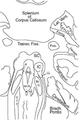 |
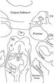
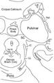
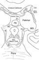
Human Brain Atlas: Brain (Coronal) - Slide 76 - Labeled Outline | Anatomical structures of the brain are identified in this labeled outline from the UCLA Interactive Neurosciences Human Brain Atlas. | UCLA Interactive Neuroscience | |
| 8 |
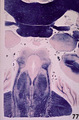 |
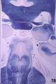
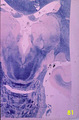
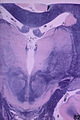
Human Brain Atlas: Brain (Coronal) - Slide 77 | Anatomical structures of the brain are depicted in this slide from the UCLA Interactive Neurosciences Human Brain Atlas. | Splenium of Fornix; Brachium Pontis; Brachium Conjunctivum; Medial Longitudinal Fasciculus; Central Tegmental Tract; Medial Lemniscus | UCLA Interactive Neuroscience |
| 9 |
 |
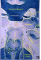
Human Brain Atlas: Brain (Coronal) - Slide 77 - Labeled | Anatomical structures of the brain are identified in this labeled slide from the UCLA Interactive Neurosciences Human Brain Atlas. | UCLA Interactive Neuroscience | |
| 10 |
 |
Human Brain Atlas: Brain (Coronal) - Slide 77 - Labeled Outline | Anatomical structures of the brain are identified in this labeled outline from the UCLA Interactive Neurosciences Human Brain Atlas. | UCLA Interactive Neuroscience | |
| 11 |
 |
Human Brain Atlas: Brain (Coronal) - Slide 78 | Anatomical structures of the brain are depicted in this slide from the UCLA Interactive Neurosciences Human Brain Atlas. | Parabrachium Pontis; Nucleus of 3; Oculomotor Nucleus; Brachium Conjunctivum; Nucleus of 4; Trochlear Nucleus; Medial Lemniscus; Central Tegmental Tract; Fimbria | UCLA Interactive Neuroscience |
| 12 |
 |
Human Brain Atlas: Brain (Coronal) - Slide 78 - Labeled | Anatomical structures of the brain are identified in this labeled slide from the UCLA Interactive Neurosciences Human Brain Atlas. | UCLA Interactive Neuroscience | |
| 13 |
 |
Human Brain Atlas: Brain (Coronal) - Slide 78 - Labeled Outline | Anatomical structures of the brain are identified in this labeled outline from the UCLA Interactive Neurosciences Human Brain Atlas. | UCLA Interactive Neuroscience | |
| 14 |
 |
Human Brain Atlas: Brain (Coronal) - Slide 79 | Anatomical structures of the brain are depicted in this slide from the UCLA Interactive Neurosciences Human Brain Atlas. | Parabrachium Pontis; Nucleus of 3; Oculomotor Nucleus; Decussation of Brachium Conjunctivum; Medial Lemniscus; Central Tegmental Tract; Fimbria; Medial Longitudinal Fasciculus; Reticular Thalamic Nucleus | UCLA Interactive Neuroscience |
| 15 |
 |
Human Brain Atlas: Brain (Coronal) - Slide 79 - Labeled | Anatomical structures of the brain are identified in this labeled slide from the UCLA Interactive Neurosciences Human Brain Atlas. | UCLA Interactive Neuroscience | |
| 16 |
 |
Human Brain Atlas: Brain (Coronal) - Slide 79 - Labeled Outline | Anatomical structures of the brain are identified in this labeled outline from the UCLA Interactive Neurosciences Human Brain Atlas. | UCLA Interactive Neuroscience | |
| 17 |
 |
Human Brain Atlas: Brain (Coronal) - Slide 80 | Anatomical structures of the brain are depicted in this slide from the UCLA Interactive Neurosciences Human Brain Atlas. | Fornical Commissure; Stria Terminalis; Decussation of Brachium Conjunctivum; Medial Lemniscus; Central Tegmental Tract; Pineal Body; Transverse Fissure; Brachium of Inferior Colliculus | UCLA Interactive Neuroscience |
| 18 |
 |
Human Brain Atlas: Brain (Coronal) - Slide 80 - Labeled | Anatomical structures of the brain are identified in this labeled slide from the UCLA Interactive Neurosciences Human Brain Atlas. | UCLA Interactive Neuroscience | |
| 19 |
 |
Human Brain Atlas: Brain (Coronal) - Slide 80 - Labeled Outline | Anatomical structures of the brain are identified in this labeled outline from the UCLA Interactive Neurosciences Human Brain Atlas. | UCLA Interactive Neuroscience | |
| 20 |
 |
Human Brain Atlas: Brain (Coronal) - Slide 81 | Anatomical structures of the brain are depicted in this slide from the UCLA Interactive Neurosciences Human Brain Atlas. | Decussation of Brachium Conjunctivum; Transverse Fissure; Cerebral Peduncle; Medial Geniculate Nucleus; Lateral Geniculate Nucleus; Habenulo Interpeduncular Tract; Reticular Thalamic Nucleus; Medial Lemniscus; Centrum Medianum | UCLA Interactive Neuroscience |
| 21 |
 |
Human Brain Atlas: Brain (Coronal) - Slide 81 - Labeled | Anatomical structures of the brain are identified in this labeled slide from the UCLA Interactive Neurosciences Human Brain Atlas. | UCLA Interactive Neuroscience | |
| 22 |
 |
Human Brain Atlas: Brain (Coronal) - Slide 81 - Labeled Outline | Anatomical structures of the brain are identified in this labeled outline from the UCLA Interactive Neurosciences Human Brain Atlas. | UCLA Interactive Neuroscience | |
| 23 |
 |
Human Brain Atlas: Brain (Coronal) - Slide 82 | Anatomical structures of the brain are depicted in this slide from the UCLA Interactive Neurosciences Human Brain Atlas. | Cingulate Gyrus; Cerebral Peduncle; Medial Lemniscus; Lateral Geniculate Nucleus; Internal Medullary Lamina; Reticular Thalamic Nucleus; Centrum Medianum; Fimbria; Habenulo Interpeduncular Tract; Red Nucleus; Ventro Basal Complex | UCLA Interactive Neuroscience |
| 24 |
 |
Human Brain Atlas: Brain (Coronal) - Slide 82 - Labeled | Anatomical structures of the brain are identified in this labeled slide from the UCLA Interactive Neurosciences Human Brain Atlas. | UCLA Interactive Neuroscience | |
| 25 |
 |
Human Brain Atlas: Brain (Coronal) - Slide 82 - Labeled Outline | Anatomical structures of the brain are identified in this labeled outline from the UCLA Interactive Neurosciences Human Brain Atlas. | UCLA Interactive Neuroscience |