The Health Education Assets Library (HEAL) is a collection of over 22,000 freely available digital materials for health sciences education. The collection is now housed at the University of Utah J. Willard Marriott Digital Library.
TO
Filters: Collection: "ehsl_heal"
| Title | Description | Subject | Collection | ||
|---|---|---|---|---|---|
| 1201 |
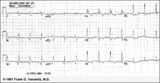 |
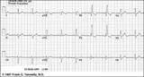
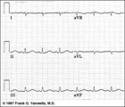
Infero-posterior MI with RBBB | This is an unusual RBBB because the initial R wave is taller than the R wave in lead V1. This is the clue for true posterior MI. The tall initial R wave in V1 is a pathologic R wave analagous to the pathologic Q wave of an anterior MI. | Knowledge Weavers ECG | |
| 1202 |
 |
Right Atrial Enlargement (RAE) & Right Ventricular Hypertrophy (RVH) | RAE is recognized by the tall (>2.5mm) P waves in leads II, III, aVF. RVH is likely because of right axis deviation (+100 degrees) and the Qr (or rSR') complexes in V1-2. | Knowledge Weavers ECG | |
| 1203 |
 |
Frontal plane: accelerated junctional rhythm and inferior MI | Frontal plane: accelerated junctional rhythm and inferior MI | Knowledge Weavers ECG | |
| 1204 |
 |
QRS axis = -30 degrees | Lead II is isoelectric; I is positive; III is negative. The axis is -30 degrees. | Knowledge Weavers ECG | |
| 1205 |
 |
Voltage criteria for LVH | Voltage criteria for LVH | Knowledge Weavers ECG | |
| 1206 |
 |
Infero-posterior MI | Infero-posterior MI | Knowledge Weavers ECG | |
| 1207 |
 |
RBBB - marquette | RBBB - marquette | Knowledge Weavers ECG | |
| 1208 |
 |
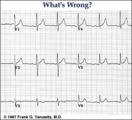
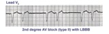
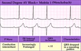
Second degree AV block, type I, with bradycardia-dependent RBBB | An interesting and unusual form of rate-dependent bundle branch block. Normal sinus rhythm at 85 bpm is present with a 3:2 and 2:1 2nd degree AV block. The progressive PR prolongation in the 3:2 block makes this a type-I or Wenckebach block.Long cycles end in RBBB; short cycles have normal QRS dur... | Knowledge Weavers ECG | |
| 1209 |
 |
Bifascicular block: RBBB + LAFB | This is the most common of the bifascicular blocks. RBBB is most easily recognized in the precordial leads by the rSR' in V1 and the wide S wave in V6 (i.e., terminal QRS forces oriented rightwards and anterior). LAFB is best seen in the frontal plane leads as evidenced by left axis deviation (-50... | Knowledge Weavers ECG | |
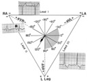
| 1210 |
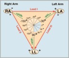 |
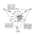
Frontal plane lead diagram | The six frontal plane leads are illustrated with their respective positive and negative poles.When forced to intersect at a center point, the six leads inscribe a 360 degree circle. The normal frontal plane axis is from -30 degrees to + 90 degrees, shaded in grey. Left axis deviation is from -30 d... | Knowledge Weavers ECG | |
| 1211 |
 |
Anteroseptal MI: fully evolved | The QS complexes, resolving ST segment elevation and T wave inversions in V1-2 are evidence for a fully evolved anteroseptal MI. The inverted T waves in V3-5, I, aVL are also probably related to the MI. | Knowledge Weavers ECG | |
| 1212 |
 |
Atrial parasystole | The evenly spaced dots indicate ectopic atrial activity from a parasystolic atrial pacemaker. Non-fixed coupled PACs are seen having a common inter-ectopic interval. One of the PACs is nonconducted. | Knowledge Weavers ECG | |
| 1213 |
 |
Ventricular tachycardia - marquette | Ventricular tachycardia - marquette | Knowledge Weavers ECG | |
| 1214 |
 |
Long QT Iinterval | Long QT Interval | Knowledge Weavers ECG | |
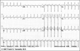
| 1215 |
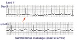 |
ECG of the century - part II: dual AV pathways | An astute cardiology fellow, yours truly, went to the patient's bedside on Day 2 and massaged the right carotid sinus as indicated by the arrow. Four beats later at a slightly slower heart rate the PR interval suddenly normalized suggesting an abrupt change from a slow AV nodal pathway to a fast AV... | Knowledge Weavers ECG | |
| 1216 |
 |
Left Atrial Enlargement & Nonspecific ST-T Wave Abnormalities | LAE is best seen in V1 with a prominent negative (posterior) component measuring 1mm wide and 1mm deep. There are also diffuse nonspecific ST-T wave abnormalities which must be correlated with the patient's clinical status. Poor R wave progression in leads V1-V3, another nonspecific finding, is als... | Knowledge Weavers ECG | |
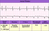
| 1217 |
 |
Atrial flutter with 3:2 AV conduction | This 12-lead ECG shows a subtle bigeminal rhythm resulting from atrial flutter with a 3:2 AV conduction ratio; RR intervals alternate by a small duration. This is uncommon! The impulses from the atrial flutter conduct through the AV junction in a Wenckebach sequence; for every 3 flutter waves the s... | Knowledge Weavers ECG | |
| 1218 |
 |
A very subtle 1st degree AV block | Where are the P waves??? They are hiding in the T waves as indicated by the arrows. How do we know? The PVC unmasked the sinus P wave, and now it is seen in the pause following the PVC. The PR interval is, therefore, about 500 ms. | Knowledge Weavers ECG | |
| 1219 |
 |
PAC's with and without aberrant conduction - marquette | PAC's with and without aberrant conduction - marquette | Knowledge Weavers ECG | |
| 1220 |
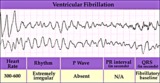 |
Ventricular fibrillation - marquette | Ventricular fibrillation - marquette | Knowledge Weavers ECG | |
| 1221 |
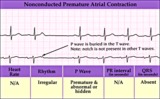 |
Nonconducted PAC - marquette | Nonconducted PAC - marquette | Knowledge Weavers ECG | |
| 1222 |
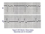 |
Trifascicular block: RBBB, LAFB, and mobitz II 2nd degree AV block | A nice example of trifascicular block: Lead V1 shows RBBB; Lead II is mostly negative with an rS morphology suggesting left anterior fascicular block. Since Mobitz II 2nd degree AV block is more often located in the bundle branch system, the only location left for this block is the left posterior ... | Knowledge Weavers ECG | |
| 1223 |
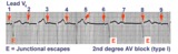 |
2nd degree AV block, type I, with junctional escapes | Junctional escapes are passive, protective events whenever the heart rate slows below that of the escape mechanism. In this example of 2nd degree AV block, type I, the escapes occur following the non-conducted P waves. Arrows indicate the position of the P waves. Note that the escape beats have a... | Knowledge Weavers ECG | |
| 1224 |
 |
Atrial flutter with 2:1 AV conduction | Atrial flutter with 2:1 AV block is one of the most frequently missed ECG rhythm diagnoses because the flutter waves are often hard to find. In this example two flutter waves for each QRS are best seen in lead III and V1. The ventricular rate at 150 bpm should always prompt us to consider atrial fl... | Knowledge Weavers ECG | |
| 1225 |
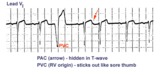 |
Identification of PVC's and PAC's | PVC's usually stick out like sore thumbs; PAC's are often difficult to see because they are hidden in the preceding ST-T wave. The PVC in this example is mostly negative in lead V1 suggesting RV origin; i.e., most of activation is moving in posterior direction towards the left ventricle. | Knowledge Weavers ECG | |
| 1226 |
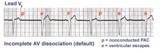 |
AV dissociation by default | The nonconducted PAC's set up a long pause which is terminated by ventricular escapes; note the wider QRS morphology of the escape beats indicating their ventricular origin. Incomplete AV dissociation occurs during the escape beats, since the atria are still under the control of the sinus node. | Knowledge Weavers ECG | |
| 1227 |
 |
Lead error: V1 and V3 are transposed! | In the precordial leads the V1 and V3 chest electrodes are interchanged. Experienced ECG interpreters should be able to spot this lead placement error. | Knowledge Weavers ECG | |
| 1228 |
 |
Infero-posterior MI & RBBB: Frontal Plane Leads + V1 | Infero-posterior MI & RBBB: Frontal Plane Leads + V1 | Knowledge Weavers ECG | |
| 1229 |
 |
WPW Type Pre-excitation: Precordial Leads | WPW Type Pre-excitation: Precordial Leads | Knowledge Weavers ECG | |
| 1230 |
 |
Acute anterior MI | Acute anterior MI | Knowledge Weavers ECG | |
| 1231 |
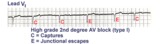 |
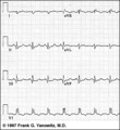
2nd degree AV block, type I with escapes and captures | Often in the setting of 2nd degree AV block the pauses caused by nonconducted P waves are long enough to enable escape pacemakers from the junction or ventricles to take over. This example illustrates junctional escapes, labled E and captures, labled C. Note that the PR intervals for the captures ... | Knowledge Weavers ECG | |
| 1232 |
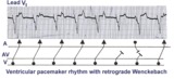 |
Ventricular paced rhythm with retrograde wenckebach | Retrograde atrial captures from a ventricular paced rhythm are occurring with increasing R-P intervals; i.e., retrograde Wenckebach. The ladder diagram indicates that after the blocked retrograde event, a single sinus P wave is seen dissociated from the ventricular rhythm. | Wenckebach AV Block | Knowledge Weavers ECG |
| 1233 |
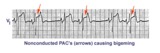 |
Nonconducted PAC's: an unusual bigeminy | Occasionally nonconducted PAC's can create interesting rhythms. In this example every other sinus beat is followed by an early, nonconducted PAC. The resulting pause sets up a bigeminal rhythm. Note the distortion of the T waves caused by the nonconducted PAC's. | Knowledge Weavers ECG | |
| 1234 |
 |
Atrial flutter with 2:1 AV conduction: leads II, III, V1 | In leads II and III, the one of the flutter waves occurs at the end of the QRS complex and might be mistaken for part of the QRS itself; i.e., the S wave. In lead V1, the two flutter waves for every QRS are more easily identified. | Knowledge Weavers ECG | |
| 1235 |
 |
Accelerated IVR with AV dissociation - marquette | Accelerated IVR with AV dissociation - marquette | Knowledge Weavers ECG | |
| 1236 |
 |
Old inferior MI | Old inferior MI | Knowledge Weavers ECG | |
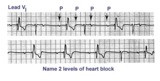
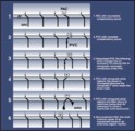
| 1237 |
 |
Diagram: type I vs. type II 2nd degree AV block | In type I 2nd degree AV block the PR progressively lengthens until a nonconducted P wave occurs. The PR gets longer by smaller and smaller increments; this results in gradual shortening of the RR intervals. The RR interval of the pause is usually less than the two preceding RR intervals. The RR i... | Knowledge Weavers ECG | |
| 1238 |
 |
PAC's with RBBB aberration | These PAC's, indicated by arrows, enter the ventricles and find the right bundle refractory. They therefore conduct with RBBB aberrancy. In most normal hearts the right bundle recovery time is longer than the left bundle's; most aberrancy, therefore, has aRBBB morphology. In some diseased hearts t... | Knowledge Weavers ECG | |
| 1239 |
 |
RBBB + LAFB = bifascicular block | The RBBB is diagnosed by the wide QRS with prominent anterior (e.g., V1) and late rightward (e.g., I, V6) forces. The LAFB is recognized by the marked left axis deviation (-75 degrees) in the frontal plane, rS complexes in II, III, aVF, and the tiny q-wave in aVL. | Knowledge Weavers ECG | |
| 1240 |
 |
Massage parlor games | When unsure of the mechanism of a supraventricular tachycardia, carotid sinus massage may help make the diagnosis. In this example, carotid sinus massage causes marked AV block which permits easy recognition of the rapid, regular atrial flutter waves. | Knowledge Weavers ECG | |
| 1241 |
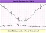 |
Wandering baseline artifact - marquette | Wandering baseline artifact - marquette | Knowledge Weavers ECG | |
| 1242 |
 |
Frontal plane QRS axis = +15 degrees | Frontal plane QRS axis = +15 degrees | Knowledge Weavers ECG | |
| 1243 |
 |
Frontal plane QRS axis = +30 degrees | Frontal plane QRS axis = +30 degrees | Knowledge Weavers ECG | |
| 1244 |
 |
Acute inferoposterior MI | Acute inferoposterior MI | Knowledge Weavers ECG | |
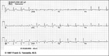
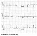
| 1245 |
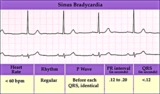 |
Sinus bradycardia | Sinus bradycardia | Knowledge Weavers ECG | |
| 1246 |
 |
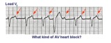
2nd degree AV block, mobitz type II, with LBBB | The wide QRS complexes in lead V1 indicates LBBB. 2nd degree AV block, Mobitz II is suggested by the two fixed PR intervals prior to the nonconducted P wave. The location of the block is most likely in the right bundle, because Mobitz II is usually a sign of bilateral bundle branch disease. | Knowledge Weavers ECG | |
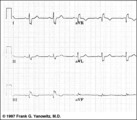
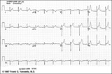
| 1247 |
 |
A very subtle atrial tachycardia with 2:1 block | Although at first glance this looks like normal sinus rhythm at 95 bpm. On closer look, there are 2 P waves for every QRS; the atrial rate is 190 bpm. Note the hidden P in the T waves. This rhythm is likely due to digitalis intoxication, as are the GI symptoms. | Knowledge Weavers ECG | |
| 1248 |
 |
Left atrial enlargement | Left atrial enlargement is illustrated by increased P wave duration in lead II, top ECG, and by the prominent negative P terminal force in lead V1, bottom tracing. | Knowledge Weavers ECG | |
| 1249 |
 |
Acute postero-lateral MI: precordial leads | Acute postero-lateral MI: precordial leads | Knowledge Weavers ECG | |
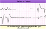
| 1250 |
 |
Pacemaker failure to pace - marquette | Pacemaker failure to pace - marquette | Knowledge Weavers ECG | |
| 1251 |
 |
Frontal plane QRS axis = 90 degrees | Frontal plane QRS axis = 90 degrees | Knowledge Weavers ECG | |
| 1252 |
 |
Junctional tachycardia - marquette | Junctional tachycardia - marquette | Knowledge Weavers ECG | |
| 1253 |
 |
Diagram: frontal plane leads | Diagram: frontal plane leads | Knowledge Weavers ECG | |
| 1254 |
 |
Inferoposterior MI | Inferoposterior MI | Knowledge Weavers ECG | |
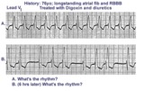
| 1255 |
 |
Digitalis intoxication: Junctional tachycardia with and without AV block | In a patient with longstanding atrial fibrillation being treated with digoxin, a regular tachycardia, as seen in A, with a RBBB suggests a junctional or supraventricular tachycardia. Group beating, in B, is likely due to a 2nd degree, Type 1, exit block below the ectopic junctional focus. This is h... | Knowledge Weavers ECG | |
| 1256 |
 |
Nonconducted PACs and junctional escapes | Although at first glance this looks like 2nd degree AV block, the P waves indicated by the arrows are premature and not sinus P waves. The pause is long enough to encourage a junctional escape focus to take over. Note the sinus P waves just before the escape beats. Had the escapes not occurred, t... | Knowledge Weavers ECG | |
| 1257 |
 |
WPW diagram | The short PR interval is due to a bypass track, also known as the Kent pathway. By bypassing the AV node the PR shortens. The delta wave represents early activation of the ventricles from the bypass tract. The fusion QRS is the result of two activation sequences, one from the bypass tract and one... | Knowledge Weavers ECG | |
| 1258 |
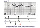 |
PVC with venticular echo | The PVC in this example retrogradely enters the AV junction and returns, usually down a different pathway, to reactivate the ventricles....a ventricular echo. This is unlikely to be an interpolated PVC because the PR interval following the PVC is too short for the sinus impulse to have entered the ... | Knowledge Weavers ECG | |
| 1259 |
 |
Left bundle branch block (LBBB) | LBBB is recognized by 1) QRS duration>0.12s; 2) monophasic R waves in I and V6; and 3) terminal QRS forces oriented leftwards and posterior. The ST-T waves should be oriented opposite to the terminal QRS forces. | Knowledge Weavers ECG | |
| 1260 |
|
PAC with RBBB aberrant conduction | PAC with RBBB aberrant conduction | Knowledge Weavers ECG | |
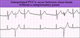
| 1261 |
 |
Interpolated PVCs - marquette | Interpolated PVCs - marquette | Knowledge Weavers ECG | |
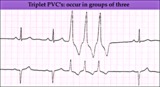
| 1262 |
 |
PVC triplet - marquette | PVC triplet - marquette | Knowledge Weavers ECG | |
| 1263 |
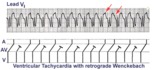 |
Ventricular tachycardia with retrograde wenckebach | Approximately 50 percent of ventricular tachycardias are associated with AV dissociation. The other 50 percent have retrograde atrial capture. This example shows ventricular tachycardia with retrograde Wenckebach. The retrograde P waves are hard to find, but the arrows are of some help. | Wenckebach AV Block | Knowledge Weavers ECG |
| 1264 |
 |
Nonconducted and aberrantly conducted PAC's | In A the slow sinus rhythm is actually caused by nonconducted PACs hidden in the ST segment. This is confirmed in B where some of the PACs are aberrantly conducted with LBBB, and some PACs are nonconducted. | Knowledge Weavers ECG | |
| 1265 |
 |
Left anterior fasicular block: frontal plane leads | Left anterior fascicular block, LAFB, is recognized by left axis deviation of -45 degrees or greater; rS complexes in II, III, aVF; and a small Q wave in I and/or aVL. | Knowledge Weavers ECG | |
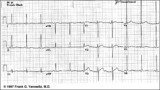
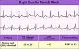
| 1266 |
 |
Right bundle branch block (RBBB) | Right bundle branch block (RBBB) | Knowledge Weavers ECG | |
| 1267 |
 |
All about premature beats | All about premature beats | Knowledge Weavers ECG | |
| 1268 |
 |
Inferoposterior MI | Inferoposterior MI | Knowledge Weavers ECG | |
| 1269 |
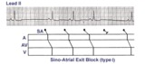 |
Sino-atrial exit block, type I or wenckebach | This example illustrates 2nd degree sino-atrial exit block. In type 1 S-A block the conduction time between sinus firing and atrial capture progressively prolong, but this cannot be seen on the ECG tracing; type I exit block is inferred if the P-P intervals gradually shorten before the pause and if... | Wenckebach AV Block; Ladder Diagram | Knowledge Weavers ECG |
| 1270 |
 |
Inferior & Anteroseptal MI + RBBB | Pathologic Q waves are seen in leads II, III, aVF (inferior MI) and in leads V1-3 (anteroseptal MI). RBBB is recognized by the wide QRS (>0.12s) and the anterior/rightwards orientation of terminal QRS forces. When an anteroseptal MI complicates RBBB (or visa versa) the rSR' complex in V1 (typical ... | Knowledge Weavers ECG | |
| 1271 |
 |
Atrial flutter with 3:2 conduction ratio: frontal plane leads | Note the subtle bigeminy in the RR intervals. The best way to identify the flutter waves in this example is to imagine what lead III would look like if the QRS complexs disappeared; what remains is a reasonable saw-tooth pattern characteristic of atrial flutter with a flutter rate of about 300 bpm... | Knowledge Weavers ECG | |
| 1272 |
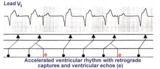 |
Accelerated ventricular rhythm with retrograde atrial capture and echo beats | Retrograde atrial captures from an accelerated ventricular focus are occurring with increasing R-P intervals, When the longer R-P occurs, the impulse traversing the AV junction finds a route back to the ventricles, and the result is a ventricular echo. | Knowledge Weavers ECG | |
| 1273 |
 |
ECG components diagram - marquette | ECG components diagram - marquette | Knowledge Weavers ECG | |
| 1274 |
 |
Postero-lateral MI: Precordial Leads | Postero-lateral MI: Precordial Leads | Knowledge Weavers ECG | |
| 1275 |
 |
Indeterminate frontal plane QRS axis | Indeterminate frontal plane QRS axis | Knowledge Weavers ECG | |
| 1276 |
 |
PAC couplet - marquette | PAC couplet - marquette | Knowledge Weavers ECG | |
| 1277 |
 |
Normal variant: Early repolarization | Early repolarization, a misnomer, describes a pattern of localized or diffuse ST segment elevation. This is especially seen in leads with prominent R waves. In this example leads I, II, V5 and V6 illustrate the early repolarization pattern. ST segments usually have a concave upwards pattern and ta... | Knowledge Weavers ECG | |
| 1278 |
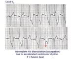 |
AV dissociation by usurpation | Normal sinus rhythm is interrupted by an accelerated ventricular rhythm whose rate is slightly faster than the sinus rhythm. Fusion QRS complexes occur whenever the sinus impulse enters the ventricles at the same time the ectopic ventricular focus initiates its depolarization. | Knowledge Weavers ECG | |
| 1279 |
 |
Long QT mischief | The long QT ECG has many causes: electrolyte abnormalities including hypo-K, hypo-Mg, and hypo-Ca; drugs including type I antiarrhythmics; CNS injury; and hereditary syndromes. Ventricular arrhythmias are thought to be caused by afterdepolarizations or triggered automaticity. | Knowledge Weavers ECG | |
| 1280 |
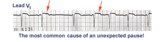 |
Nonconducted and conducted PAC's | The pause in this example is the result of a nonconducted PAC, as indicated by the first arrow. The second arrow points to a conducted PAC. The most common cause of an unexpected pause in rhythm is a nonconducted PAC. | Knowledge Weavers ECG | |
| 1281 |
 |
Atypical LBBB with Q waves in leads I and aVL | In typical LBBB, there are no initial Q waves in leads I, aVL, and V6. If Q waves are present in 2 or more of these leads, myocardial infarction is present. | Knowledge Weavers ECG | |
| 1282 |
 |
Old inferior MI, PVCs, and atrial fibrillation | Old inferior MI, PVCs, and atrial fibrillation | Knowledge Weavers ECG | |
| 1283 |
 |
Frontal plane QRS axis = -45 degrees | Frontal plane QRS axis = -45 degrees | Knowledge Weavers ECG | |
| 1284 |
 |
LVH & PVCs: Precordial Leads | LVH & PVCs: Precordial Leads | Knowledge Weavers ECG | |
| 1285 |
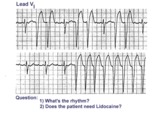 |
Atrial flutter with variable AV block and rate-dependent LBBB | The basic rhythm is atrial flutter with variable AV block. When 2:1 conduction ratios occur there is a rate-dependent LBBB. Don't be fooled by the wide QRS tachycardia on the bottom strip. It's not ventricular tachycardia, but atrial flutter with 2:1 conduction and LBBB. Lidocaine is not needed ... | Knowledge Weavers ECG | |
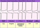
| 1286 |
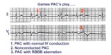 |
three fates of PAC's | As illustrated, PAC's can have three fates: PAC-1enters the ventricles and encounters no conduction delays, therefore causing a narrow QRS; PAC-2 occurs a little earlier and can't get through the AV junction, therefore beingnonconducted; PAC-3 seen inlead V1 makes it into the ventricles but encounte... | Knowledge Weavers ECG | |
| 1287 |
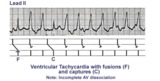 |
Ventricular tachycardia with AV dissociation, captures, and fusions | Approximately 50 percent of ventricular tachycardias are associated with AV dissociation. In these cases atrial impulses can enter the ventricles and either fuse with a ventricular ectopic beat or completely capture the ventricles. This ladder diagram illustrates these events. | Knowledge Weavers ECG | |
| 1288 |
 |
Frontal plane QRS axis = +75 degrees | Since there is no isoelectric lead in this ECG, the two closest leads are I and aVL. If I were isoelectric, the axis would be +90 degrees; if aVL were isoelectric, the axis would be +60 degrees. A nice compromize is +75 degrees. (The two closest leads are always 30 degrees apart.) | Knowledge Weavers ECG | |
| 1289 |
 |
Right axis deviation: QRS axis = +130 degrees | Lead aVR is almost isoelectric; lead I is mostly negative, and lead III is very positive. The QRS axis, therefore, is +130 degrees. Note that the slightly more positive AVR moves the axis slightly beyond +120 degrees; i.e., closer to the + pole of the aVR lead. | Knowledge Weavers ECG | |
| 1290 |
 |
Extensive anterior/anterolateral MI: precordial leads | Extensive anterior/anterolateral MI: precordial leads | Knowledge Weavers ECG | |
| 1291 |
 |
LVH: strain pattern + left atrial enlargement | LVH: strain pattern + left atrial enlargement | Knowledge Weavers ECG | |
| 1292 |
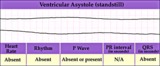 |
Ventricular asystole - marquette | Ventricular asystole - marquette | Knowledge Weavers ECG | |
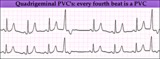
| 1293 |
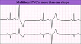 |
Multifocal PVC's - marquette | Multifocal PVC's - marquette | Knowledge Weavers ECG | |
| 1294 |
 |
Atrial flutter with 2:1 AV conduction | In this example of atrial flutter with 2:1 AV conduction the flutter waves are very hard to see. Atrial flutter with 2:1 block must be considered, however, because the heart rate is about 150 bpm. A careful look at V1 shows the two flutter waves for each QRS complex complex. One flutter wave imme... | Knowledge Weavers ECG | |
| 1295 |
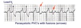 |
Ventricular parasystole | In ventricular parasystole, non-fixed coupled PVC's occur at a common inter-ectopic interval. Fusion beats, indicated by arrows, are often seen. Fusions occur when the sinus impulse entering the ventricles find the ventricles already partially depolarized by the parasystolic focus. | Knowledge Weavers ECG | |
| 1296 |
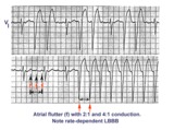 |
Atrial flutter with 2:1 and 4:1 conduction and rate dependent LBBB | In this example of atrial flutter with variable AV conduction, the faster rates are associated with rate-related LBBB. Don't confuse this for ventricular tachycardia. | Knowledge Weavers ECG | |
| 1297 |
 |
2nd degree AV block, type I (Wenckebach) | 2nd degree AV block, type I (Wenckebach) | Knowledge Weavers ECG | |
| 1298 |
 |
Diagram: stages of acute Q-wave MI | Diagram: stages of acute Q-wave MI | Knowledge Weavers ECG | |
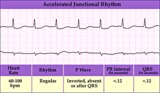
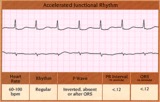
| 1299 |
 |
Accelerated junctional rhythm | Accelerated junctional rhythm | Knowledge Weavers ECG | |
| 1300 |
 |
PVC's - marquette | PVC's - marquette | Knowledge Weavers ECG | |
| 1301 |
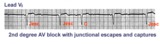 |
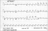
2nd degree AV block with junctional escapes and captures | Second degree AV block is present; conducted beats are identified by those QRS's that terminate shorter cycles than the junctional escape cycle; i.e., the 3rd and probably the 4th QRS's are captures; the other QRS's are junctional escapes. | Knowledge Weavers ECG | |
| 1302 |
 |
Atrial fibrillation with moderate ventricular response - Marquette | Atrial fibrillation with moderate ventricular response - Marquette | Knowledge Weavers ECG | |
| 1303 |
 |
Ventricular pacing in atrial fibrillation - marquette | Ventricular pacing in atrial fibrillation - marquette | Knowledge Weavers ECG | |
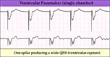
| 1304 |
 |
Ventricular pacemaker: demand mode functioning | Ventricular pacemaker: demand mode functioning | Knowledge Weavers ECG | |
| 1305 |
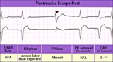 |
Ventricular escape beat - marquette | Ventricular escape beat - marquette | Knowledge Weavers ECG | |
| 1306 |
 |
LAFB: frontal plane leads | LAFB: frontal plane leads | Knowledge Weavers ECG | |
| 1307 |
 |
Old infero-posterior MI | Old infero-posterior MI | Knowledge Weavers ECG | |
| 1308 |
 |
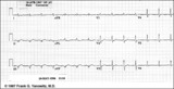
2nd degree AV block, type I | The 3 rules of classic AV Wenckebach are: 1. decreasing RR intervals until pause; 2. the pause is less than preceding 2 RR intervals; and 3. the RR interval after the pause is greater than the RR interval just prior to pause. Unfortunately, there are many examples of atypical forms of Wenckebach wh... | Wenckebach AV Block | Knowledge Weavers ECG |
| 1309 |
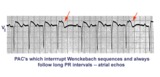 |
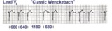
Atrial echos | In this example a typical Wenckebach sequence is interrupted by what looks like a PAC - indicated by red arrows. Atrial echos are more likely, however, because the preceding beat has a long PR interval, a condition that facilitates reentry and echo formation. | Knowledge Weavers ECG | |
| 1310 |
 |
Anteroseptal MI with RBBB: precordial leads | Anteroseptal MI with RBBB: precordial leads | Knowledge Weavers ECG | |
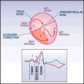
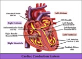
| 1311 |
 |
Cardiac conduction system diagram - marquette | Cardiac conduction system diagram - marquette | Knowledge Weavers ECG | |
| 1312 |
 |
ST segment depression: precordial leads | ST segment depression: precordial leads | Knowledge Weavers ECG | |
| 1313 |
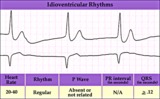 |
Idioventricular escape rhythm | Idioventricular escape rhythm | Knowledge Weavers ECG | |
| 1314 |
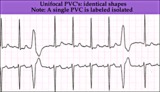 |
Unifocal PVCs - marquette | Unifocal PVCs - marquette | Knowledge Weavers ECG | |
| 1315 |
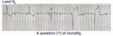 |
Two Wrongs Sometimes Make a Right | The question mark is over a normal looking QRS that occurs during 2:1 AV block with RBBB. Following this QRS a ventricular escape rhythm takes over. The normal looking beat is actually a fusion beat resulting from simultaneous activation of the ventricles; the sinus impulse enters the left ventric... | Knowledge Weavers ECG | |
| 1316 |
 |
WPW type preexcitation | Note the short PR and the subtle delta wave at the beginning of the QRS complexes. The delta wave represents early activation of the ventricles in the region where the AV bypass tract inserts. The rest of the QRS is derived from the normal activation sequence using the bundle branches. | Knowledge Weavers ECG | |
| 1317 |
 |
Long QT interval | The QT interval duration is greater than 50% of the RR interval, a good indication that it is prolonged in this patient. Although there are many causes for the long QT, patients with this are at risk for malignant ventricular arrhythmias, syncope, and sudden death. | Knowledge Weavers ECG | |
| 1318 |
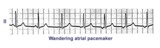 |
Wandering atrial pacemaker | Wandering atrial pacemaker is a benign rhythm change where the pacemaker site shifts from the sinus node into the atrial tissues. P-wave morphology varies with the pacemaker site. | Knowledge Weavers ECG | |
| 1319 |
|
QRS axis = +60 degrees | Lead aVL is isoelectric; leads II and III are mostly positive. The QRS axis, therefore, is +60 degrees. | Knowledge Weavers ECG | |
| 1320 |
 |
Electronic ventricular pacemaker rhythm - marquette | Electronic ventricular pacemaker rhythm - marquette | Knowledge Weavers ECG | |
| 1321 |
 |
High lateral wall MI (seen in aVL) | High lateral wall MI (seen in aVL) | Knowledge Weavers ECG | |
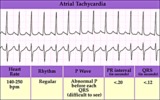
| 1322 |
 |
Atrial tachycardia - marquette | Atrial tachycardia - marquette | Knowledge Weavers ECG | |
| 1323 |
 |
LVH with Strain | LVH with Strain | Knowledge Weavers ECG | |
| 1324 |
 |
PAC's with RBBB aberrant conduction | PAC's are identified by the arrows. Note that the PP interval surrounding the PAC is less than 2x the basic sinus cycle indicating that the sinus node has been reset by the ectopic P wave. The pause after the PAC, therefore, is incomplete. | Knowledge Weavers ECG | |
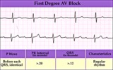
| 1325 |
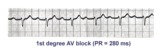 |
1st degree AV block | The normal PR interval is 0.12 - 0.20 sec, or 120 -to- 200 ms. 1st degree AV block is defined by PR intervals greater than 200 ms. This may be caused by drugs, such as digoxin; excessive vagal tone; ischemia; or intrinsic disease in the AV junction or bundle branch system. | Knowledge Weavers ECG | |
| 1326 |
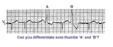 |
Sore thumbs | Two funny looking premature beats are seen in this rhythm strip. Beat A is preceded by a PAC which distorts the T wave, making this an aberrantly conducted PAC. Beat B is a PVC. The notch on the down slope of the QRS complex clearly identifies this as a PVC and not aberrancy. | Knowledge Weavers ECG | |
| 1327 |
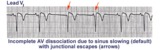 |
AV dissociation by default | If the sinus node slows too much a junctional escape pacemaker may take over as indicated by arrows. AV dissociation is incomplete, since the sinus node speeds up and recaptures the entricles. | Knowledge Weavers ECG | |
| 1328 |
 |
Atrial tachycardia with 2:1 AV block: a manifestation of digitalis intoxication | Atrial tachycardia with 2:1 AV block: a manifestation of digitalis intoxication | Knowledge Weavers ECG | |
| 1329 |
 |
Accelerated junctional rhythm - marquette | Accelerated junctional rhythm - marquette | Knowledge Weavers ECG | |
| 1330 |
 |
First degree AV block - marquette | First degree AV block - marquette | Knowledge Weavers ECG | |
| 1331 |
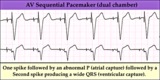 |
AV sequential pacemaker - marquette | (Summary) | Knowledge Weavers ECG | |
| 1332 |
 |
Postero-lateral MI: Fully Evolved | The true posterior MI is recognized by pathologic R waves in leads V1-2. These are the posterior equivalent of pathologic Q waves (seen from the perspective of the anterior leads). Tall T waves in these same leads are the posterior equivalent of inverted T waves in this fully evolved MI. The loss o... | Knowledge Weavers ECG | |
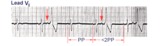
| 1333 |
 |
Atrial tachycardia with 3:2 and 2:1 AV block | The ectopic atrial rate is 150 bpm. Some of the ectopic P waves are easily seen and indicated by the arrows. Other P waves are burried in the T waves and not so easily identified. Atrial tachycardia with AV block is often a sign of digitalis intoxication. 3:2 and 2:1 AV block is seen in this examp... | Knowledge Weavers ECG | |
| 1334 |
 |
RBBB with primary ST-T wave abnormalities | RBBB is recognized by 1) rR' in V1; 2) QRS duration>0.12s; 3) terminal QRS forces oriented rightwards and anterior. In RBBB the ST-T waves should be oriented opposite to the terminal QRS forces. In this example there areprimary ST-T wave abnormalitiesin leads I, II, aVL, V5, V6. In these leads th... | Knowledge Weavers ECG | |
| 1335 |
 |
What are those funny looking beats???? | The differential diagnosis of funny-looking-beats, or FLB's, primarily considers beats of supraventricular origin with aberrant conduction and ventricular ectopic beats. In this example the two FLB's have an easily seen ectopic P wave before them; therefore these are PAC's with RBBB aberration. | Knowledge Weavers ECG | |
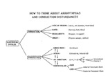
| 1336 |
 |
Conceptual framework: aArrhythmias and conduction abnormalities | Conceptual framework: aArrhythmias and conduction abnormalities | Knowledge Weavers ECG | |
| 1337 |
 |
Atrial flutter with variable AV block - marquette | Atrial flutter with variable AV block - marquette | Knowledge Weavers ECG | |
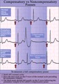
| 1338 |
 |
Compensatory vs. non-compensatory pauses - marquette | Compensatory vs. non-compensatory pauses - marquette | Knowledge Weavers ECG | |
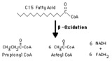
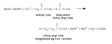
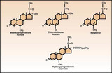
| 1339 |
 |
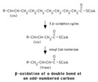
Propionyl CoA production from odd-chain fatty acids | Beta-oxidation of fatty acids with an odd number of carbons inthe chain yields the three-carbon propionyl CoA as the final fragment. | Biosynthesis | Knowledge Weavers Fatty Acids |
| 1340 |
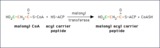 |
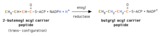
Transfer of a malonyl group to the acyl carrier peptide | During fatty acid synthesis the incoming two carbon fragment is introduced as the three-carbon malonyl group. It is added to the -SH group of the acyl carier peptide domain of fatty acid synthase. In a subsequent reaction the carbon shown in green will be lost as bicarbonate ion. | Knowledge Weavers Fatty Acids | |
| 1341 |
 |
Transport of fatty acyl CoA into mitochondria by the carnitine shuttle | Fatty acyl CoA cannot cross the inner mitochondrial membrane, so it is carried in the form of fatty acyl carnitine. | Knowledge Weavers Fatty Acids | |
| 1342 |
 |
Hydroxymethylglutaryl CoA lyase reaction | In this mitochondrial process hydroxymethylglutaryl CoA is converted to acetoacetate, a ketone body. Acetyl CoA is another product. | Knowledge Weavers Fatty Acids | |
| 1343 |
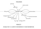 |
Acetyl CoA metabolism -- overview | Major metabolic sources of acetyl CoA and some of the processes in which it serves as a substrate. | Knowledge Weavers Fatty Acids | |
| 1344 |
 |
Oleic acid structure | Oleic acid is a typical monounsaturated fatty acid. | Knowledge Weavers Fatty Acids | |
| 1345 |
 |
methylmalonyl CoA mutase reaction | In the metabolism of propionyl CoA L-methylmalonyl CoA is converted to succinate by methylmalonyl CoA mutase. Succinate can then be metabolized throgh the tricarboxylic acid cycle. | Knowledge Weavers Fatty Acids | |
| 1346 |
 |
Mammalian fatty acyl synthase dimer | This schematic diagram is intended to show the sequence of enzyme activities in the two subunits of a mammalian fatty acyl synthase dimer. It is not intended to imply anything about the detailed spatial relationships of the activities. | Coenzyme A Synthetases | Knowledge Weavers Fatty Acids |
| 1347 |
 |
Hydration of an enoyl CoA | Hydration of the double bond is catalyzed by enoyl CoA hydratase. The product is an L-3-hydroxyacyl CoA. This reaction is a step in the beta-oxidation of fatty acids. | Knowledge Weavers Fatty Acids | |
| 1348 |
 |
Acetyl CoA structure | Acetyl CoA Structure. The thioester bond linking the acetyl group to CoA is ahigh energybond. | Knowledge Weavers Fatty Acids | |
| 1349 |
 |
Use of greek letters to designate carbons in fatty acids | The carbon next to the -COOH group is the alpha carbon; the next one is the beta carbon, and so forth. The carbon most distant from the -COOH group is designated omega. Carbons close to the omega carbon may be designated in relation to it. E.g., the third carbon from the end is omega - 3, and the... | Nomenclature | Knowledge Weavers Fatty Acids |
| 1350 |
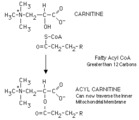 |
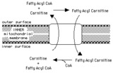
Acylation of carnitine by a long chain fatty acyl CoA | Long chain fatty acyl CoA cannot cross the inner mitochondrial membrane to participate in beta-oxidation. The fatty acyl group is therefore transferred to a carrier, carnitine, in a reversible reaction catalyzed by carnitine acyl transferase I. The resulting fatty acyl carnitine crosses the membra... | Knowledge Weavers Fatty Acids | |
| 1351 |
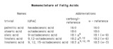 |
Four systems for denoting fatty acids | There are four commonly used ways of designating fatty acids. The first two columns show samples of names, and the last two columns show systems of abbreviating these names. | Nomenclature | Knowledge Weavers Fatty Acids |
| 1352 |
 |
Reduction of 2-enoyl acyl carrier peptide | A 2-enoyl acyl group on the acyl carrier peptide is reduced by NADPH in a reaction catalyzed by enoyl acyl carrier peptide reductase. | Knowledge Weavers Fatty Acids | |
| 1353 |
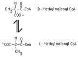 |
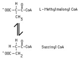
methylmalonyl CoA racemase reaction | In the metabolism of propionyl CoA, D-methylmalonyl CoA is produced by a carboxylase reaction. This product must be converted to L-methylmalonyl CoA in order to be metabolized further. The conversion is catalyzed by methylmalonyl CoA racemase. | Knowledge Weavers Fatty Acids | |
| 1354 |
 |
Oxidation of a polyunsaturated fatty acid -- part I | The cycles of beta-oxidation prior to the one involving the original Delta-12 double bond act to get past the Delta-9 double bond. | Knowledge Weavers Fatty Acids | |
| 1355 |
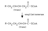 |
enoyl CoA isomerase reaction | Enoyl CoA isomerase converts a trans-3-enoyl CoA to a trans-2-enoyl CoA. Thus a Delta-9 fatty acyl CoA or the product of the 2,4-dienoyl CoA reductase reaction can proceed through beta-oxidation. | Enoyl CoA Isomerase Reaction | Knowledge Weavers Fatty Acids |
| 1356 |
 |
Structures of the ketone bodies | These are the structures of the ketone bodies. Acetoacetate and beta-hydroxybutyrate are important physiological substrates. Acetone is a byproduct, and is not metabolized further. It is excreted in the urine and in expired air. | Knowledge Weavers Fatty Acids | |
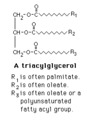
| 1357 |
 |
Triacylglycerol structure | The structure of a typical triacylglycerol (triglyceride). | Knowledge Weavers Fatty Acids | |
| 1358 |
 |
Dehydrogenation of fatty acyl CoA | Fatty acyl CoA is dehydrogenated by FAD in a reaction catalyzed by one of the acyl CoA dehydrogenases. Note that the dehydrogenation occurs between the alpha- and beta-carbons. | FAD | Knowledge Weavers Fatty Acids |
| 1359 |
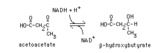 |
Reduction of acetoacetate | Acetoacetate is reduced by NADH in a reversible reaction catalyzed by beta-hydroxybutyrate dehydrogenase. This reaction is the source of beta-hydroxybutyrate in the blood of individuals with ketosis. | Knowledge Weavers Fatty Acids | |
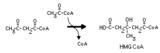
| 1360 |
 |
Hydroxymethylglutaryl CoA synthase reaction | This irreversible reaction occurs in the mitochondria, where it is the first step in ketone body synthesis. It also occurs in the cytoplasm, where it leads to isoprenoid and steroid synthesis. | Biosynthesis | Knowledge Weavers Fatty Acids |
| 1361 |
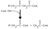 |
Thiolase reaction | Thiolase (3-ketoacyl CoA thiolase) cleaves a long chain fatty acyl CoA, forming acetyl CoA and a long chain fatty acyl CoA that is two carbons shorter. | Knowledge Weavers Fatty Acids | |
| 1362 |
 |
Beta-oxidation of a delta-9 fatty acyl CoA | Enoyl CoA isomerase is required to move the double bond in a Delta-9 fatty acyl CoA to a position where it can continue in beta-oxidation. | Knowledge Weavers Fatty Acids | |
| 1363 |
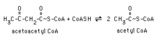 |
Thiolase reaction with acetoacetyl CoA | Thiolase (3-ketoacyl CoA thiolase) cleaves acetoacetyl CoA, forming two molecules of acetyl CoA. | Knowledge Weavers Fatty Acids | |
| 1364 |
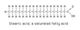 |
Stearic acid structure | Stearic acid is a typical long chain saturated fatty acid. | Knowledge Weavers Fatty Acids | |
| 1365 |
 |
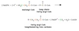
Fatty acid elongation in mitochondria | This shows the overall reaction of fatty acid elongation in mitochondria. The process is essentially a reversal of beta-oxidation, except that one NADPH and one NADH are required (beta-oxidation yields two NADH). Mitochondrial fatty acid elongation acts primarily on fatty acyl CoA substrates short... | Knowledge Weavers Fatty Acids | |
| 1366 |
 |
Fatty acyl CoA elongation in the endoplasmic reticulum | This shows the overall process of fatty acyl elongation in the endoplasmic reticulum. The process resembles that catalyzed by fatty acyl synthase, but the individual activities appear to be on separate enzymes. | Knowledge Weavers Fatty Acids | |
| 1367 |
 |
Propionyl CoA carboxylase reaction | Propionyl CoA is metabolized by a process that first converts it to D-methylmalonyl CoA. The reaction is catalyzed by propionyl CoA carboxylase,and requires energy in the form of ATP. | Biosynthesis | Knowledge Weavers Fatty Acids |
| 1368 |
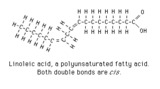 |
Linoleic acid structure | Linoleic acid is a typical polyunsaturated fatty acid. | Knowledge Weavers Fatty Acids | |
| 1369 |
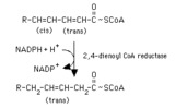 |
Reduction of a 2,4-dienoyl CoA | The reduction of 2,4-dienoyl CoA by NADPH is a step in the oxidation of polyunsaturated fatty acids. It is ironic that a reduction reaction is a required step in a process that is oxidative. | Knowledge Weavers Fatty Acids | |
| 1370 |
 |
Stearic, oleic and linoleic acid structures | Stearic, oleic and linoleic acid structures | Knowledge Weavers Fatty Acids | |
| 1371 |
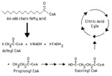 |
Complete oxidation of an odd-chain fatty acid -- overview | This diagram indicates production of propionyl CoA from an odd-chain fatty acid and the subsequent conversion of propionyl CoA to succinyl CoA, which can be metabolized through the citric (tricarboxylic) acid cycle. | Biosynthesis | Knowledge Weavers Fatty Acids |
| 1372 |
 |
Initiation of beta-oxidation | An acetyl group is transferred from acetyl CoA to the -SH group of the condensing enzyme domain of fatty acyl synthase, forming acetyl-CE. The reaction is catalyzed by the acyltransferase activity of fatty acyl synthase. | Knowledge Weavers Fatty Acids | |
| 1373 |
 |
Fatty acid metabolism -- schematic overview | Fatty acids are taken up by cells, where thy may serve as precursors in the synthesis of other compounds, as fuels for energy production, and as substrates for ketone body synthesis. Ketones bodies may then be exported to other tissues, where they can be used for energy production. | Biosynthesis | Knowledge Weavers Fatty Acids |
| 1374 |
 |
Condensation of an acyl group with a malonyl group | The acetyl group displaces the carboxyl of the malonyl group, forming a beta-ketoacyl group. This reaction is catalyzed by beta-ketoacyl Acyl Carrier Peptide synthase. The carboxyl released in the form of bicarbonate regenerates the bicarbonate used earlier in the acetyl CoA carboxylase reaction. | Knowledge Weavers Fatty Acids | |
| 1375 |
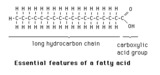 |
Essential features of a fatty acid | The essential features of a fatty acid are a long hydrocarbon chain terminating in a carboxylic acid group. | Knowledge Weavers Fatty Acids | |
| 1376 |
 |
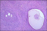
Corpus luteum cysts exhibit a convoluted lining with luteinized granulosa and theca cells. | Corpus luteum of the ovary, medium power. The granulosa cells have undergone proliferation and alteration to lutein cells that produce progesterone. The lutein cells are large and polyhedral and the cytoplasm is foamy and eosinophilic. | Knowledge Weavers Human Reproduction | |
| 1377 |
 |
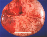
Mucinous cystadenoma | These tumors comprise 20% of all neoplasms, and 50% of ovarian neoplasms found in women less than 20 years of age. They are frequently multiloculated. The favored hypothesis for their origin is metaplastic surface epithelium as they have a predominance of endocervical gland type epithelium. | Knowledge Weavers Human Reproduction | |
| 1378 |
 |
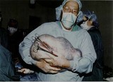
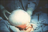
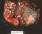
Ovarian tumor | A large vertical incision allows delivery and excision of an ovarian neoplasm. Pelvic washings for cytology and a frozen section rule out a malignancy. The cyst is excised carefully to avoid spilling contents into the abdomen. | Knowledge Weavers Human Reproduction | |
| 1379 |
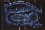 |
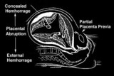
Ultrasound of placental abruption | The fetal body and bladder are labeled FB, and BL, respectively. | Knowledge Weavers Human Reproduction | |
| 1380 |
 |
Uterine rupture | Uterine rupture | Knowledge Weavers Human Reproduction | |
| 1381 |
 |
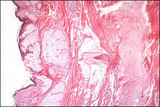
Brenner tumor | Solid and partially cystic epithelial nests are surrounded by a stroma composed of bundles of tightly-packed spindle shaped cells. The epithelial cells are polygonal and of the squamoid type, with pale, eosinophilic cytoplasm and oval nuclei having distinct nuclei and longitudinal grooving, a coffee... | Knowledge Weavers Human Reproduction | |
| 1382 |
 |
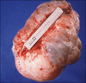
Mucinous cystadenoma | This tumor weighed 35 pounds. | Knowledge Weavers Human Reproduction | |
| 1383 |
 |
Brenner tumor | Brenner tumors are comprised of solid to partly cystic epithelial nests surrounded by stroma composed of bundles of tightly packed spindle-shaped cells. The epithelial cells are polygonal and of squamoid type, with pale, eosinophilic cytoplasm and oval nuclei with distinct nucleoli and longitudinal ... | Knowledge Weavers Human Reproduction | |
| 1384 |
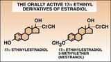 |
Estradiol | The structure of ethinyl estradiol and mestranol. | Knowledge Weavers Human Reproduction | |
| 1385 |
 |
Mucinous cystadenoma | Neglected mucinous cystadenoma may become extremely large. | Knowledge Weavers Human Reproduction | |
| 1386 |
 |
Mature cystic teratoma - Microscopic | Ectodermal tissue is usually most abundant and represented by: squamous epithelium and appendages, brain tissue, glia, retina, choroid plexus, and ganglia. Mesodermal tissue is represented by bone and cartilage. Endodermal tissue is represented by gastrointestinal and bronchial epithelium and glands... | Knowledge Weavers Human Reproduction | |
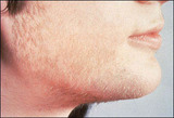
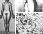
| 1387 |
 |
Hirsutism | This woman has polycystic ovarian syndrome and hyperandrogenemia resulting in hirsutism. | Knowledge Weavers Human Reproduction | |
| 1388 |
 |
Structures of acetoxy progestins (pregnane steroids) | From C. Matthew Peterson, M.D. Progestogens, Progesterone Antagonists, Progesterone, and Androgens: Synthesis, Classification and Uses, Clinical Obstetrics and Gynecology, 1995; 38: 813-20. The structures of acetoxy progestins (pregnane steroids). | Knowledge Weavers Human Reproduction | |
| 1389 |
 |
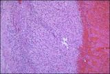
Serous cystadenoma | These tumors comprise 20% of all ovarian neoplasms. The epithelium is a single layer of regular cuboidal epithelium, with basal nuclei and rare mitoses. | Knowledge Weavers Human Reproduction | |
| 1390 |
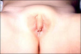 |
Congenital adrenal hyperplasia and clitoromegaly | Congenital adrenal hyperplasia and clitoromegaly | Knowledge Weavers Human Reproduction | |
| 1391 |
 |
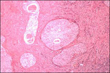
Mature cystic teratoma-torsed with adenexal structures | Elements of all three germ layers are present: endoderm, mesoderm, and ectoderm. They are classified as germ cell tumors and are thought to arise through parthenogenesis. They are found in the path of migration of the germ cells from the yolk sac to the primitive gonad. | Pathogenesis | Knowledge Weavers Human Reproduction |
| 1392 |
 |
Total placenta previa: placenta accreta in uterine specimen | Placenta accreta or percreta may be associated with placenta previa especially if the patient has had a previous cesarean section. | Knowledge Weavers Human Reproduction | |
| 1393 |
 |
Ovarian follicular scanning | Ovarian follicular scanning. Ovaries are scanned transvaginally to determine the size of the developing follicles. | Knowledge Weavers Human Reproduction | |
| 1394 |
 |
Total placenta previa: schematic of complete placenta previa | The placenta completely covers the internal os of the cervix. | Knowledge Weavers Human Reproduction | |
| 1395 |
 |
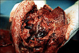
Abruptio placenta | Abruptio placenta | Knowledge Weavers Human Reproduction | |
| 1396 |
 |
Contraception for women in their later years | Contraception for women in their later reproductive years. | Knowledge Weavers Human Reproduction | |
| 1397 |
 |
Pelvic mass | A pelvic mass may be present with abdominal distention from the mass, ascities, or both. | Abdominal Distention | Knowledge Weavers Human Reproduction |
| 1398 |
 |
Androgen insensitivity | Adapted with permission from Jaffe R.B. Disorders of Sexual Differentiation. In Yen SSC and Jaffe RB, eds, Reproductive Endocrinology, W.B. Saunders Co., Philadelphia, 1986, p 300. An inability to respond to testosterone through a target organ receptor defect in 60-70% and a post-receptor signalling... | Knowledge Weavers Human Reproduction | |
| 1399 |
 |
Klinefelter's syndrome | Adapted with permission from Grumbach M.M. and Conte F.A.,Disorders of Sexual Differentation. In Williams R.H. ed, Textbook of Endocrinology, W. B.Squanders Co., Philadelphia, 1981, p 451. Males with an XXY karyotype characteristically have long extremities, aeunuchoidal habitus, and gynecomastia. | Knowledge Weavers Human Reproduction | |
| 1400 |
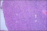 |
Polycystic ovaries | Polycystic ovary, high power view: Prominent layer of theca interna cells with extensive luteinization. Atretic follicles are more abundant in the cortex. | Knowledge Weavers Human Reproduction |