A collection of videos relating to the diagnosis and treatment of eye movement disorders. This collection includes many demonstrations of examination techniques.
Dan Gold, D.O., Associate Professor of Neurology, Ophthalmology, Neurosurgery, Otolaryngology - Head & Neck Surgery, Emergency Medicine, and Medicine, The Johns Hopkins School of Medicine.
A collection of videos relating to the diagnosis and treatment of eye movement disorders.
NOVEL: https://novel.utah.edu/
TO
1 - 25 of 24
| Title | Description | Subject | ||
|---|---|---|---|---|
| 1 |
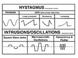 |
A Comparison of Nystagmus and Saccadic Intrusions/Oscillations | Nystagmus can be classified into pendular and jerk waveforms, where both are generated by a slow, pathologic phase. Corrective phase (the position reset mechanism) differs. In pendular nystagmus, the eyes move back and forth with about the same velocity and amplitude, similar to that of a pendulum... | Jerk Nystagmus; Flutter; Pendular Nystagmus; Square Wave Jerks; Opsoclonus |
| 2 |
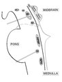 |
Brainstem Ocular Motor Machinery | Seen here is a sagittal view of the brainstem. The medulla has a significant role in gaze-holding, and the nucleus prepositus hypoglossi (NPH, along with the medial vestibular nucleus ) is the horizontal neural integrator. The abducens (6th) nucleus is located in the dorsal pons, and sends off the 6... | Medulla OMS; Pons OMS; Mesencephalon OMS; Dorsal Midbrain OMS |
| 3 |
 |
Central Anatomy of the Fourth Nerve | The IVth or trochlear nucleus is located ventral to the central periaqueductal grey matter, dorsal to the medial longitudinal fasciculus (MLF) and medial to the oculosympathetic tract at the level of the inferior colliculus. The fascicles of the IVth nerve travel dorsally and caudally around the cen... | Mesencephalon OMS |
| 4 |
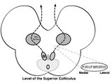 |
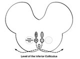
Central Anatomy of the Third Nerve | Seen here is an axial section of the midbrain at the level of the superior colliculus. The paired nuclei are located ventral to the periaqueductal grey, and the midline central caudal nucleus (CCN) is located between the right and left nuclei. The CCN sends projections to bilateral levator palpebrae... | Mesencephalon; Third Nerve Palsy |
| 5 |
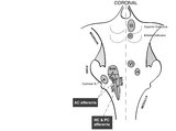 |
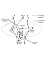
Coronal Section of the Brainstem Showing Ocular Motor Nuclei and Anatomy of the Vestibular Nucleus (with SCC Inputs) | (A) Seen here is a coronal view of the brainstem showing the locations of the ocular motor nuclei (IIIrd, IVth, VIth) as well as the nuclei of VII and VIII (vestibular and cochlear). The vestibular nucleus (VN) is divided into the inferior, lateral, medial, and superior subnuclei, and the medial ves... | Pons; Medullar OMS; Mesencephalon OMS; Figures |
| 6 |
 |
Eyelid Anatomy | Seen here are the major muscles of eyelid opening and closure. The levator palpebrae, which is innervated by the oculomotor nerve, inserts on the tarsus via the levator aponeurosis and directly on the skin of the upper eyelid. The superior tarsal muscle (also known as Muller's muscle, which is inner... | Figures |
| 7 |
 |
Medullary Structures Relevant to Upbeat Nystagmus | This is an axial section of the medulla, slightly more caudal as compared to (please refer to figure "medullary structures relevant to the ocular motor and vestibular consequences of the lateral medullary (Wallenberg) syndrome). Again seen are the inferior cerebellar peduncle (ICP) and caudal aspect... | Medulla OMS; Figures |
| 8 |
 |
Medullary Structures Relevant to the Ocular Motor and Vestibular Consequences of Lateral Medullary (Wallenberg) Syndrome | This is an axial section of the medulla showing the structures that, when damaged, are responsible for the vestibular and ocular motor features of the lateral medullary or Wallenberg syndrome. The nucleus prepositus hypoglossi (NPH) and medial vestibular nucleus (MVN) complex is important for horizo... | Medulla OMS; Figures |
| 9 |
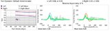 |
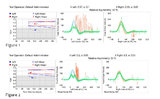
Neuro-Ophthalmic Features and Pseudo-MG Lid Signs in Miller Fisher Syndrome (Figure 1) | This is a 51-year-old woman who presented with imbalance, acute onset dizziness and diplopia that developed over three days following two weeks of upper respiratory infection and bacterial conjunctivitis. When she was initially seen as an outpatient, nystagmus was noted to the right and left, and a ... | Abnormal VOR; Miller Fisher Syndrome; Myasthenia Gravis; Acute Vestibular Syndrome; Jerk Nystagmus; Gaze Evoked Nystagmus |
| 10 |
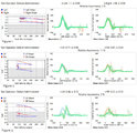 |
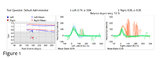
Ocular Motor & Vestibular Features of the MLF Syndrome (Figures 1, 2, and 3) | This 61-year-old woman with HTN and DM presented for evaluation of acute onset diagonal diplopia. Adduction OS was about 60% of normal while medialization OS improved with convergence. In right gaze, dissociated abducting nystagmus was present OD, and there was a clear adduction lag when asking he... | INO; Jerk Nystagmus; Torsional Nystagmus; Gaze-Evoked Nystagmus |
| 11 |
 |
Pons: 6th and 7th Nerve Anatomy and the Central Segmental Tract | From this cross-section of the pons, the proximity of the 6th nucleus to the 7th nerve fascicles is apparent. This is the basis of the so-called facial colliculus syndrome, where an ipsilesional horizontal gaze palsy from a nuclear 6th lesion (usually related to stroke or demyelination) can be seen ... | Sixth Nerve Palsy; Inter Nuclear Ophthalmoplegia; INO; One-and-a-Half; Horizontal Gaze Palsy; OMS Pons; Facial Nerve; Oculopalatal |
| 12 |
 |
Pons: 6th, 7th, 8th, and Middle Cerebellar Peduncle Anatomy | From this cross-section of the pons, the proximity of the 7th and 8th fascicles can be appreciated, and a lateral inferior pontine syndrome (anterior inferior cerebellar artery territory), which could involve both of these fascicles, could cause acute prolonged vertigo accompanied by a + ipsilateral... | Sixth Nerve Palsy; OMS Pons; Facial Nerve; VOR Normal; VOR Abnormal |
| 13 |
 |
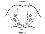
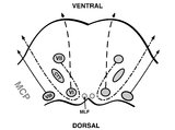
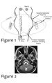
Relationship Between Semicircular Canals and Extraocular Muscles | Figure 1: When stimulated, each of the 6 angular acceleration detecting semicircular canals (3 on the right and 3 on the left) responds with a conjugate eye movement, with the vector(s) indicated below. PC=posterior canal; HC=horizontal (also known as lateral) canal; AC=anterior (also known as super... | Extraocular Muscles |
| 14 |
 |
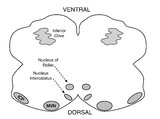
Saccadic Pathways in the Brainstem and Cerebellum & Mechanism for Saccadic Dysmetria in Wallenberg Syndrome - Abnormal Function of the Brainstem/Cerebellar Saccadic Pathways with a Left Wallenberg Syndrome | The end result of a lesion involving the climbing fibers within the left lateral medulla is deficient rightward saccades (contralesional hypometric saccades), and over-active leftward saccades (ipsilesional hypermetric saccades), and ipsilesional ocular lateropulsion given this baseline imbalance. M... | Medulla OMS; Normal Saccades; Abnormal Saccades; Figures |
| 15 |
 |
Saccadic Pathways in the Brainstem and Cerebellum & Mechanism for Saccadic Dysmetria in Wallenberg Syndrome - Normal Function of the Brainstem/Cerebellar Saccadic Pathways | The inferior cerebellar peduncle (ICP) carries climbing fibers to the dorsal vermis, and these fibers have an inhibitory influence over the Purkinje cells. These Purkinje cells normally inhibit the ipsilateral fastigial nucleus, and the fastigial nucleus projects to the contralateral inhibitory burs... | Medulla OMS; Normal Saccades; Abnormal Saccades; Figures |
| 16 |
 |
Sagittal Section of the Brainstem Showing Structures Related to Normal Eyelid Function | Seen here is a sagittal view of the brainstem, with the structures relevant to normal eyelid function highlighted. The M-group, which can be found medial to the riMLF (coordinates eye and lid movements), has (weak) projections to the facial nucleus for frontalis muscle contraction, and (strong) proj... | Figures; Mesencephalon OMS |
| 17 |
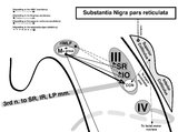 |
Sagittal Section of the Midbrain Showing Structures Related to Normal Eyelid Function | 𝗢𝗿𝗶𝗴𝗶𝗻𝗮𝗹 𝗗𝗲𝘀𝗰𝗿𝗶𝗽𝘁𝗶𝗼𝗻: During a vertical saccade, the rostral interstitial nucleus of the medial longitudinal fasciculus (riMLF) is activated, which excites the superior rectus (SR) and inferior oblique (IO) (IIIrd nerve) subnuclei. Additionall... | Figures; Mesencephalon OMS |
| 18 |
 |
Subtle Torsional Pendular Nystagmus in Oculopalatal Tremor (OPT) (Figure 1) | This is a 50-year-old woman who presented with imbalance, and MRI demonstrated a right cerebellar cavernous malformation. She underwent surgery to resect the malformation, and post-operatively experienced right hemiparesis and ataxia. Six months after the surgery, balance worsened and vision became ... | Pendular Nystagmus; Oculopalatal Tremor |
| 19 |
 |
The Utriculo-Ocular Motor Pathways - Physiologic and Pathologic Ocular Tilt Reaction: OTR Diagram Pathologic EOMs Labelled (Figure 3) | A skew deviation is a non-paralytic vertical ocular misalignment that is due to imbalance in the utriculo-ocular motor pathways. While vestibular jerk nystagmus is a consequence of static semicircular canal pathway imbalance (e.g., left-beating nystagmus due to acute right vestibular hypofunction fr... | Skew Deviation |
| 20 |
 |
The Utriculo-Ocular Motor Pathways - Physiologic and Pathologic Ocular Tilt Reaction: Pathologic OTR (Figure 2) | A skew deviation is a non-paralytic vertical ocular misalignment that is due to imbalance in the utriculo-ocular motor pathways. While vestibular jerk nystagmus is a consequence of static semicircular canal pathway imbalance (e.g., left-beating nystagmus due to acute right vestibular hypofunction fr... | Skew Deviation |
| 21 |
 |
The Utriculo-Ocular Motor Pathways - Physiologic and Pathologic Ocular Tilt Reaction: Physiologic Ocular Tilt Reaction (OTR) (Figure 1) | A skew deviation is a non-paralytic vertical ocular misalignment that is due to imbalance in the utriculo-ocular motor pathways. While vestibular jerk nystagmus is a consequence of static semicircular canal pathway imbalance (e.g., left-beating nystagmus due to acute right vestibular hypofunction fr... | Skew Deviation |
| 22 |
 |
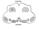
Triangle of Guillain-Mollaret | Seen here is a schematic representation of the Gullain-Mollaret triangle (Figure 1), also referred to as the dentato-olivary pathway, reflecting the 3 points of this imaginary triangle - 1) dentate nucleus, 2) red nucleus, and 3) inferior olivary nucleus. The olive sends decussating climbing fibers ... | Pendular Nystagmus; Oculopalatal Tremor |
| 23 |
 |
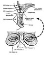
Using Video Head Impulse Testing to Unmask Covert Saccades in Compensated Vestibular Neuritis (Figures 1 and 2) | This is a 30-year-old woman who experienced the acute vestibular syndrome (prolonged vertigo for >24 hours, nausea, unsteadiness, spontaneous nystagmus, head motion intolerance) and was diagnosed with vestibular neuritis. This diagnosis was based on a positive head impulse test to the left (see Figu... | VOR Normal; VOR Abnormal |
| 24 |
 |
Vestibular Neuritis with + Head Impulse Test and Unidirectional Nystagmus (Figure 1) | Vestibular neuritis is the most common cause of the acute vestibular syndrome, which is characterized by continuous vertigo and spontaneous nystagmus lasting days. It may be mimicked by central causes, including stroke, but in the hands of subspecialists, the HINTS+ (Head Impulse, Nystagmus, Test of... | Jerk Nystagmus; Acute Vestibular; Vestibular Nystagmus |
1 - 25 of 24