The Health Education Assets Library (HEAL) is a collection of over 22,000 freely available digital materials for health sciences education. The collection is now housed at the University of Utah J. Willard Marriott Digital Library.
TO
| Title | Description | Subject | Collection | ||
|---|---|---|---|---|---|
| 651 |
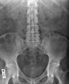 |
X-ray of Normal Kidney Ureter Bladder (Male) | Normal kidney, bladder, and ureter (KUB) of a male | Anatomy | HEAL Reviewed Collection |
| 652 |
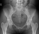 |
X-ray of Normal Pelvis (Female) | Normal anteroposterior (AP) radiograph of the female pelvis | Anatomy | HEAL Reviewed Collection |
| 653 |
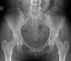 |
X-ray of Normal Pelvis (Female) | Normal anteroposterior (AP) radiograph of the female pelvis | Anatomy | HEAL Reviewed Collection |
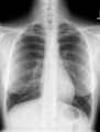
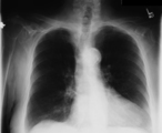
| 654 |
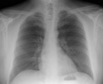 |
X-ray of Normal Chest (Male) | Normal posteroanterior (PA) view of the chest in a middle-aged male | Anatomy; Chest X-ray; CXR | HEAL Reviewed Collection |
| 655 |
 |
X-ray of Normal Chest (Female) | Normal posteroanterior (PA) view of the chest in a young female | Anatomy; Chest X-ray; CXR | HEAL Reviewed Collection |
| 656 |
 |
X-ray of Normal Chest (Male) | Normal posteroanterior (PA) view of the chest in an older male | Anatomy; Chest X-ray; CXR | HEAL Reviewed Collection |
| 657 |
 |
X-ray of Normal Chest (Male) | Normal posteroanterior (PA) view of the chest in a young male | Anatomy; Chest X-ray; CXR | HEAL Reviewed Collection |
| 658 |
 |
X-ray of Normal Chest (Male) | Normal posteroanterior (PA) view of the chest in an older male | Anatomy; Chest X-ray; CXR | HEAL Reviewed Collection |
| 659 |
 |
X-ray of Normal Chest (Male) | Normal lateral view of the chest in a middle-aged male | Anatomy; Chest X-ray; CXR | HEAL Reviewed Collection |
| 660 |
 |
X-ray of Normal Chest (Female) | Normal lateral view of the chest in a young female | Anatomy; Chest X-ray; CXR | HEAL Reviewed Collection |
| 661 |
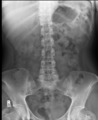 |
X-ray of Normal Kidney Ureter Bladder (Female) | Normal kidney, bladder, and ureter (KUB) of a young female | Anatomy | HEAL Reviewed Collection |
| 662 |
 |
X-ray of Normal Pelvis (Male) | Normal anteroposterior (AP) radiograph of the male pelvis | Anatomy | HEAL Reviewed Collection |
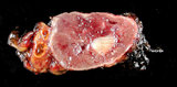
| 663 |
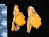 |
Adrenal Adenoma in Clinical Conn's Syndrome | Adrenal adenoma, aldosterone producing associated with Conn's syndrome. Gross photograph showing 2 contiguous slices of adrenal gland with cortical adenoma. Patient had hyperaldosteronism (Conn's syndrome). | Adrenal Adenoma; Conn Syndrome | HEAL Reviewed Collection |
| 664 |
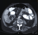 |
Intussusception | This image depicts Intussusception. Intussusception can be found in an abdominal CT. The CT may show nothing if early in the disease or may show a perforation, an obstruction, or a soft tissue mass. | Intestinal Invagination; Intussuscipiens; Intussusceptum; Lead Point; Concentric Rings | HEAL Reviewed Collection |
| 665 |
 |
Pancoast Tumor | This radiograph depicts Pancoast Tumor. Pancoast Tumors can be difficult to detect, if in early stages, on an x-ray. Interpreting overlying shadows at the apices is complex. | Lung Cancer; Superior Sulcus Tumor; Homer Syndrome; Bone Destruction | HEAL Reviewed Collection |
| 666 |
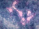 |
Parotid, benign mixed tumor (pleomorphic adenoma) 20X | parotid, benign mixed tumor (pleomorphic adenoma) 20X showing hyaline/mucoid matrix with benign epithelial nests. | BMT; Benign mixed tumor | HEAL Reviewed Collection |
| 667 |
 |
Micrognathia | This is a photograph of an 11 year old girl with pronounced micrognathia as a result of bilateral temporo-mandibular arthritis in longstanding juvenile rheumatoid arthritis. The inflammation has led to growth arrest in the mandible. | HEAL Reviewed Collection | |
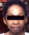
| 668 |
 |
Internal Capsule | This image depicts the Internal Capsule and includes a cross reference key. The ventricles are enlarged which is consistent with atrophy of the brain. | HEAL Reviewed Collection | |
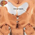
| 669 |
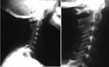 |
Cervical Spine Involvement in Juvenile Rheumatoid Arthritis | Involvement of the cervical spine with obliteration of apophyseal articulations and eventual fusion of the posterior articulations of C2 and C3 is commonly seen in polyarticular or systemic onset disease. | HEAL Reviewed Collection | |
| 670 |
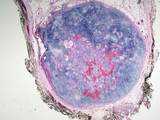 |
Parotid gland with benign mixed tumor (pleomorphic adenoma) | Low power image (2X) of a BMT of the parotid gland showing the hyalin/myxoid matrix and benign epithelial nests. | BMT; Benign mixed tumor | HEAL Reviewed Collection |
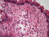
| 671 |
 |
Benign mixed tumor of parotid gland | Medium power image (10X) of a BMT of the parotid gland showing the hyalin/myxoid matrix and benign epithelial nests. | BMT; Benign mixed tumor | HEAL Reviewed Collection |
| 672 |
 |
Duodenum amyloid deposition | Duodenum with amyloid deposition in lamina propria. Amyloid etiology unknown. Amyloid shows up as homogenous pink material in lamina propria and around blood vessels 40X. | Small Bowel | HEAL Reviewed Collection |
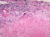
| 673 |
 |
Eosinophilic granuloma mastoid | High power view (40X) of a neoplastic proliferation of Langerhan's cells (Histiocytosis) in mastoid bone. | HEAL Reviewed Collection | |
| 674 |
 |
Adrenal gland with granulomatous reaction (gumma) to syphilis | This high power image (20X) shows a granuloma with a giant cell in the adrenal gland. The granuloma is a gumma formed during tertiary syphilis | gumma | HEAL Reviewed Collection |
| 675 |
 |
Adrenal gland with pheochromocytoma | Patient with clinical symptoms of hypertension. Image shows adjacent normal adrenal gland with paraganglioma (pheochromocytoma) involving adrenal medulla. | Adrenalin; Noradrenalin | HEAL Reviewed Collection |