A collection of videos relating to the diagnosis and treatment of eye movement disorders. This collection includes many demonstrations of examination techniques.
Dan Gold, D.O., Associate Professor of Neurology, Ophthalmology, Neurosurgery, Otolaryngology - Head & Neck Surgery, Emergency Medicine, and Medicine, The Johns Hopkins School of Medicine.
A collection of videos relating to the diagnosis and treatment of eye movement disorders.
NOVEL: https://novel.utah.edu/
TO
| Title | Description | Type | ||
|---|---|---|---|---|
| 1 |
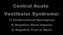 |
Central Acute Vestibular Syndrome Due to Posterior Fossa Hemorrhage | This is a patient presenting with the acute vestibular syndrome (AVS, e.g., acute prolonged vertigo, spontaneous nystagmus) whose HINTS (Head Impulse, Nystagmus, Test of Skew) testing indicated a central etiology based on negative (normal) head impulse testing (HIT). Nystagmus was unidirectional and... | Image/MovingImage |
| 2 |
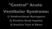 |
Acute Vestibular Syndrome with Ocular Tilt Reaction Due to Bacterial Labyrinthitis | This is a patient who initially presented with the acute vestibular syndrome (AVS, e.g., acute prolonged vertigo, spontaneous nystagmus) and right sided hearing loss, and was diagnosed with bacterial labyrinthritis. Her HINTS (Head Impulse, Nystagmus, Test of Skew) testing indicated a central etiolo... | Image/MovingImage |
| 3 |
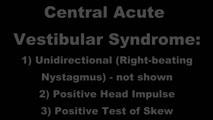 |
Acute Vestibular Syndrome With Skew Deviation and Positive Head Impulse Test Due to a Demyelinating Lesion | This is a patient who initially presented with the acute vestibular syndrome (AVS, e.g., acute prolonged vertigo, spontaneous nystagmus). ; See https://collections.lib.utah.edu/details?id=187730 for additional history. ; Her HINTS (Head Impulse, Nystagmus, Test of Skew) testing indicated a central e... | Image/MovingImage |
| 4 |
 |
Oculogyric Crisis | 𝗢𝗿𝗶𝗴𝗶𝗻𝗮𝗹 𝗗𝗲𝘀𝗰𝗿𝗶𝗽𝘁𝗶𝗼𝗻: This is a patient with neuroleptic-induced oculogyric crisis. 𝗡𝗲𝘂𝗿𝗼-𝗼𝗽𝗵𝘁𝗵𝗮𝗹𝗺𝗼𝗹𝗼𝗴𝘆 𝗮𝗻𝗱 𝗡𝗲𝘂𝗿𝗼-𝗼𝘁𝗼𝗹𝗼𝗴𝘆 𝗧𝗲𝘅𝘁𝗯�... | Image/MovingImage |
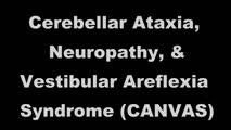
| 5 |
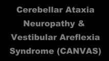 |
Cerebellar Ataxia, Neuropathy, & Vestibular Areflexia Syndrome (CANVAS): Impaired Visually-Enhanced VOR and Abnormal Head Impulse Testing | A 67 year old woman presented with 1 year of progressive numbness, gait instability, and oscillopsia when walking or with head movements. Examination showed excessive square-wave jerks, bilateral horizontal gaze-evoked nystagmus, impairment of the visually-enhanced vestibular ocular reflex (vVOR - s... | Image/MovingImage |
| 6 |
 |
Abnormal Visually-Enhanced VOR in Cerebellar Ataxia, Neuropathy, Vestibular Areflexia Syndrome (CANVAS) | A 67 year old woman presented with 1 year of progressive numbness, gait instability, and oscillopsia when walking or with head movements. Examination showed excessive square-wave jerks, bilateral horizontal gaze-evoked nystagmus, impairment of the visually-enhanced vestibular ocular reflex (vVOR - s... | Image/MovingImage |
| 7 |
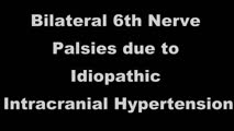 |
Bilateral 6th Nerve Palsies Due to Idiopathic Intracranial Hypertension | This is a 25-year-old woman who presented with diplopia and blurry vision. On exam, she was found to have papilledema and bilateral 6th nerve palsies. Her opening pressure was >40 cm of water with a normal CSF analysis, and neuroimaging was unremarkable aside from subtle findings that have been asso... | Image/MovingImage |
| 8 |
 |
Paraneoplastic Downbeat Nystagmus and Cerebellar Ataxia Due to Small Cell Lung Carcinoma | 𝗢𝗿𝗶𝗴𝗶𝗻𝗮𝗹 𝗗𝗲𝘀𝗰𝗿𝗶𝗽𝘁𝗶𝗼𝗻: This is a 61-year-old woman (non-smoker) who developed a gait disorder, dizziness and oscillopsia that was progressive over 2 months. Exam demonstrated spontaneous downbeat nystagmus with side pocket nystagmus in lateral ... | Image/MovingImage |
| 9 |
 |
Testing for Adduction Lag in Partial INO Using an Optokinetic Stimulus | In this patient we demonstrate the use of an optokinetic stimulus to elicit an internuclear ophthalmoplegia (INO). Occasionally adduction appears to be normal with an INO, and an adduction lag with horizontal saccades should be sought as a confirmatory sign. Optokinetic tape is an easy way to assess... | Image/MovingImage |
| 10 |
 |
Demonstration of HINTS Examination in a Normal Subject | In the acute vestibular syndrome - consisting of acute prolonged vertigo, spontaneous nystagmus, imbalance, nausea/vomiting, head motion intolerance which is typically due to vestibular neuritis or posterior fossa stroke - a 3 step test of ocular motor and vestibular function known as HINTS, has hig... | Image/MovingImage |
| 11 |
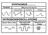 |
A Comparison of Nystagmus and Saccadic Intrusions/Oscillations | Nystagmus can be classified into pendular and jerk waveforms, where both are generated by a slow, pathologic phase. Corrective phase (the position reset mechanism) differs. In pendular nystagmus, the eyes move back and forth with about the same velocity and amplitude, similar to that of a pendulum... | Image |
| 12 |
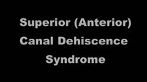 |
Superior Canal Dehiscence | 𝗢𝗿𝗶𝗴𝗶𝗻𝗮𝗹 𝗗𝗲𝘀𝗰𝗿𝗶𝗽𝘁𝗶𝗼𝗻: This is a 60-yo-man who complained of autophony (eg, hearing his own heartbeat, noting that his own voice sounded too loud) and dizziness triggered with loud noises and straining at times. With pinched-nose Valsalva maneu... | Image/MovingImage |
| 13 |
 |
Examples of Patients with Saccadic Intrusions (Square Wave Jerks) | Seen here are patients with saccadic intrusions that do have an intersaccadic interval. Square wave jerks are commonly seen in degenerative conditions, mainly involving the posterior fossa (e.g., cerebellar degeneration) and basal ganglia (e.g., progressive supranuclear palsy). | Image/MovingImage |
| 14 |
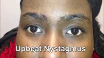 |
Upbeating and Gaze-evoked Nystagmus, V-pattern Esotropia from Bilateral 4th Nerve Palsies | Video example of a patient with upbeating and gaze-evoked nystagmus, V-pattern esotropia from bilateral 4th nerve palsies. | Image/MovingImage |
| 15 |
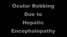 |
Ocular Bobbing Due to Hepatic Encephalopathy | 𝗢𝗿𝗶𝗴𝗶𝗻𝗮𝗹 𝗗𝗲𝘀𝗰𝗿𝗶𝗽𝘁𝗶𝗼𝗻: This is a 55-year-old man presented with hepatic encephalopathy, and found to have ocular bobbing. Head CT did not show any acute changes. Ocular bobbing almost always localizes to the pons, although cerebellar pathology ... | Image/MovingImage |
| 16 |
 |
One-and-a-Half Syndrome Due to Pontine Hemorrhage | This is a 50-year-old woman who, while exercising in the gym, suddenly experienced vertigo, nausea, vomiting, tingling in the left arm, and diplopia. MRI demonstrated a brainstem hemorrhage that involved the right greater than left pons. Examination demonstrated a right horizontal gaze palsy due to ... | Image/MovingImage |
| 17 |
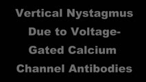 |
Upbeat and Downbeat Nystagmus Due to Anti-VGCC Antibodies | Seen here are two patients who presented with imbalance and vertical oscillopsia, the first with upbeat nystagmus, and the second with downbeat nystagmus. Both patients were found to have voltage-gated calcium channel antibodies in serum without evidence of systemic malignancy. The UBN patient had m... | Image/MovingImage |
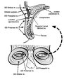
| 18 |
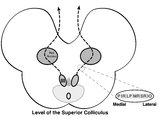 |
Central Anatomy of the Third Nerve | Seen here is an axial section of the midbrain at the level of the superior colliculus. The paired nuclei are located ventral to the periaqueductal grey, and the midline central caudal nucleus (CCN) is located between the right and left nuclei. The CCN sends projections to bilateral levator palpebrae... | Image |
| 19 |
 |
Sagittal Section of the Brainstem Showing Structures Related to Normal Eyelid Function | Seen here is a sagittal view of the brainstem, with the structures relevant to normal eyelid function highlighted. The M-group, which can be found medial to the riMLF (coordinates eye and lid movements), has (weak) projections to the facial nucleus for frontalis muscle contraction, and (strong) proj... | Image |
| 20 |
 |
Pendular Nystagmus and Vision Loss | Three patients are presented here, each with poor vision (counting fingers or worse) related to retinitis pigmentosa in one patient (Usher's syndrome) and optic neuropathy in two patients, each of whom developed pendular nystagmus after vision loss developed. Visually mediated movements normally pre... | Image/MovingImage |
| 21 |
 |
Saccadic Pathways in the Brainstem and Cerebellum & Mechanism for Saccadic Dysmetria in Wallenberg Syndrome - Abnormal Function of the Brainstem/Cerebellar Saccadic Pathways with a Left Wallenberg Syndrome | The end result of a lesion involving the climbing fibers within the left lateral medulla is deficient rightward saccades (contralesional hypometric saccades), and over-active leftward saccades (ipsilesional hypermetric saccades), and ipsilesional ocular lateropulsion given this baseline imbalance. M... | Image |
| 22 |
 |
Chronic Progressive External Ophthalmoplegia (CPEO) and Cerebellar Signs | This is a 60-yo-woman who initially presented with imbalance and ophthalmoparesis. Initially, there was mild horizontal gaze limitation with mild gaze-evoked nystagmus and slow saccades, and over the years, gait ataxia and dysarthria (mainly a scanning quality to her speech) developed, and her ophth... | Image/MovingImage |
| 23 |
 |
Post-infectious Ocular Flutter and Myoclonus Syndrome | 𝗢𝗿𝗶𝗴𝗶𝗻𝗮𝗹 𝗗𝗲𝘀𝗰𝗿𝗶𝗽𝘁𝗶𝗼𝗻: This is a 35-yo-woman presenting with oscillopsia following a viral illness. She described being easily startled, with "shakiness" of the head/neck and body. She had myoclonus and ocular flutter, with the latter evident w... | Image/MovingImage |
| 24 |
 |
Chiari Malformation Causing Downbeat Nystagmus in Lateral Gaze | This is a 20-yo-man who presented with oscillopsia in lateral gaze from downbeat nystagmus (DBN). In primary gaze, very subtle DBN was only noted with ophthalmoscopy, but in lateral gaze, prominent DBN was present. Other central ocular motor signs included gaze-evoked nystagmus (GEN) vertically, in ... | Image/MovingImage |
| 25 |
 |
Eyelid Anatomy | Seen here are the major muscles of eyelid opening and closure. The levator palpebrae, which is innervated by the oculomotor nerve, inserts on the tarsus via the levator aponeurosis and directly on the skin of the upper eyelid. The superior tarsal muscle (also known as Muller's muscle, which is inner... | Image |