John A. Moran Eye Center Neuro-Ophthalmology Collection: A variety of lectures, videos and images relating to topics in Neuro-Ophthalmology created by faculty at the Moran Eye Center, University of Utah, in Salt Lake City.
NOVEL: https://novel.utah.edu/
TO
| Title | Description | Type | ||
|---|---|---|---|---|
| 1 |
 |
Benign Episodic Unilateral Mydriasis | Presentation covering benign episodic mydriasis. | Text |
| 2 |
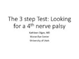 |
The 3 Step Test: Looking for a 4th Nerve Palsy | Description of the three step test (3 step test) used when looking for a 4th nerve palsy. | Text |
| 3 |
 |
The Electro-oculogram: Clinical Applications | The electrooculogram measures the potential that exists between the cornea and Bruch's membrane at the back of the eye. The potential produces a dipole field with the cornea approximately 5 millivolts positive compared to the back of the eye, in a normally illuminated room. Although the origin of th... | Text |
| 4 |
 |
The Electroretinogram and Electro-oculogram: Clinical Applications | The global or full-field electroretinogram (ERG) is a mass electrical response of the retina to photic stimulation. The ERG is a test used worldwide to assess the status of the retina in eye diseases in human patients and in laboratory animals used as models of retinal disease. | Text |
| 5 |
 |
The Multifocal Electroretinogram: Clinical Applications | The most important development in ERGs is the multifocal ERG (mfERG). Erich Sutter adapted the mathematical sequences called binary m-sequences creating a program that can extract hundreds of focal ERGs from a single electrical signal. This system allows assessment of ERG activity in small areas of ... | Text |
| 6 |
 |
Visually Evoked Potentials | Detailed explanation of visually evoked potentials. The terms visually evoked potential (VEP), visually evoked response (VER) and visually evoked cortical potential (VECP) are equivalent. They refer to electrical potentials, initiated by brief visual stimuli, which are recorded from the scalp overl... | Text |
| 7 |
 |
Amsler Grid Testing | Demonstration of Amsler Grid examination. | Text |
| 8 |
 |
Fusional Vergence Amplitudes | Demonstration of fusional vergence amplitudes examination. Incluudes: a. Convergence Amplitudes b. Divergence Amplitudes c. Vertical Ampitudes | Text |
| 9 |
 |
Exophthalmometry | Demonstration of exophthalmometry examination. | Text |
| 10 |
 |
Pupil Exam | Demonstration of pupil examination. | Text |
| 11 |
 |
Color Vision Testing | Demonstration of color vision examination. | Text |
| 12 |
 |
Stereoacuity Testing | Demonstration of examination for stereoacuity. | Text |
| 13 |
 |
Cone Dystrophy | PPT covering Cone Dystrophy - An inherited degeneration that presents between 10 - 30 years of age. Symptoms are decreased visual acuity, poor color vision, and sometimes light sensitivity. | Text |
| 14 |
 |
Hydroxychloroquine Maculopathy (Plaquenil) | An overview of Chloroquine Maculopathy. | Text |
| 15 |
 |
Macula | Overview of the structure and viewing of the macula. | Text |
| 16 |
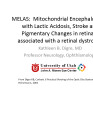 |
MELAS and RP | MELAS; Mitochondrial Encephalopathy with Lactic Acidosis, Stroke and Pigmentary Changes in retina-associated with a retinal dystrophy. This 53 year old man had seizures, encephalopathy and lactic acidosis typical of MELAS. His fundus examination showed granularity and some slight pigmentary changes ... | Text |
| 17 |
 |
Normal Eye Movements | This is an examination of a person with normal eye movements. Notice the patient has normal excursions. He has normal pursuit and saccades (horizontally and vertically). | Text |
| 18 |
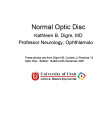 |
Normal Optic Disc | Overview of the structure and function of the normal optic disc. | Text |
| 19 |
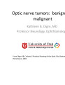 |
Optic Nerve Tumors Benign and Malignant | Discussion of optic nerve tumors including meningioma and glioma. | Text |
| 20 |
 |
Retinitis Pigmentosa Disease of Rods | Discussion of retinitis pigmentosa which is a retinal/choroidal degeneration caused by various genetic defects. | Text |
| 21 |
 |
Stargardt's Disease | Discussion of Stargardt's disease, an inherited maculopathy which frequently presents with a loss of central vision. | Text |
| 22 |
 |
Basic Headache | Presentation covering an overview of headache and migraine. | Text |
| 23 |
 |
Retino-choroidal Vessels or Optociliary Veins or Ciliary Shunt | Overview of retino-choroidal collaterals, which are potential telangiectatic connections between the retina and choroidal circulation. Although sometimes called "shunts", these collaterals are between the retinal venous circulation and the choroidal venous circulation. | Text |
| 24 |
 |
Glaucoma: The Basics | Glaucoma is the most common optic neuropathy. Progressive cupping of the optic disc due to increased intraocular pressure together with visual field abnormalities and local disc susceptibility factors characterize this neuropathy. This PowerPoint lecture covers the basics of Glaucoma and includes ma... | Text |
| 25 |
 |
Optic Disc Pallor Pseudo and Real | Discussion of the causes of optic disc pallor. | Text |