A collection of videos relating to the diagnosis and treatment of eye movement disorders. This collection includes many demonstrations of examination techniques.
Dan Gold, D.O., Associate Professor of Neurology, Ophthalmology, Neurosurgery, Otolaryngology - Head & Neck Surgery, Emergency Medicine, and Medicine, The Johns Hopkins School of Medicine.
A collection of videos relating to the diagnosis and treatment of eye movement disorders.
NOVEL: https://novel.utah.edu/
TO
Filters: Collection: "ehsl_novel_gold"
| Title | Description | Type | ||
|---|---|---|---|---|
| 1 |
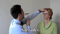 |
Saccades | 𝗢𝗿𝗶𝗴𝗶𝗻𝗮𝗹 𝗗𝗲𝘀𝗰𝗿𝗶𝗽𝘁𝗶𝗼𝗻: The examiner should note: conjugacy (a lag of the adducting eye may be seen with an INO); accuracy (posterior fossa lesions commonly produce dysmetria (overshooting or undershooting); velocity (if slow, may suggest a lesi... | Image/MovingImage |
| 2 |
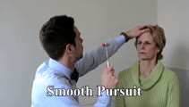 |
Smooth Pursuit | 𝗢𝗿𝗶𝗴𝗶𝗻𝗮𝗹 𝗗𝗲𝘀𝗰𝗿𝗶𝗽𝘁𝗶𝗼𝗻: A pursuit deficit in one direction suggests an ipsilesional localization, but beware of a superimposed spontaneous nystagmus; a pursuit deficit in all directions is commonly seen with cerebellar lesions. 𝗡𝗲𝘂𝗿�... | Image/MovingImage |
| 3 |
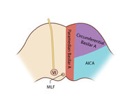 |
Figure 61: Vascular Distribution and Anatomy (Including 6th, 7th, 8th Nerves, MLF) of the Pons (Supplement) | Image | |
| 4 |
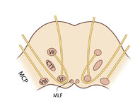 |
Figure 61: Vascular Distribution and Anatomy (Including 6th, 7th, 8th Nerves, MLF) of the Pons (Supplement) | Image | |
| 5 |
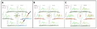 |
CANVAS (Cerebellar Ataxia, Neuropathy, and Vestibular Areflexia Syndrome) Video Head Impulse Test (vHIT) Figure | CANVAS (Cerebellar Ataxia, Neuropathy, and Vestibular Areflexia Syndrome) is a genetic condition consisting of slowly progressive late-onset ataxia, bilateral vestibulopathy, sensory neuropathy, chronic cough, and autonomic dysfunction. While the term vestibular areflexia typically implies bilateral... | Image |
| 6 |
 |
Abnormal Visually-enhanced Vestibulo-ocular Reflex (vVOR) in Cerebellar Ataxia, Neuropathy, Vestibular Areflexia Syndrome (CANVAS) | 𝗢𝗿𝗶𝗴𝗶𝗻𝗮𝗹 𝗗𝗲𝘀𝗰𝗿𝗶𝗽𝘁𝗶𝗼𝗻: This patient complained of chronic (unexplained cough), progressive numbness in the legs and feet, gait instability, and oscillopsia when walking or with head movements. Examination showed excessive square-wave jerks, bil... | Image/MovingImage |
| 7 |
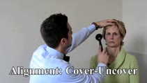 |
Ocular Alignment | 𝗢𝗿𝗶𝗴𝗶𝗻𝗮𝗹 𝗗𝗲𝘀𝗰𝗿𝗶𝗽𝘁𝗶𝗼𝗻: These tests allow for detection of eso-, exo- or hyperdeviations (phorias (one eye viewing) or tropias (both eyes viewing) that can be seen with ocular motor palsy, skew deviation, or with cerebellar disease (commonly eso... | Image/MovingImage |
| 8 |
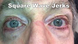 |
Saccadic Intrusions (Square Wave Jerks, SWJ) | 𝗢𝗿𝗶𝗴𝗶𝗻𝗮𝗹 𝗗𝗲𝘀𝗰𝗿𝗶𝗽𝘁𝗶𝗼𝗻: Seen here are SWJ, which is the most common example of a saccadic intrusion. Here the patient is fixating on the camera, and all of the sudden a saccade takes the eyes off the fixation target, there's a brief intersaccadi... | Image/MovingImage |
| 9 |
 |
An Optokinetic Stimulation Home Exercise Program | 𝗢𝗿𝗶𝗴𝗶𝗻𝗮𝗹 𝗗𝗲𝘀𝗰𝗿𝗶𝗽𝘁𝗶𝗼𝗻: A plainly written program of optokinetic exercise intended for patient use at home. 𝗡𝗲𝘂𝗿𝗼-𝗼𝗽𝗵𝘁𝗵𝗮𝗹𝗺𝗼𝗹𝗼𝗴𝘆 𝗮𝗻𝗱 𝗡𝗲𝘂𝗿𝗼-𝗼𝘁𝗼𝗹𝗼𝗴𝘆 ... | Text |
| 10 |
 |
Expanded Acute Onset Persistent Vision Loss Differential | Text | |
| 11 |
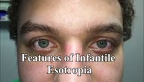 |
Eye Signs in Infantile Esotropia - Latent Nystagmus and Inferior Oblique Overaction | 𝗢𝗿𝗶𝗴𝗶𝗻𝗮𝗹 𝗗𝗲𝘀𝗰𝗿𝗶𝗽𝘁𝗶𝗼𝗻: This is a 25-yo-man with a history of amblyopia and intermittent eye crossing. On exam, he had a comitant 25 prism diopter esotropia, and other features of infantile (or congenital) esotropia including: latent nystagmus (... | Image/MovingImage |
| 12 |
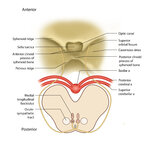 |
Figure 51: Lateral Medullary Lesion Causing Saccadic Dysmetria (Supplement) | Image | |
| 13 |
 |
Figure 51: Lateral Medullary Lesion Causing Saccadic Dysmetria (Supplement) | Image | |
| 14 |
 |
Figure 53: Vascular Distribution and Anatomy Relevant to the Lateral Medullary (Wallenberg) Syndrome (Supplement) | Image | |
| 15 |
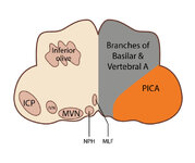 |
Figure 53: Vascular Distribution and Anatomy Relevant to the Lateral Medullary (Wallenberg) Syndrome (Supplement) | Image | |
| 16 |
 |
Figure 64: The Course of the 3rd (III) Nerve (Supplement) | Image | |
| 17 |
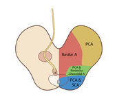 |
Figure 65: Vascular Distribution and Anatomy (Including 3rd Nerve) of the Rostral Midbrain (Supplement) | Image | |
| 18 |
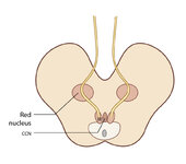 |
Figure 65: Vascular Distribution and Anatomy (Including 3rd Nerve) of the Rostral Midbrain (Supplement) | Image | |
| 19 |
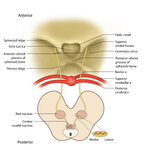 |
Figure 68: The Course of the 4th (IV) Nerve (Supplement) | Image | |
| 20 |
 |
Figure 69: Vascular Distribution and Anatomy (Including 4th Nerve) of the Caudal Midbrain (Supplement) | Image | |
| 21 |
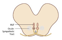 |
Figure 69: Vascular Distribution and Anatomy (Including 4th Nerve) of the Caudal Midbrain (Supplement) | Image | |
| 22 |
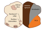 |
Figure 80: Vascular Distribution and Anatomy Relevant to the Medial Medullary Syndrome (Supplement) | Image | |
| 23 |
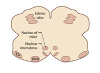 |
Figure 80: Vascular Distribution and Anatomy Relevant to the Medial Medullary Syndrome (Supplement) | Image | |
| 24 |
 |
Head Movement Independent ('Sitting') Oscillopsia - A Common Symptom of Nystagmus and Saccadic Intrusions/Oscillations | 𝗢𝗿𝗶𝗴𝗶𝗻𝗮𝗹 𝗗𝗲𝘀𝗰𝗿𝗶𝗽𝘁𝗶𝗼𝗻: This video is an example of what a patient with spontaneous nystagmus or saccadic intrusions/oscillations experiences visually during the abnormal eye movements - i.e., oscillopsia (illusion of movement of the stationary ... | Image/MovingImage |
| 25 |
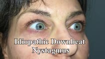 |
Idiopathic Downbeat Nystagmus Exacerbated with Positional Maneuvers - Part 2: Patient is Now on 4-Aminopyridine | This is a 45-yo-woman presented in "Idiopathic downbeat nystagmus exacerbated with positional maneuvers". This video was taken after the patient had been on 4-aminopyridine for 3 months. There was marked improvement in subjective oscillopsia and objective downbeat nystagmus. The strong positional co... | Image/MovingImage |