Home
Browse
Ask Us
Chat
Harmful Language Statement
Log in
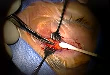
NOVEL - Neil R. Miller Collection
Advanced Search
About
The Neil R. Miller Collection covers a broad range of neuro-ophthalmologic conditions and syndromes in videos, images, and lecture presentations.
Year
2024
TO
2024
Type
Image
19
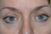
Image/MovingImage
5
Format
image/jpeg
19
video/mp4
5
Collection
NOVEL - Neil R. Miller Collection
24
Filters:
Collection:
"ehsl_novel_nrm"
Subject:
"Surgery"
1
-
25
of
24
Gallery view
Number of results to display per page
10
25
50
100
200
Sort by Relevance
Sort by Title A-Z
Sort by Title Z-A
Sort by Date Ascending
Sort by Date Descending
Sort by Last Modified Ascending
Sort by Last Modified Descending
Title
Date
Type
1
Cannulation of a Dilated Superior Ophthalmic Vein in a Patient with an Ipsilateral Direct (Traumatic) Carotid-cavernous Sinus Fistula
2024-07-10
Image
2
Cannulation of the Superior Ophthalmic Vein in a Patient with an Ipsilateral Carotid-cavernous Sinus Fistula Prior to Embolization
2024-07-10
Image
3
CCF - Surgery - SOV9
2024-07-10
Image
4
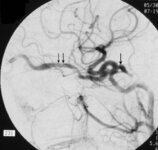
Common Carotid Angiogram, Lateral View
2024-07-10
Image
5
Common Carotid Angiogram, Lateral View, Following Successful Transvenous Embolization with Platinum Coils Using the Superior Ophthalmic Vein Approach
2024-07-10
Image
6
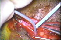
Exposure of an Enlarged Superior Ophthalmic Vein (on a Right-angle Muscle Hook) in a Patient with a Carotid-cavernous Sinus Fistula
2024-07-10
Image
7
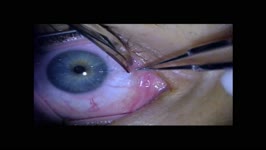
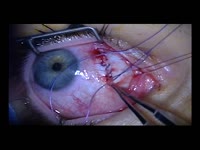
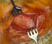
External Appearance of the Lid Crease Incision Site Used to Access the Superior Ophthalmic Vein for Transvenous Embolization of a Dural or Direct Carotid-cavernous Sinus Fistula
2024-07-10
Image
8
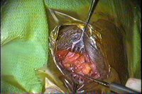
Isolation of a Dilated Superior Ophthalmic Vein Prior to Transvenous Embolization in a Patient with a Direct Carotid-cavernous Sinus Fistula
2024-07-10
Image
9
Isolation of a Segment of an Enlarged Superior Ophthalmic Vein in a Patient with an Ipsilateral Carotid-cavernous Sinus Fistula Prior to Transvenous Embolization
2024-07-10
Image
10
Isolation of a Segment of an Enlarged Superior Ophthalmic Vein Using Silk Sutures Passed Through Small Feeding Tubes in a Patient with a Carotid-cavernous Sinus Fistula Prior to Attempted Transvenous Embolization
2024-07-10
Image
11
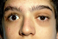
Marked Left Eye Proptosis, Chemosis, and Arterialization of Conjunctival and Episcleral Vessels in a Patient with a Direct (Traumatic) Carotid-cavernous Sinus Fistula
2024-07-10
Image
12
Optic Nerve Sheath Decompression: Nasal Approach (Narrated)
2024-06
Image/MovingImage
13
Optic Nerve Sheath Decompression: Nasal Route
2024-06
Image/MovingImage
14
Optic Nerve Sheath Fenestration Technique
2024-06
Image/MovingImage
15
Patient Immediately Following Successful Transvenous Embolization of a Left-sided Direct Carotid-cavernous Sinus Fistula Via the Superior Ophthalmic Vein
2024-07-10
Image
16
Patient with Left-sided Proptosis and Orbital Swelling from an Ipsilateral Direct Carotid-cavernous Sinus Fistula Prior to Transvenous Embolization
2024-07-10
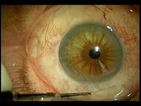
Image
17
Persistent Post-cataract Pupillary Mydriasis
2024-07-10
Image
18
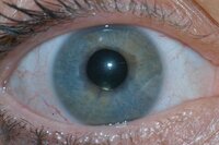
Pupillary Mydrasis OD After Cataract Surgery (Magnified View)
2024-07-10
Image
19
Pupillary Mydriasis After Cataract Surgery
2024-07-10
Image
20
Pupillary Mydriasis OD After Cataract Surgery
2024-07-10
Image
21
Trans-cutaneous Exposure of a Dilated, Arterialized Superior Ophthalmic Vein in a Patient with an Ipsilateral Direct (Traumatic) Carotid-cavernous Sinus Fistula
2024-07-10
Image
22
Transcutaneous Approach for Endovascular Occlusion of a Dural Carotid-cavernous Sinus Fistula (Narrated by NRM)
2024-06
Image/MovingImage
23
Transcutaneous Approach to the Superior Ophthalmic Vein for Treatment of a Dural Carotid-cavernous Sinus Fistula
2024-06
Image/MovingImage
24
Transcutaneous Approach to the Superior Ophthalmic Vein in a Patient with an Ipsilateral Carotid-cavernous Fistula Prior to Transvenous Embolization
2024-07-10
Image
1
-
25
of
24