| OCR Text |
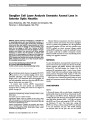
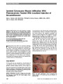
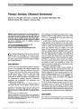
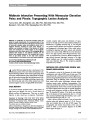
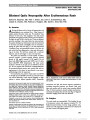
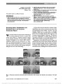
Show Williams F. Hoyt, MD The North American Neuro-Ophthalmology Society, in conjunction with the American Academy of Ophthalmology, established the annual Hoyt Lecture in 2001 in honor of William Fletcher Hoyt, MD, whose contributions to neuro-ophthalmology have spanned seven decades. A fellow of Frank Walsh, MD, the grandfather of clinical neuro-ophthalmology, Dr. Hoyt co-authored the 3rd edition of Clinical Neuro- Ophthalmology, the "bible" of our specialty. A faculty member of the departments of Ophthalmology, Neurology and Neurosurgery at the University of California San Francisco since 1958, Dr. Hoyt is world-renowned as a clinician, scholar and edu-cator. He has published more than 300 scientific contributions and has trained more than 100 fellows, many of whom are senior professors in their own right, training the next generations of neuro-ophthalmologists on six continents. Optical Imaging of the Optic Nerve: Beyond Demonstration of Retinal Nerve Fiber Layer Loss Mark J. Kupersmith, MD Journal of Neuro-Ophthalmology 2015;35:210-219 doi: 10.1097/WNO.0000000000000248 © 2015 by North American Neuro-Ophthalmology Society Iam deeply honored to be included among the neuro-ophthalmologists who were previously honored to give this lecture. Then I thought about it and I realized I must be getting old! This is my one opportunity to pontificate a bit or at least impart what I consider a few ruminations, hopefully of use for those of you planning high level careers, and hope-fully long before my retirement. I was not trained by William Hoyt, but I was fortunate that he helped me early in my career, taking my calls, and guiding me to think through clinical problems. I hope to emulate what I consider one of his greatest attributes-the ability to admit he was in error or appreciate concepts that contradicted his theories or prevailing wisdom if new or better research provided scientific evidence. Dr. Hoyt continually evolved as he fostered the career of countless motivated physicians. I must acknowledge two of my mentors, at NYU Medical Center/School of Medicine who shaped my career. The neurosurgeon Dr. Joseph Ransohoff (See Supplemental Dig-ital Content, Figure E1, http://links.lww.com/WNO/A154) understood the complexity of neurosurgical problems and was never reluctant to ask for help and other opinions. Dr. Albert Goodgold (See Supplemental Digital Content, Figure E2, http://links.lww.com/WNO/A155), a brilliant neurologist, who questioned every unsubstantiated neuro-logical concept, pressed to apply new knowledge, and was a master of uncovering iatrogenic-induced disease. From these two individuals, I learned that in order to advance clinical care, we have to read critically and question pre-vailing concepts if supporting evidence is lacking. Do not be afraid to admit you do not know. Have a "golden ro-lodex" of individuals you respect and who have knowledge complementary to your own to call for help. Now, let us turn to the applied science of optical imaging of the pathological changes in the optic nerve. Fortuitously, this topic grew out of findings reported by Dr. Hoyt and one of his fellows, Lars Frisén, MD, demonstrating retinal nerve fiber layer (RNFL) loss in eyes with multiple sclerosis (MS) and, recovered optic neuritis (ON) (1). For clinical trial design for the Neuro-Ophthalmology Research Disease Investigator Consortium, we researched what type of optical imaging would be the best to study new therapies for 3 optic neuropathies: ON, nonarteritic arterior ischemic optic neuropathy (NAION) and papilledema. When we began our projects, except for evaluating the RNFL in ON, few prospective studies had been done. We also were limited New York Eye and Ear Infirmary, Mount Sinai Roosevelt Hospital, Icahn School of Medicine at Mount Sinai, New York, New York. Supported by U10 EY017281-01A1, U10 EY017281-01A1S1, ECRIP Center Grant at New York Eye and Ear Infirmary. The author reports no conflicts of interest. Supplemental digital content is available for this article. Direct URL citations appear in the printed text and are provided in the full text and PDF versions of this article on the journal's Web site (www.jneuro-ophthalmology.com). Address correspondence to Mark J. Kupersmith, MD, 1000 10th Avenue, New York, NY 10019; E-mail: mkuper@chpnet.org 210 Kupersmith: J Neuro-Ophthalmol 2015; 35: 210-219 Hoyt Lecture Copyright © North American Neuro-Ophthalmology Society. Unauthorized reproduction of this article is prohibited. because RNFL thickness, the major optical imaging parameter in use to determine axonal loss, may not be normal due to optic nerve head (ONH) swelling and may not have be a suitable baseline to determine future RNFL thinning. HISTORICAL BACKGROUND OF RETINAL NERVE FIBER LAYER ASSESSMENT Frisén and Hoyt had photographically demonstrated nerve fiber layer defects in eyes with glaucoma and recovered ON (1,2). This predated by many years the realization that attacks of MS often caused permanent loss of axons even if visual acuity recovered. However, it took the development of optic coherence tomography (OCT) and scanning laser polarimetry (SLP) to provide quantitative measurement of RFNL thinning in a variety of optic nerve disorders. These and other techni-ques can show injury to peripapillary retinal axons and ganglion cells before RNFL thinning can be demonstrated and assist in clinical decision making. OCT can demonstrate dynamic shape changes in the ONH due to glaucoma, papilledema, and optic disc drusen. Importantly, optic imaging can be used to address pathophysiologic mechanisms affecting the optic nerve. RETINAL NERVE FIBER LAYER THINNING AFTER ACUTE OPTIC NEURITIS: BASELINE AND THE IMPORTANCE OF 1 MONTH FINDINGS OCT evaluation performed several months or longer after the onset of ON revealed that reduction in the global mean RNFL thickness was more prevalent in eyes with persistent visual field deficits (3). In order to investigate potential mechanisms of early neural injury or peripapillary change as well as the rate of structural alteration or loss over time, we studied patients with acute ON at presentation, typically within 14 days of vision loss. One month had been reported to be the earliest time point to predict vision outcome from an attack of ON (4). In a study of 40 eyes with acute ON, evaluating the OCT measured RNFL thickness by compar-ison with the normal fellow eye revealed that axonal swell-ing was much more common than is seen clinically (5). RNFL swelling was found in 80% of eyes when compared with normal fellow eye and in 33% of when compared with control eyes. Thus, OCT can show swelling in the ONH region at a higher rate than seen on ophthalmoscopy. No RNFL thinning was seen at presentation. By 1 month, using interocular comparison of clock hour sectors, RNFL thickness revealed thinning in 54% of affected eyes. How-ever, RNFL swelling of some amount was detected in 82% of affected eyes at 1 month (Fig. 1). Without the persistent swelling, it is possible that more global or regional axonal loss might have been demonstrated. As a cautionary note, sector analysis, due to variability, must be carefully applied and requires precise alignment of images. RETINAL NERVE FIBER LAYER CHANGES IN OPTIC NEURITIS OVER 6 MONTHS We and others suggested that OCT evaluation of the RNFL changes over time to 6 months could provide structural correlates to complement the Optic Neuritis Treatment Trial results, which demonstrated that the 6-month vision performance is an accurate measure of functional outcome for a given episode (6). We also questioned whether FIG. 1. Optic neuritis RNFL thickness compared with fellow normal eye shows swelling (expressed as percent greater than fellow eyes) across all sectors at presentation (diamonds) which diminishes at 1 month (solid squares). Control eyes (open circles) in subjects with no disease were similar to the unaffected fellow eyes (open squares). *Average value. RNFL, retinal nerve fiber layer. Kupersmith: J Neuro-Ophthalmol 2015; 35: 210-219 211 Hoyt Lecture Copyright © North American Neuro-Ophthalmology Society. Unauthorized reproduction of this article is prohibited. measurement of neuronal structural loss can be used to monitor the effects of therapy, if OCT would be able to show a time when the majority of RNFL thinning occurs, and if there was significant further thinning of the RNFL thickness after 3 months. Additionally, we hypothesized that eyes with significant RFNL thinning at 1 month, a key time point if functional vision recovery does not occur (4), would also be associated with a poor visual outcome (Fig. 2). Follow-up of the 40 eyes with ON reported above (5) showed RNFL loss by OCT at 3 months occurred in approximately 60% of affected eyes. The thinning occurred in all quadrants and was not selective. Further evaluation of 25 of these eyes showed the 1 month OCT RNFL loss strongly correlated (r = 0.58, P = 0.001) with the amount of OCT thinning at 6 months (7). Furthermore, the amount of average OCT RNFL thinning at 6 months cor-related with the 6-month visual field mean deviation (r = 0.48, P = 0.03). No significant further RNFL thinning occurred at 1 year. These data support previous OCT and SLP reports and the results of the Optic Neuritis Treatment Trial that the 6-month functional and structural outcomes are appropriate to use in investigations of acute ON. SCANNING LASER POLARIMETRY USE IN ACUTE OPTIC NEUROPATHY WITH OPTIC DISC SWELLING OCT shows RNFL thickening in eyes with ONH swelling from many causes, but in this setting, it does not provide information on acute axonal disruption or injury. SLP reflects polarization due to intact RNFL axons (8,9) and does not consistently demonstrate ONH swelling in disor-ders like papilledema or NAION (10). We hypothesized that optical imaging using SLP and OCT together might reveal the status of axon integrity in eyes with acute RNFL swelling. In a prospective study, we evaluated eyes with ONH swelling due to papilledema, ON, and NAION (8). We determined whether regional RNFL values were reduced using criteria of having the OCT or SLP thickness measure of a quadrant be less than the fifth percentile of controls. At presentation, the average RNFL thickness of OCT was similar for eyes with papilledema (213 ± 100 mm) and NAION (214 ± 76 mm, P = 0.97), and less for ON (141 ± 21 mm). In contrast, the average RNFL thickness by SLP was less often increased. It was similar for papilledema (59 ± 7 mm) and ON (55 ± 5 mm) but was reduced for NAION (48 ± 9 mm, P = 0.02) eyes (Fig. 3). The RNFL by SLP was significantly reduced in at least 1 quadrant in 1 of the 24 eyes with papilledema, 1 of the 13 eyes with ON, and in 13 of the 21 eyes with NAION. In NAION eyes, quadrants with reduced SLP had corresponding regional visual field loss that did not recover at 1 or 6 months. By 1 month, eyes with NAION showed RNFL thinning by OCT (41%, mean 111 ± 49 mm) and by SLP (88%, mean 43 ± 9 mm) in contrast to ON (by OCT, 0%, mean 127 ± 16 mm, P = 0.006); and by SLP (9%,mean 52 ± 7 mm, P = 0.0004). The results confirm that OCT and SLP measure different aspects of RNFL changes associated with ONH swelling. OCT reveals thickening, due to edema. SLP reveals a decrease in retardance in eyes with axonal injury associated with visual field loss, which is unlikely to recover. SLP results may be predictive of regions of permanent axon dysfunction and visual field loss in eyes with optic disc edema. OCT shows anatomical thickening of the peripapillary RNFL no matter what the etiology of the ONH swelling, which is useful for quantifying the degree of edema at presentation and over time. The disadvantage of OCT is that it is relatively insensitive to acute axon damage at the time of presentation and even 1 month later. SLP does not directly measure the dimension of each retinal layer. RNFL birefringence induces delay in 1 of 2 orthogonal beams of polarized light passing through the bundles of nerve fibers. This slowing measured in the reflected light is termed retardance. The retardance is used to calculate RNFL thickness, which is proportional to the bifrefringence. The SLP retardance is primarily dependent on the integrity of the parallel structural organization of axon plasma mem-branes, intracellular microtubules, and neurofilaments, which produce birefringence (11,12). Therefore, SLP would not be expected to reveal an increase in RNFL thickness to the same extent as OCT in conditions of optic disc edema having intact axons with retained intra-axonal cellular organization (5,10). SLP showed a decrease in retardance or birefringence (resulting in a decrease in the derived RNFL thickness) in a large num-ber of the NAION eyes at presentation and at 1 month while optic disc edema was still present. The importance of this finding was that OCT did not show RNFL thinning or loss in any of the affected quadrants for any of the 3 disorders at presentation. The greatest disparity between OCT and SLP FIG. 2. Optic neuritis eyes with severe vision loss (less than 20/50) at 1 month shows more severe RNFL loss at 6 months than eyes with good vision (20/50 or better) at 1 month. RNFL, retinal nerve fiber layer. 212 Kupersmith: J Neuro-Ophthalmol 2015; 35: 210-219 Hoyt Lecture Copyright © North American Neuro-Ophthalmology Society. Unauthorized reproduction of this article is prohibited. FIG. 3. Examples of OCT and SLP comparisons for eyes with swollen optic nerve head. A. Papilledema eyes have significant thickening of the entire RNFL by OCT (left) and no thickening by SLP (right). B. Optic neuritis with severe visual field loss at pre-sentation in the left eye shows RNFL thickening (right side of plot) by OCT (left) and slight thickening (right side of plot) in comparison with normal right eye by SLP (right). C. NAION in right eye with inferior altitudinal field defect shows RNFL thickening (left side of plot) by OCT (left) and a reduced RNFL thickness in the superior quadrant (left side of plot) by SLP (right). OCT, optical coherence tomography; SLP, scanning laser polarimetry; RNFL, retinal nerve fiber layer; NAION, nonarteritic arterior ischemic optic neuropathy. Kupersmith: J Neuro-Ophthalmol 2015; 35: 210-219 213 Hoyt Lecture Copyright © North American Neuro-Ophthalmology Society. Unauthorized reproduction of this article is prohibited. values for RNFL at presentation was seen in eyes with NAION, suggesting that in the setting of ischemia, optic disc edema, and visual field loss, SLP may be unique in revealing early disruption of intra-axonal microtubular and neurofilament structure, which may be an early sign of potentially permanent axon loss. Conversely, regional areas that are normal by SLP in acute NAION may not have yet developed the axonal changes associated with inevitable injury and might indicate potential location for recovery from ischemia. This delay in evolution to a more degenerated state might be an explanation for why not all eyes with NAION examined with SLP showed RNFL reduction at presentation. Similarly, since significant permanent vision and axon loss are not as common in papilledema and acute ON, this may explain why SLP-derived RNFL measurements were infre-quently thinned on presentation for these 2 disorders. In a report of 25 eyes with acute NAION, we correlated the SLP findings with visual field threshold and RNFL values organized into Garway-Heath inferior and superior disc sec-tors and corresponding superior and inferior field regions (9). At presentation, no eyes had reduced RNFL thickness by OCT. Eyes with abnormal field regions had corresponding SLP sectors thinner (P = 0.003) than for sectors with normal field regions. During the acute phase, the SLP-derived sector correlated with presentation (r = 0.59, P = 0.02) and with at least 3 month post presentation (r = 0.44, P = 0.02) corre-sponding superior and inferior field thresholds (Fig. 4). Since the visual field deficits often show no significant recovery, SLP may be an early marker for axonal injury and used to assess recovery potential at RNFL locations with respect to new treatments for NAION. GIVEN THE RESULTS IN NONARTERITIC ANTERIOR ISCHEMIC OPTIC NEUROPATHY, WE STUDIED SLP IN ACUTE OPTIC NEURITIS Using the OCT and SLP RNFL data in 10 eyes with acute ON, we calculated a relative birefringence for each time point. This was derived using a ratio of percent of thickness change using the OCT and the percent retardance change using the SLP. The RNFL relative birefringence was approximately 217.7% at presentation and gradually re-bounded over 6 months (13) (Fig. 5). RNFL swelling due to ON (5) appears to be principally due to increased water content. This is due to intra-axonal edema from axoplasmic flow stasis, which then becomes extracellular as the process progresses. OCT-derived RNFL thickness would be ex-pected to reflect both intracellular and extracellular compo-nents. In contrast, SLP does not directly measure the actual dimension of each retinal layer but measures retardance that is primarily dependent on the integrity of the parallel struc-tural organization of axon plasma membranes (11). The relative birefringence reduction in eyes with ON at presentation could reflect permanent injury, but it is transient in ON. The acute calculated drop in FIG. 4. At onset of NAION, SLP measurement of the supe-rior and inferior quadrants (x axis) correlates with the mean deviation of the regional visual field (y axis) (closed circles, broken fit curve) and at 3 months (open circles, solid fit curve). NAION, nonarteritic anterior ischemic optic neurop-athy; RNFL, retinal nerve fiber layer; SLP, scanning laser polarimetry. FIG. 5. For acute optic neuritis, the relative birefringence (closed circles; calculated using percent of the normalized SLP/OCT ratio) was reduced at presentation and normalized over 6 months. In contrast, the percent SLP [closed triangle, solid line] and OCT [open squares, non-uniform dash line] were increased at presentation and became reduced over 6 months. SLP, scanning laser polarimetry; OCT, optical coherence tomography. 214 Kupersmith: J Neuro-Ophthalmol 2015; 35: 210-219 Hoyt Lecture Copyright © North American Neuro-Ophthalmology Society. Unauthorized reproduction of this article is prohibited. birefringence may result from reduction in microtubule density due to intra-axonal swelling, which dilates axons or microtubular alterations in response to injury or from extracellular edema (14-16). RETINAL GANGLION CELL LAYER MEASUREMENT IN ACUTE OPTIC NEUROPATHY AND/OR PAPILLEDEMA Since ONH swelling can prevent accurate OCT evaluation of early structural axonal loss, we sought another region of the optic nerve to study. Measurement of the retinal ganglion cell layer (GCL) in the macula was a potential candidate for demonstrating permanent neuronal injury that would be less susceptible to the effects of ONH swelling. In 38 eyes with acute NAION and 29 eyes with acute ON, we prospectively used spectral domain optical coherence tomography (SD-OCT) to image the macula and applied 2 methods to measure the GCL plus inner plexiform layer (IPL) thickness (17,18). One method, developed by the University of Iowa, used 3 dimensional (3D) layer segmentation (19), and the second method used the OCT machine software, a 2D seg-mentation algorithm. At presentation, affected eyes did not demonstrate significant thinning of either the RNFL or GCL + IPL thickness by 3D segmentation. The mean macula GCL + IPL values, 80 ± 8.1 mm for NAION and 83 ± 8.9 mm for ON eyes, were not different from unaffected fellow eyes (83 ± 6.4 mm and 82 ± 7.0 mm), using 3D segmentation. In 17 of 57 eyes with NAION (14) or ON (3), the 2D method (46 ± 9 mm) values for GCL + IPL were markedly less than through the 3D method (83 ± 7 mm; P = 0.001) values at presentation (See Supplemental Digital Content, Figure E3, http://links.lww.com/WNO/A156). All 17 eyes had 2D method GCL + IPL values #20 mm than the values for the 3D method. The initial lower GCL + IPL values from the 2D method rebounded and were thicker by more than 10 mm in these eyes at the next visit (1 month) as the ONH swelling decreased. At 1 month, GCL + IPL thinning developed in 84%of all affected eyes, and the decrease was greater in NAION eyes. For the 3D segmentation method, the mean change was 217.2 ± 12.3 mm and 29.0 ± 6.2 mm, for NAION and ON eyes, respectively (P = 0.001). At 1 month, GCL + IPL thinning correlated with the 1-month LogMar acuity (r = 0.36, P = 0.04) and visual field mean deviation (r = 0.61, P = 0.001) for NAION but not for ON eyes, due to recovery of function in these latter eyes. In contrast, although there was reduced RNFL swelling and/or thickening, the RNFL thickness was less than controls in only 11% of eyes with both disorders. Reduc-tion of GGL + IPL thickness occurred rapidly in both NAION and ON, but it was more profound in NAION eyes. GCL thinning occurred before RNFL loss, making it a better bio-marker of early structural loss in both NAION and ON. The 2D method for determining GCL thickness fre-quently fails when the peripapillary RNFL or macula are considerably thickened, presumably due to edema or factors that distort normal retinal layer architecture. This algorithm failure caused the average GCL + IPL thickness at presenta-tion to appear significantly less. The 2D method uses an algorithm that assumes a quantitative relationship between the internal limiting membrane and the other layers of the retina. Thus, this method would be more susceptible to failure with any process, such as edema due to swelling of the peripapillary RNFL and adjacent retina, which disrupts the regular retinal layer position or shape or boundaries. In contrast, the 3D segmentation method uses an algorithm that incorporates 3D contextual information into the optimiza-tion process which in general helps to reduce failures due to local distortions in retinal layers. It is clinically important to carefully evaluate algorithm performance in OCT scans, since failures may lead to false interpretations of data and may adversely influence clinical decisions. SPECIFICS FOR OPTICAL IMAGING OF PAPILLEDEMA Commercially available algorithms, typically using 2D segmentation methodology, also frequently fail to accurately measure the degree of RNFL thickening when ONH swelling due to severe papilledema (20) (Fig. 6). We at-tempted to improve the imaging to measure the effects of papilledema and enhance the reliability and accuracy of the OCT images to be collected in the Idiopathic Intracranial Hypertension Treatment Trial (IIHTT). Our pilot work demonstrated that multiple OCT measures of swelling in the ONH region can be used to follow papilledema and uncovered a dynamic alteration of the ONH shape from factors outside of the globe such as increased pressure in the perioptic subarachnoid space. OPTIC NERVE HEAD SHAPE CHANGES The deformity of ONH structures, particularly the peri-papillary Bruch membrane (BM) and retinal pigment epithelium (RPE) layers, due to papilledema was first noted by Patrick Sibony, MD, as we began applying various programs to reliably image and quantify the effects of papilledema. This observation leads us to conduct studies using SD-OCT to examine the biomechanical deformation of load bearing structures of the ONH resulting from raised intracranial pressure. We postulated that elevated intracra-nial pressure would create forces in the retrolaminar sub-arachnoid space that could cause the observed deformity. As a pilot project, we compared ONH shape and RNFL findings in eyes with papilledema (with a wide range of duration and vision loss) due to raised intracranial pressure to findings in eyes with optic disc swelling due to acute ON and NAION, conditions without intracranial hypertension (21,22). The RPE/BM complex at the temporal and nasal borders of the neural canal opening was noted to be deflected Kupersmith: J Neuro-Ophthalmol 2015; 35: 210-219 215 Hoyt Lecture Copyright © North American Neuro-Ophthalmology Society. Unauthorized reproduction of this article is prohibited. inward in 20 of 30 eyes (67%) in the 15 patients with pap-illedema (Fig. 7). In contrast, only 1 of 8 ON eyes (12%) and 1 of 12 NAION eyes (8%) had inward deflection of either border. Of the 22 eyes with papilledema that had less RNFL thickening over time, 17 had less inward deflection of the RPE/BM borders. In papilledema, the inward deflection RPE/BM neural canal border appears to reflect the pressure changes in the retrobulbar subarachnoid space. In subsequent work, we showed that interventions that reduce the elevated intracranial pressure have the effect of reducing a "U" shaped RPE/BM displacement more toward the normal "V" shape (23). This was most significant in patients undergoing ventric-ular shunt procedures. Further application of shape analysis of the neural canal borders shows that OCT can provide non-invasive information of improved or worse elevation of intra-cranial hypertension even in eyes with atrophic papilledema that are not capable of showing significant RNFL or ONH swelling (Fig. 8). Our findings are consistent with experimental studies in dogs. Using confocal scanning laser tomography, Morgan et al (24,25) have shown that intracranial hypertension displaces the optic disc surface and lamina cribosa anteriorly, whereas ocular FIG. 6. When papilledema is severe, the OCT 2D commercial algorithm fails to measure the amount of RNFL thickening. OCT, optical coherence tomography; RNFL, retinal nerve fiber layer. 216 Kupersmith: J Neuro-Ophthalmol 2015; 35: 210-219 Hoyt Lecture Copyright © North American Neuro-Ophthalmology Society. Unauthorized reproduction of this article is prohibited. hypertension displaces the disc surface posteriorly. An increase in cerebrospinal fluid (CSF) pressure resulted in greater anterior displacement of the disc surface than the posterior displacement induced by a corresponding increase in intraocular pressure. The normal lamina cribosa is already slightly bowed backwards (as is the RPE/BM in our controls). This makes it difficult to demonstrate a further increase in the bowing backward until the pressure gradient increases significantly and damages sup-port structures of the ONH as in untreated glaucoma. In the case of raised intracranial pressure, a reversal in the direction of this bowing is seen and may be easier to detect. Additionally, the retrolaminar tissue pressure was shown to be determined by the CSF pressure, and the translaminar pressure gradient was related to the difference between intraocular and cerebrospinal fluid pressures across a wide range of both intraocular pressure and CSF pressures (26,27). OPTICAL COHERENCE TOMOGRAPHY MEASURES OF OPTIC NERVE HEAD SWELLING DUE TO PAPILLEDEMA OCT has been used to investigate papilledema in single site, mostly retrospective studies. In a prospective multisite clinical trial, we demonstrated that SD-OCT, which provides thickness and volume measurements of the optic nerve and peripapillary retina, can reliably demonstrate structural changes due to papilledema (28,29). At entry, 126 subjects in the IIHTT with mild visual field loss had optic disc and macula region scans collected using a single brand of SD-OCT and software. The images were analyzed using proprietary commercial 2D and custom 3D segmen-tation algorithms to calculate the RNFL, total retinal thick-ness (TRT), ONH volume, and GCL + IPL thickness. As expected in papilledema, 90% of eyes had average RNFL thickness greater than 95th percentile of control eyes. The RNFL, TRT, and ONH volume showed strong (r . 0.8) correlations for interocular comparisons. Variability for repeated testing of OCT parameters was low for both 2D and 3D methods, and intraclass correlations were greater than 0.9 except for the 2D method for measuring GCL thickness. In addition, the 2D algorithm derived RNFL, TRT, and GCL + IPL thickness measurements had failure rates of 10%, 16%, and 20% for papilledema eyes, respectively, whereas algorithm failure was uncommon with 3D segmentation-derived meas-urements (See Supplemental Digital Content, Figure E4, http://links.lww.com/WNO/A157). Only 7% of eyes had GCL + IPL thinning (by 3D segmentation) less than the fifth percentile of normal age-matched control eyes. The RNFL, TRT, and ONH volume strongly correlated (r . 0.67, P = 0.0001) with the Frisén grade of papil-ledema (30). The results suggest that when 3D segmen-tation is used, all measurements that reflect ONH swelling, RNFL, TRT, ONH volume (latter 2 measures are not available with commercial algorithms on current OCT machines) can be accurately measured in eyes with papilledema. Furthermore, it is uncommon to detect FIG. 7. This case of papilledema demonstrates the utility and the dynamic nature of OCT neural canal border shape (arrowheads) changes. At presentation, the RPE/BM deflection is relatively neutral (A) but with increasing swelling the deflection becomes inward (B). The patient had worsening visual fields and increased inward deflection (C). Within 1 week of ventricular shunting, the deflection is mostly neutral (D), and at 1 month postoperatively, the deflection is more outward (E). OCT, optical coherence topography; RNFL, retinal nerve fiber layer; BM, Bruch membrane. Kupersmith: J Neuro-Ophthalmol 2015; 35: 210-219 217 Hoyt Lecture Copyright © North American Neuro-Ophthalmology Society. Unauthorized reproduction of this article is prohibited. a reduction in retinal ganglion cells in eyes with papille-dema and mild visual field loss at presentation. Given the reliability of these OCT measures, we evaluated them all to determine whether they change over time and which might be best at showing the effects of each treatment during the IIHTT. The RNFL, TRT, ONH volume, and GCL + IPL measurements were derived using the 3D seg-mentation methods. At study entry, OCT values were similar in both treatment groups. At 6 months, the acetazolamide plus weight management group had greater reduction than the placebo plus weight management group for the RNFL (175 vs 89 mm, P = 0.001), TRT (220 vs 113 mm, P = 0.001), and ONH volume (4.9 vs 2.1 mm3, P = 0.001). The RNFL (P = 0.01), TRT (P = 0.003), and ONH volume (P = 0.002) also showed less swelling in subjects in both treatment groups who lost at least 6 percent of the weight at study entry. GCL + IPL thinning was minimal in either the ACZ (3.6 mm) or placebo (2.1 mm) group. The RNFL, TRT, and ONH volume demonstrated moderate correla-tions (r = 0.48-0.59, P # 0.0001) with Frisén grade. At 6 months, the GCL + IPL thickness was less than the fifth percentile of controls in 14 eyes, and these eyes had worse perimetric mean deviation (P = 0.001) than study eyes without GCL + IPL thinning. Reduced swelling as shown by reduction in the RNFL, TRT, andONH volume meas-urements in IIH provides structural data to confirm the effectiveness of acetazolamide and weight loss in patients with mild vision loss at presentation. Retinal ganglion cell atrophy or loss appears to be uncommon in treated patients. SUMMARY Although we are still early in the evolution of optical imaging of the optic nerve, the available techniques already play an important role in clinical decision making. I would summarize our findings to date as follows: For acute ON: Presentation: OCT shows RNFL swelling, normal GCL + IPL by OCT; 1 month: OCT and SLP show RNFL thinning and swell-ing, GCL + IPL thinning by OCT; 3 months or later: OCT and SLP show RNFL thinning, further mild GCL thinning by OCT; 6 months: RNFL and GCL + IPL thinning finished. For acute NAION: Presentation: OCT shows RNFL swelling and SLP shows loss of birefringence, normal GCL + IPL by OCT; 1 month: RNFL swelling and thinning by OCT and thin-ning by SLP, GCL + IPL thinning by OCT; 3 months or later: RNFL and further mild GCL + IPL thinning; 6 months: RNFL and GCL + IPL thinning finished. FIG. 8. OCT reveals neural canal border changes in pa-tients with optic atrophy where the RNFL is so reduced, and it may not show significant thickening with intracranial hypertension. A. The patient had a ventricular shunt and known optic atrophy with significant visual field loss for 1 year. Note the neutral RPE/BM borders. B. Six months later, the patient had vague symptoms suggestive of shunt failure. Note the inward defection of the RPE/BM borders. C. One month after shunt revision, the borders are neutral. D. The RPE/BM borders are shown diagram-matically. OCT, optical coherence tomography; RNFL, retinal nerve fiber layer; RPE, retinal pigment epithelium; BM, Bruch membrane. 218 Kupersmith: J Neuro-Ophthalmol 2015; 35: 210-219 Hoyt Lecture Copyright © North American Neuro-Ophthalmology Society. Unauthorized reproduction of this article is prohibited. For IIH Papilledema with mild vision loss: Presentation: OCT shows swelling of RNFL, TRT, and ONH volume; Presentation: OCT shows normal GCL + IPL; Presentation: OCT shows neural canal border inward deflection; 6 months: OCT shows structural shape changes reflecting the effectiveness of treatment. ACKNOWLEDGMENTS I wish to thank a number of individuals who provided critical support and expertise for the research studies described in this Hoyt Lecture. They include Randy Kardon, MD, PhD, Mary Durbin, PhD, Mona Garvin, PhD, Jui-Kai Wang, MS, Patrick Sibony, MD, and John Keltner, MD. REFERENCES 1. Frisén L, Hoyt W. Insidious atrophy of retinal nerve fiber layers in multiple sclerosis. Fundoscopic identification in patients with and without visual complaints. Arch Ophthalmol. 1974;92:91-97. 2. Hoyt W, Frisén L, Newman N. Fundoscopy of nerve fiber layer defects in glaucoma. Invest Ophthalmol. 1973;12:814-829. 3. Costello F, Coupland S, Hodge W, Lorello G, Koroluk J, Pan I, Freedman M, Zackon D, Kardon R. Quantifying axonal loss after optic neuritis with ocular coherence tomography. Ann Neurol. 2006;59:963-969. 4. Kupersmith M, Gal R, Beck R, Xing D, Miller N; The Optic Neuritis Study Group. Visual function at baseline and one month in acute demyelinating optic neuritis predicts visual outcome. Neurology. 2007;69:508-514. 5. Kupersmith M, Mandel G, Anderson S, Meltzer D, Kardon R. Baseline and one month changes in the peripapillary retinal nerve fiber layer in acute optic neuritis: relation to baseline vision and MRI. J Neurol Sci. 2011;308:117-123. 6. Beck RW, Cleary PA, Anderson MM, Keltner JL, Shults WT, Kaufman DI, Buckley EG, Corbett JJ, Kupersmith MJ, Miller MR, Savino PJ, Guy JR, Trobe JD, McCrary JA, Smith CH, Chrousos GA, Thompson HS, Katz BJ, Brodsky MC, Goodwin JA, Atwell CW, and the Optic Neuritis Study Group. A randomized, controlled trial of corticosteroids in the treatment of acute optic neuritis. N Engl J Med. 1992;326:581-588. 7. Kupersmith M, Anderson S, Kardon R. Predictive value of 1 month retinal nerve fiber layer thinning for deficits at 6 months after acute optic neuritis. Mult Scler. 2013;19:1743-1748. 8. Kupersmith M, Kardon R, Durbin M, Horne M, Schulman J. Scanning laser polarimetry reveals status of axon integrity in areas of optical coherence tomography revealed thickened retinal nerve fiber layer (RNFL) with optic nerve head swelling. Invest Ophthalmol Vis Sci. 2012;53:1962-1970. 9. Kupersmith M, Anderson S, Durbin M, Kardon R. Scanning laser polarimetry, but not optical coherence tomography predicts permanent visual field loss in nonarteritic anterior ischemic optic neuropathy. Invest Ophthalmol Vis Sci. 2013;54:5414-5419. 10. Banks M, Robe-Collignon N, Rizzo J, Pasquale L. Scanning laser polarimetry of edematous and atrophic optic nerve heads. Arch Ophthalmol. 2003;121:484-490. 11. Zhou Q, Knighton R. Light scattering and form of parallel cylindrical arrays that represent cellular organelles of the retinal nerve fiber layer. Appl Opt. 1997;36:2273-2285. 12. Huang XR, Bagga H, Greenfield D, Knighton R. Variation of peripapillary retinal nerve fiber layer birefringence in normal human subjects. Invest Ophthalmol Vis Sci. 2004;45:3073-3080. 13. Kupersmith M, Zhou Q, Mandel G, Atkinson V, Anderson S, Kardon R. Retinal nerve fiber layer birefringence: acute reduction and recovery in optic neuritis. Invest Ophthalmol Vis Sci. 2009;50:E-abstract 5664. 14. Tso M, Fine B. Electron microscopic study of human papilledema. Am J Ophthalmol. 1976;82:424-434. 15. Hayreh S. Pathogenesis of optic disc oedema in raised intracranial pressure. Trans Ophthalmol Soc U K. 1976;96:404-407. 16. Hayreh S. Evolution and early diagnosis of optic disc oedema in raised intracranial pressure. Trans Ophthalmol Soc U K. 1976;96:408-411. 17. Kupersmith M, Garvin M, Wang JK, Kardon R. Early retinal ganglion cell layer thinning due to acute NAION and optic neuritis. Invest Ophthalmol Vis Sci. 2013;54:E-abstract 3233. 18. Wang JK, Kupersmith MJ, Garvin MK, Karden R. Retinal ganglion cell layer thinning and vision outcome in NAION and optic neuritis over 6 months. Invest Ophthalmol Vis Sci. 2014;55:E-abstract 5780. 19. Garvin M, Abràmoff M, Kardon R, Russell S, Wu X, Sonka M. Intraretinal layer segmentation of macular optical coherence tomography images using optimal 3-D graph search. IEEE Trans Med Imaging. 2008;27:1495-1505. 20. Mandel GM, Durbin M, Kupersmith MJ, Kardon RH. Spectral domain OCT parameters to measured papilledema due to intracranial hypertension. Invest Ophthalmol Vis Sci. 2010;51: E-abstract 555. 21. Kupersmith M, Sibony P, Mandel G, Durbin M, Kardon R. Optical coherence tomography of the swollen optic nerve head: deformation of the peripapillary RPE layer in papilledema. Invest Ophthalmol Vis Sci. 2011;52:6558-6564. 22. Sibony P, Kupersmith M, Rohlf FJ. Geometric morphometrics of the peripapillary SD-OCT: shape analysis of the RPE layer in papilledema and ischemic optic neuropathy. Invest Ophthalmol Vis Sci. 2011;52:7987-7995. 23. Sibony P, Honkanen R, Kupersmith M, Rolf FJ, Torab-Parhiz A. Effects of lowering cerebrospinal fluid pressure on the shape of the peripapillary retina in intracranial hypertension. Invest Ophthalmol Vis Sci. 2014;55:8180-8188. 24. Morgan WH, Chauhan BC, Yu DY, Cringle SJ, Alder VA, House PH. Optic disc movement with variations in intraocular and cerebrospinal fluid pressure. Invest Ophthalmol Vis Sci. 2002;43:3236-3242. 25. Morgan WH, Yu DY, Balaratnasingam C. The role of cerebrospinal fluid pressure in glaucoma pathophysiology: the dark side of the optic disc. J Glaucoma. 2008;17:408-413. 26. Bellezza AJ, Rintalan CJ, Thompson HW, Downs JC, Hart RT, Burgoyne CF. Deformation of the lamina cribrosa and anterior scleral canal wall in early experimental glaucoma. Invest Ophthalmol Vis Sci. 2003;44:623-637. 27. Killer HE, Laeng HR, Flammer J, Groscurth P. Architecture of arachnoid trabeculae, pillars, and septa in the subarachnoid space of the human optic nerve: anatomy and clinical considerations. Br J Ophthalmol. 2003;87:777-781. 28. Wang J, Kardon R, Kupersmith M, Garvin M. Automated quantification of volumetric optic disc swelling in papilledema using spectral-domain optical coherence tomography. Invest Ophthalmol Vis Sci. 2012;53:4069-4075. 29. OCT Sub-Study Committee for the NORDIC Idiopathic Intracranial Hypertension Study Group. Baseline OCT measurements in the idiopathic intracranial hypertension treatment trial: part I: quality control, comparisons and variability. Invest Ophthalmol Vis Sci. 2014;55:8173-8179. 30. OCT Sub-Study Committee for the NORDIC Idiopathic Intracranial Hypertension Study Group. Baseline OCT measurements in the idiopathic intracranial hypertension treatment trial: part II: correlations and relationship to clinical features. Invest Ophthalmol Vis Sci. 2014;55:8180-8188. Kupersmith: J Neuro-Ophthalmol 2015; 35: 210-219 219 Hoyt Lecture Copyright © North American Neuro-Ophthalmology Society. Unauthorized reproduction of this article is prohibited. |