| OCR Text |
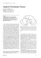
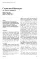
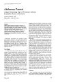
Show ]. Clin. Neuro-ophthalmol. 2: 33-37, 1'182. Cryptococcal Meningitis An Atypical Presentation PHILIP CUSTER, M.D. LENORE A. BREEN, M.D. RONALD M. BURDE, M.D. Abstract A previously healthy 23-year-old white female presented with a history of horizontal diplopia prior to any other symptoms. Physical examination revealed bilateral sixth cranial nerve palsies, papilledema, right Homer's syndrome, and right hemiparesis. Cryptococcus neoformans was isolated from the cerebrospinal fluid. This is the first documented case of Homer's syndrome associated with cryptococcal meningitis. Introduction Cryptococcus neoformans, a saprophytic budding yeast, has a strong affinity for the central nervous system and is the most common agent isolated in fungal meningitis. Neuro-ophthalmologic signs and symptoms are common, but often appear late in the disease and are seldom the presenting complaint. We present the following atypical case of a previously healthy woman with cryptococcal meningitis. She developed diplopia prior to any other symptom, and was later found to have bilateral sixth cranial nerve palsies, bilateral papilledema, transient right hemiparesis, and a transient right Horner's syndrome. Case Report A previously healthy 23-year-old white fem,lle was referred to the Neuro-Ophthalmologv Consult Office at Washington Universitv MediC'll Center for evaluation of diplopia. Sh(' W,h ,1 resident of southern Missouri and livpd 111 d mobile home Chickens, ducks, dogs, and other domestic ,1nim,11., were kept in close proximity to the' dwellin~. She experienced the sudden onset of bil10culM horizontal diplopia while driving her car The dl'- From the O<,partm<,nt, "f Orhth.llnwlogy (I'l.I.I\K.I{MI\I. N"II rology and Neuroloj;IC<I' '>ur~ery fRMHI. WJ,h,ngt,.n Un,vl'lsity School of Med,cine, St. Lou", M,,,.,.,,n March 1982 viation remained constant, and 1 week later she noted the onset of a global headache. The headache was constant, pounding, and seemed to be more severe in the late afternoon and early morning. She experienced intermittent periods of nausea, malaise, and anorexia, as well as occasional chills and night sweats over the last 6 months. In addition, she had reported a 10-lb. weight loss over that same period. The patien~ consulted her optometrist on July 28, 1981, 1 week after the onset of diplopia. Examination at that time showed a best-corrected visual acuity of 20/20 in both eyes, and a 16-prism diopter esotropia at distance. Sinusitis was suspected, and the patient was started on Ornade Spansules and Ceclor by her internist. There was no improvement in her symptoms, so the patient consulted an ophthalmologist on August 4. 1981. Esotropia was again documented, and papilledema was noted. The patient was referred to Washington University Medical Center. Examination in the Neuro-Ophthalmology Consult Office on August 6, 1981, showed a visual acuity of 20/20 in the right eye and 20/25 in the left eye with a myopic correction. A I-mm right ptosis was present. Pupillary diameters were () mm on the right and 6.5 mm on the left. Extraocular motility examination showed an incomitant esotropia, greater for distance than near. Mild underaclion of the right lateral rectus muscle and moderate underaction of the left lateral rectus muscle \yere present. Kinetic visual fields (Gl)ldm.lnn perimeter) were unrem,lrk,lblc except for sli~hlh' enl,H~ed blind .,pe)1s Small. feLl I l1ucle.H C1t,11',1l-t, were Illlted bilJter,llly I"undlhc()pl( c\.,lmin,lll,)n rl've, lleJ l',HI\' p.lpillcdem,l ,111d Ill) sp,)nt,lI1el)U, v('nOli, pUI."ltl"l1~ in bl)th eve, (ri~. I) The il1still,llion l)1 5% '-')l ,1il1e n,.,ultcd in pllpill,lrv di.lnlC'ters "f:; mill lll1 tlV' I'i~ht ,111d S Illln l)n the left. f\ mild right 11l'mip.lresis W,lS t,)und lll1 l1eurologic examin. ll'lll1_ An intnnsil' posteri,)f fOSS,l mJSS lesion was "lIspec!ed, ,1I1d the patient was admitted to the Iwspit.ll. Cl)lllpllterized tomographic scan was nor- 33 CryptOCllCC.ll M{'l1il1~itis (0) Figure 1. Ri~hl ,a) .md left ,I>I d", dL'I1"'I1"lr~lil1~ trut:' p.,pllkdt:'ln.l .,t the lime of ~dmission, mal. Repeat examination the following d.1y showed resolution of the ptosis, anisocoria, and right hemiparesis. Lumbar puncture was performed with .1n opening pressure of 320 mm H~O. The cerebrospinal fluid (CSF) contained 200 white blood cells with 74% lymphocytes, 23% segmented leukocytes, two macrophages, and one monocyte. Cerebrospindl fluid protein was 107, .1nd CSF glucose W.1S 10 (·.imllit,lIH' lI 11" ,,('rurn gluCt)s('. 06). Indi.1 ink ex- .lmination demonstrated multiple, encapsulated, nonbudding yeast. Cerebrospinal fluid cryptococ(. 11 antigen was 1: 16. Following the diagnosis of cryptococcal meningitis, combination therapy with amphotericin-B .1nd flucytosine was initiated. Forty-eight hours later, the diplopia and headache had resolved. Cerebrospinal fluid cultures subsequently grew out Cryptococcus neoformans. Blood and urine cul- Journal of Clinical Neuro-ophthalmology Custer, Breen, Burde TABLE 1. Cerebrospinal Fluid Findings in Cryptococcal Meningitis n.w nt l;llInl~t' Pnlh'in Whih> Blood Indl.1 In" Cryptllltlll.ll Tn.'.ltnll'nt Cell <. Illlnt Llo",llHlll,llillll Antl~l'll 0 10 107 200 + I: 1(, oJ 4.~ 44 + J :2 I~ 45 33 47 + 1:1 J2 4b 35 27 Neg.JlIve tures for the ye.lst were negative. The patient was given a total of 831 mg of amphotericin-B over 42 d.tys, and 20 days of flucytosine (2000 mg, 4 times per day). The CSF findings gradually improved, and ne.H the conclusion of treatment the cryptococcal .1I1tigen was negative, and no encapsulated veast could be found (Table 1). All signs of papilledema had resolved prior to discharge from the hospital (Fig. 2). Discussion Infection with Cryptococcus neoformans has traditionally been associated with an environmental exposure to pigeons. However, the organism has also been isolated in the habitats of various other wild and domestic animals and fow!.1 Man appears to have a high resistance to the fungus, and manto- man infection has not been described. The respiratory tract is the usual portal of entry. Pulmonary infection may cause cough, sputum production, chest pain, weight loss, or fever, although 32% of patients are asymptomatic." Hematologic dissemination from a pulmonary focus can spread the fungus to any organ system in the body. There appears to be a predilection for the central nervous system, with resultant meningitis, cerebritis, or meningoencephalitis. Immunosuppressed states are evident in onethird to one-half of the patients with central nervous system infection. Common predisposing conditions include diabetes mellitus, sarcoidosis, collagen vascular diseases, neoplastic diseases, and corticosteroid therapy.:l-fi Cryptococcal meningitis typically has an insidious onset and a progressive course. A study of 40 patients demonstrated a prevalence of white, middle- aged patients. Males are more commonly affected than females. Headache is the most frequent presenting symptom.7 Mental changes, nuchal pain and rigidity, malaise, nausea and vomiting, and low-grade fever may soon become evident. Neurologic signs and symptoms are usually present. These may be related to the elevated intracranial pressure, basilar arachnoiditis causing cranial nerve palsies and communicating hydrocephalus, cryptococcal granulomas (torulomas), angiitis, and mycotic embolic lesions." March 1982 Okun and Butler reported visual symptoms in 44% of patients with cryptococcal meningitis. Blurred vision was the most frequent complaint, followed by retrobulbar pain, diplopia, and photophobia.!' Ophthalmologic findings described in cryptococcal meningitis include decreased vision, papilledema, optic atrophy, anisocoria, internal ophthalmoplegia, and ocular motor .dysfunction from supranuclear, internuclear, and infranuclear lesions.'" H. III Pathologic examination has indicated that optic atrophy and decreased vision may be more frequently related to direct invasion of the optic nerves by the fungus than to the effects of chronic papilledema. II Uveitis, chorioretinitis, and endophthalmitis may result either from a direct extension of the infection via the subarachnoid space, or from secondary cryptococcemia.';' II, I:' The diagnosis of cryptococcal meningitis is confirmed by isolating the organism from the cerebrospinal fluid. Repeat lumbar punctures can be negative, and a cisternal tap may be required for positive identification of the fungus when the diagnosis is suspected. I:! The CSF typically shows an elevated protein, low glucose, and pleocytosis, with a mononuclear predominance. Cryptococcal capsular polysaccharide can be detected in the cerebrospinal and other body fluids using the latex agglutination method. Recent studies indicate that the ELISA titer is more sensitive th.lI1 latex agglutination, and provides earlier detection of the crvptococcal antigen in body fluids. 11 Prior to the introduction of amphl)tericin-B, cryptococcal meningitis was uniformly fata!. This agent can be administered either intravenously or by intrathecal injection, .1I1d has produced .1 60-83% survival rate. t', Flucytosine can be taken orally, with 80°lc, of p.ltients responding to treatment. 11; Combination therapy with both flucytosine and amphotericin-B results in a more rapid sterilization of the CSF, while avoiding m.lI1Y of the toxic side effects of prolonged amphotericin-B treatment and the developing of resistance reported with flucytosine.1 7 Disseminated infection carries a poor prognosis. Failure to respond to treatment and death are invariably associated with the presence of a serious underlying disease. 7 . 1o, I" Relapse occurs in one-third to one-half of patients treated, and tends to occur in the first year. 7 The likelihood 35 (0 ) Figure 2. Ri~ht 1.1) .1IH-i left It> I dis, .,t the time "f d",h.H~e d€'n1l'n'tr.'tin~ Jlm",t totJI rC'",olutic..)n of di ...( ",w('lIin~. of relapse cannot be predicted from the duration of the illness, the amount of amphoteriein-B .1dministered, the presence of coexisting dise.lse. or the spinill fluid findings ilt the conclusion of tre.ltment. 7 However, thC' failure of thC' cryptoeoeeal antigen titC'r to decre.1se during tre.1tmC'nt indie.ltes .1n incn·.l"f'd risk (If treatment failure. I " w,· I. 1\',' d,·..,rib",J .1 LISe (If (rylll«)(eal men-ingitis in .1 previously heillthy 23-year-old white fem.lle. The diagnosis of fungill meningitis was not initi.llly suspected beciluse of her atypical presentation. She had the sudden onset of diplopia 1 week prior to developing other symptoms. Although she eventually reported headache, anorexia, malilise, and weight loss, these were all mild and nonspecific symptoms. The sixth cranial nerve Journal of Clinical Neuro-ophthalmology palsies were certainly rl'latC'd to the dev,lted intr,lcranial pressure, ,15 th('y r('stllvrd rapidly ,1fter the di,lgnostic lumb,u puncturrs ,md initi,ltion of tre,ltment. To our knowledge, thr ,1ssoci,ltitlll tlf HClI"ner's syndrome with cryptococcal mrningitis h,ls not been previously rl'portC'd. The tr,1I1sient n,lture of this finding could br e,pl,lined by ,1n end,lrteritis with local ischemi,l. Conye,l ,1I1d Heilm,1I1 described ,1 p,ltient with cryptclCOCC,ll meningitis whtl developed ,1 transient right internucle.H ophth,llmoplegia. ltl P,lthologic C",lmin,ltion denwnstrated ,111 end,lrteritis with inf,udion of the right lat('ral br,lin stem nH.'sC'nceph,1It1l1, ,1dj,lCl'nt ttl the mrdi,1I1 k1ngitudin,11 f.1sciculus. Our p,ltient h,lS responded well to combination therapy with ,1mphotericin-B and flucytosine. Although she must be followed closely for the possibility of rel,lpse, she probably has been cured of ,1 previously fatal disease. Acknowledgment Supported in part by a grant from Research To Prevent Blindness, Inc., New York, New York (Department of Ophthalmology). References 1. Denton, JF. and DiSalvo, A.F.: The prevalence of Cryptococcus neoformans in various natural habitats. Sabouraudia. 6: 213-217, 19b8. 2. Campbell. GD.: Primary pulmonary cryptococcosis. Am. Rev. Respir. Dis. 94: 236-243, 196b. 3. Cogan, D.G. Immunosuppression and eye disease. Am.]. Ophtha/mo/. 83: 777-788, 1977. 4. Review: Treatment of cryptococcosis. Lancet 2: 132-133, 1979. 5. Lesser, R.L., Simon, R.M., Leon, H., and Siegel, N.: Cryptococcal meningitis and internal ophthalmoplegia. Am.]. Ophtha/mo/. 87: b82-687, 1979. 6. Condon, P.I, Terry, 5.1, and Falconer, H.: Cryptococcal eye disease. Doc. Ophtha/mol. 44: 49 -5b, 1977. 7. Butler, W.T., Alling, D.W., Spickard, A, and Utz, I: Diagnostic and prognostic value of clinical and laboratory finhdings in cryptococcal menmigitis. N. Eng/. ]. Med. 270: 59-67, 1964. March 1982 Custer, Breen, Burde tI. Littm,ln, M.L.: Cryptococcosis (torulosis). Am. ]. Med. 27: 07t,-001\, 1959. Q. Ul..un, L, ,1Ild Butler, W.T.: Ophthalmic com plicaticlI1s of C ryptocon,ll meningitis. Arch. Ophtha/mo/. 71: 52-57, 1964. 10. Conye,l, LF., ,lIld Heilman, K.M.: Neuro-ophthalmic ,lspeds of centr,ll nervous system C ryptococcosis. Arch. Opth,l/mol. 87: 164-lbl\, IQ72. II. Kupfer, C, and McCrane, L.: A possible cause of decreased vision in cryptococlal meningitis. Invest. Ophth,Jinwl. 13: 1\01-1\04, 1974. 12. Shields, I.A, Wright, D.M, Augsburger, 1.1., and Wc,ll..owicz, M.I: CryptolOllal chorioretinitis. Am. I. Ophth,llmo/. 89: 210-218, 1980. 13. Edwards, V.E., Sutherland, I.M., and Tyrer, IH.: Cryptococcosis of the central nervous system. ]. Neurol. Neur05urg. Psychiatr. 33: 415-425, 1970. 14. Scott, LN., Muchmore, H.G., and Felton, F.G.: Comparison of enzyme immunoassay and latex agglutination methods for detection of Cryptococcus neoform, lIIs antigen. Am. /. Gin. Pdtho/. 73: 790-794, 191\0. 15. Spickard, A, Butler, W.T., Andnole, U., and Utz, J.P.: The improved prognosis of cryptococcal meningitis with amphotericin B therapy. Ann. Intern. Med. 58: bb-1\3, 19b3. 16. Utz, IP., Tynes, BS, Shadomy, H.J. Duma, R.I., Kannan, M.M., and Mason, K.N.: 5-Fluorocytosine in human cryptococcosis. Antimicrob. Agents Chemother. 1968: 344-346, 1969. 17. Bennett, I.E., Dismukes, W.L, Duman, R.J., Medoff, G., Sande, M.A, Gall is, H., Leonard, I, Fields, B.T., Bradshaw, M, Haywood, H., McGee, Z.A, Cate, T.R., Cobbs, CG., Warner, J.F., and Alling, D.W.: A comparison of amphotericin B alone and combined with flucytosine in the treatment of cryptococcal meningitis. N. Engl.]. Med. 301: 120-131, 1979. 18. Utz, J.P., Garriques, IL. Sande, M.A., Warner. I-F. Mandell, G.L., McGehee, R.F., Dum,1l1. R.I. and Shadomy, 5.: Therapy of cryptococcosis with J combination of flucytosine and amphotericin B. I. Infect. Dis. 132: 368-373, 1975. 19. DiJmond, RD., Jnd Bennett. IE .. Prc'gnostic betl'rs in cryptococcal meningitis. A.nn. Intern. !'.fed. 80: 17b-II\I,1 974. Write for reprints tl" Ronald M. Burde. M.D. Department of Ophth,llmoILlgv, Bll' 1\000,000 Slluth Euclid Avenue, St. Louis, Missouri b31 10 37 |