| OCR Text |
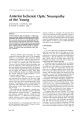
Show ]. Clin. Neuro-ophthalmof. 3: 137-146, 1983. Anterior Ischemic Optic Neuropathy of the Young JONATHAN J. DUTTON, M.D. RONALD M. BURDE, M.D. Abstract Anterior ischemic optic neuropathy is a well-recognized syndrome usually occurring in an older adult population. It is unusual for these patients to experience a second attack in an eye already affected. We describe three young patients with anterior ischemic optic neuropathy who have experienced repeat attacks in one or both eyes, leaving the patients severely disabled, without evidence of other ocular or systemic disease. We believe they may represent a new, but rare, syndrome. Anterior ischemic optic neuropathy is a wellrecognized syndrome that occurs as an idiopathic entity or in association with numerous disease conditions, all leading to anterior optic nerve anoxia and infarction. It was first reported by Cowers] in 1879 in a patient with acute blood loss. However, the first detailed clinical description was given by Uhthoff in 1924.2 Subsequently, the disease was defined in Europe by Kurz·l and Fran~ois et al. 4 Its first description in the American literature was by Miller and Smith in 1966." Since then, several additional studies have further delimited the clinical spectrum of the conditionfi - 9 or reviewed its major features. 10-14 Anterior ischemic optic neuropathy is a disease that typically affects patients over 40 years of age with acute onset of painless visual loss, and pallid edema of the optic nerve head progressing to optic atrophy. The idiopathic variety is characteristically associated with altitudinal visual field defects. 7 . 9 Most patients retain some degree of permanent loss of vision. The syndrome in older patients is usually associated with systemic disorders, most commonly arteriosclerotic vascular disease,lo. 12 hypertension and diabetes mellitus, and less frequently with temporal arteritis 7 . IS-17 When seen in the younger patient, anterior ischemic optic neuropathy is almost always related to a systemic disease such as From the Departments of Ophthalmology (JJD, RMB), Neurology, and Neurological Surgery (RMB), Washington University School of Medicine, 51. Louis, Missouri. June 1983 diabetes mellitus or vasculitis. We present three cases most consistent with the idiopathic variety of anterior ischemic optic neuropathy in patients less than 30 years of age, without evidence of associated ocular or systemic disease. Their courses, including repeat attacks in each eye, are so similar that they may define a new syndrome. Case Reports Case 1 A 27-year-old female complained of headache, dizziness, and fuzzy spots in the right eye in June 1969. She was seen by an ophthalmologist who undertook an extensive evaluation including blood chemistries, a glucose tolerance test, skull films, and a brain scan, all of which were normal. No treatment was given. Over the next 3 years she experienced three similar episodes. The patient was first referred to us on October 13, 1972, with 3 days of blurred vision and subjective field loss in the right eye. Visual acuities were 20/25 in the right eye and 20/20 in the left eye; there was no afferent pupillary defect. Both optic discs were pale without spontaneous venous pulsations, and mild disc edema was noted in the left eye (Figs. 1 a and 1 b). Kinetic perimetry showed inferior altitudinal field cuts bilaterally (Fig. 2). She was hospitalized for evaluation. Her physical examination and studies including multiple systemic blood pressures, complete blood chemistries, collagen-vascular workup, thyroid battery, radioactive brain scan, electroencephalography, and pneumoencephalography were all normal. A fluorescein angiogram revealed late staining of the left optic disc. She was believed to have anterior ischemic optic neuropathy of uncertain etiology. The patient did well until October 1977, when she noted decreased vision and pain in the right eye of 3 weeks duration. Vision was finger-counting in the right eye and 20/20 in the left eye with a marked right afferent pupillary defect. The right optic disc was atrophic with swelling in the inferior half. The left disc showed atrophy in its superior half (Figs. 3 a and 3 b ). Kinetic perimetry revealed an inferior altitudinal field cut in the left eye and more extensive superior field loss in the right eye 137 Optic Neuropathy of the Young lb) Figur'?s la and lb. Case 1. (d) Left optic disc at the time of initial examination demonstrating mild edema with superior pallor. (b) Right optic disc demonstrating altitudinal pallor above. than was seen previously (Fig. 4). Skull films, computerized axial tomographic scan, and pneumoencephalography were normal, as were all cerebrospinal fluid studies for chemistries and immunoglobulins, repeat blood chemistries, and repeat evaluation for collagen vascular diathesis. The patient remained stable until August 1979, when vision in the right eye deteriorated to bare light perception with no color appreciation and pallid disc edema (Fig. 5). Vision in her left eye 138 remained 20/20. The visual field in the left eye remained stable. She was given pulsed corticosteroid therapy with intravenous methylprednisolone, 1,000 mg every 12 hours, without improvement. Repeat laboratory blood and cerebrospinal fluid studies were negative. In November 1979, she noted decreased vision in the left eye. Visual acuity was 20/20 with pale swelling in the superior portion of the optic disc and further constriction of the visual field (Fig. 6). Journal of Clinical Neuro-ophthalmology Dutton, Burde LEFT RIGHT Figure 2. Case 1. Kinetic visual fields demonstrating bilateral inferior altitudinal scotomas, worse in the left eye. Figures 3,. and 3b. (,,) Superior disc pallor of the left eye with capillary network and nerve fiber bundles remaining inferiorly. (b I Disc swelling of the right optic nerve head involving the previously spared inferior tissue (see Fig. 1b). Since that time she has remained stable without further recurrence. Case 2 A 23-year-old male first noted painless blurring of vision in the right eye in 1976. In 1979, he again June 1983 noted blurring of vision in the right eye with constriction of the right visual field. A skull series was obtained, the results of which were normal. He remained stable until March 1982, when he complained of sudden painless loss of the superior field in his left eye. On examination he had visual 139 Optic Neuropathy of the Young Figure 3. (Cont.) (b) LEFT RIGHT Figure 4. Case 1. Kinetic visual fields demonstrating further 1055 of central and peripheral isopters in a relative altitudinal fashion. acuities of 20/40 in the right eye and 20/20 in the left eye. There was an afferent pupillary defect on the right side with a relative brightness deficit and color desaturation compared to the left eye. Fundus examination revealed a segmental optic atrophy in a butterfly pattern in the right eye and pallid disc edema with an inferior arcuate nerve fiber bundle infarct in the left eye (Figs. 7 a and 7 b). Kinetic perimetry showed a horizontal hourglass constric- 140 tion in the right eye and a superior arcuate defect in the left eye (Fig. 8). The patient's general medical examination was entirely normal. Routine laboratory evaluation included complete blood chemistries, blood count, erythrocyte sedimentation rate, serum protein electrophoresis, cryoglobulins, serum complement titers, and a lumbar puncture for cerebrospinal fluid chemistries, cytology, and immunoglobulins. All Journal of Clinical Neuro-ophthalmology Dutton, Burde Figure 5. Pallid disc edema of the right eye of case 1. LEFT RIGHT Figure 6. November 1979. Kinetic visual field demonstrating further constriction in an altitudinal fashion. studies were within normal limits. Immunologic workup consisted of antinuclear antibodies, antidouble- stranded DNA antibodies, rheumatoid factor, erythrocyte sedimentation rate, and Raji-ceII immune complex, all of which were normal. A fluorescent treponemal antibody absorption test was nonreactive. A diagnosis of probable ischemic optic neuropathy was made, and he was begun on a tapering dose of prednisone beginning with 100 mg daily. June 1983 By May 1982, his vision was 20/25 in the right eye and 20/20 in the left. The left optic disc was flat with an area of segmental atrophy inferiorly. Kinetic perimetry revealed a persistent superior arcuate field defect. Case 3 A 17-year-old male without prior ocular disease first complained of intermittent blurred vision in 141 Optic Neuropathy of the Young 1"1 Figures 7d dnd 7b. Cdse 2. (d) Left optic disc demonstrdtes pdllid swelling without nerve fiber bundle hemorrhdges. (b) Right optic disc demonstrdtes butterfly pdllor with complete loss of nerve fibN bundle IdyerS ophthdlmoscopicdlly between 8 dnd 10:00. the left eye in May 1976. By June 1976, the patient was hospitalized when he could no longer read with his left eye. Examination revealed visual acuity of 20/50 with optic atrophy in the right eye and only finger-counting with disc edema in the left eye. A diagnosis of papillitis was made, and he was treated with intravenous ACTH. Vision improved to 20/30 in the left eye but remained unchanged in the right. Complete physical and neurologic ex- 142 aminations were unremarkable. Evaluation included skull series and CT scan, electroencephalogram, complete blood chemistries, erythrocyte sedimentation rate, antinuclear antibody (ANA), LE cell prep, serum complement, serum protein electrophoresis, cerebrospinal fluid chemistries, and immunoglobulin levels, all of which were normal. One month later, the patient noted decreased vision in his right eye associated with pale disc Journal of Clinical Neuro-ophthalmoJogy Dutton, Burde LEFT RIGHT Figure 8. Case 2. Kinetic visual fields demonstrating d peculiar hourglass loss in the right eye and a relative arcuate bundle defect in the left. edema. He was again treated with ACTH with improvement of vision to 20/25 in both eyes. Repeat blood chemistries and immunologic workup remained negative. He was stable until May 1980, when he noted blurring in the inferior field of the left eye. Examination revealed segmental edema in the superior half of the left optic disc with an inferior altitudinal field defect on kinetic perimetry. He was treated with prednisone, 80 mg daily, rapidly tapered over 10 days with return of vision to 20/25 bilaterally. Once again repeat blood and cerebrospinal fluid laboratory studies were all negative. Because a brother, age 30 years, had presented recently with optic atrophy and diffuse neurologic symptoms, our patient underwent a complete neurologic workup including electromyography, nerve conduction velocities, visual-evoked response, electroencephalography, lumbar puncture for cerebrospinal fluid protein, glucose, cytology and immunoglobulins, and a brain stem audio-evoked response. As expected, the patient had an abnormal visualevoked response, consistent with optic nerve dysfunction. Because of an absent qualitative arylsulfatase A on one test, the patient was thoroughly evaluated for possible metachromatic leukodystrophy, including urine and leukocyte arylsulfatase, galactosidase, glucouronidase, mannosidase, fucosidase, and hexosaminidase, all of which were normal. The patient remained stable until May 1981, when he noted visual blurring in the right eye. Vision was reduced to 20/50 in his right eye, and the optic disc again showed mild pallid edema. June 1983 Vision in his left eye was 20/25, but his visual field demonstrated inferior altitudinal loss. He was started on prednisone, 100 mg daily, on tapering doses. The patient's vision slowly improved. He was first seen by us on June 3, 1981. Vision at that time was 20/20 in both eyes without an afferent pupillary defect. The anterior segments were normal as were intraocular pressures. A moderate relative brightness deficit was noted in the left eye as well as marked color desaturation in both eyes. Kinetic perimetry revealed superior and nasal field loss in the right eye and more extensive severe inferior altitudinal and nasal field loss in the left eye (Fig. 9). Ophthalmoscopic examination of the right eye revealed a pale disc with capillary and nerve fiber bundle absence everywhere except superior nasally. Similarly, the left disc was pale except for some residual vascularization temporally. General medical and neurologic examinations were entirely normal. There were no carotid bruits, and systemic blood pressures were consistently within normal limits. Ophthalmodynamometry was normal. Repeat collagen-vascular evaluation, including ANA, LE prep, antidoublestranded DNA, erythrocyte sedimentation rate, and serum complement levels, as well as a fluorescent treponema I antibody absorption test (FTAabs), were negative. He has been followed since without further recurrence. Discussion The syndrome of anterior ischemic optic neuropathy appears to result from impaired perfusion 143 Optic Neuropathy of the Young LEFT RIGHT Figure 9. Case 3. Visual field obtained on a Goldmann bowl perimeter. Left eye: Relative inferior altitudinal field defect with an additional involvement of the inferior arcuate bundle producing a superior nasal defect. Right eye: Marked constriction of the smaller isopter (I~e) with absolute loss of the superior nasal field. in the microcirculation to the optic diSc. lU , 15, 18-24 The etiologies of such vascular embarrassment are numerous, most commonly arteriosclerotic vascular disease and temporal arteritis, both of which typically affect older patients. The disease is also associated with hypertension,2'. embolic phenomena/ Ii diabetes mellitus,2'. 28 chronic papilledema,29 migraine, I" :111 syphilis, I, postoperative intraocular pressure elevation,:ll severe preeclampsia,'J2 the various collagen vascular diseases,:J:J severe anemia,:J:1 severe blood loss/2 and trauma,:14 Many of the latter etiologies may affect younger individuals, Clinically anterior ischemic optic neuropathy usually presents as an acute loss of vision, although rarely it may progress slowly over days or weeks, 14, :]:, Males and females are affected equally, usually in the fourth to sixth decades of life with a peak incidence between 55 and 64 years,li, Y Typically the disease is initially unilateral, although 25-65% of patients will eventually have involvement of the fellow eye,'" I;, H, Y. 12, I:J, 2;, The average time course for bilateral involvement is 2-4 years.-'· Y A second attack in an already affected eye has been considered to be a rare occurrence,Ii,9 Recently Repka et al. reported a recurrence rate of 2% in 200 patients followed for 20 years:JIi (Repka, M" Savino, P, J., Schatz, N. J" Sergott, R.: Personal communication, February 1983). Hayreh reports two cases in which recurrence was seen in the same eye," In addition, he states, "Recurrence of AION [anterior ischemic optic neuropathy) .. , was seen , , , in a number of other cases in our clinic, however it is [aJ very uncommon. , , occurrence, , . ," 144 Visual acuity remains in the 20/20-20/100 range in at least 50% of cases, with relatively few patients sustaining severe permanent loss of vision,5. 8, 9.13 Field loss is the rule, most commonly an inferior altitudinal defect, although arcuate and central scotomas may also occur,t!· 9,11-1:1 Hayreh18-24,:1I dem-onstrates convincingly the pathogenesis of this field loss in anterior ischemic optic neuropathy is due to occlusion of one of the posterior ciliary arteries that supply the optic disc in segmental fashion. Ophthalmoscopically the optic disc shows pallid edema in the acute stage, frequently with a few flame-shaped hemorrhages at the disc margin. Eventually this progresses to segmental or more complete optic atrophy within several weeks to months. Our three patients present with rapid painless loss of vision over several days with typical pallid edema of the optic nerve head, progressing to segmental optic atrophy. In all three cases the disease has recurred several times, eventually involving both eyes over a period of several years. At the time of presentation with acute ischemia in one eye, all three patients already had evidence of atrophy in the fellow optic disc. This is also true in the majority of bilateral cases of anterior ischemic optic neuropathy discussed by Boghen and Glaser6 As would be expected, during sequential attacks only those regions of the optic disc not demonstrating optic atrophy have the propensity to swell. Of the six involved eyes discussed here, only one shows total loss of vision at this time. Although the clinical picture in these cases is not Journal of Clinical Neuro-ophthalmology completely typical of anterior ischemic optic neuropathy as seen in the older population, particularly in the frequent recurrence of attacks and recovery of vision, their history, physical findings, and negative laboratory findings are most consistent with this diagnosis. Complete physical examination and laboratory investigations fail to reveal any specific etiology for anterior ischemic optic neuropathy in our cases, either ocular or systemic. Although echography is not available on these patients, careful cardiac evaluation reveals no evidence of a "floppy mitral valve syndrome" or atrial myxoma, and the patients certainly show no other evidence of systemic embolization. The causes of microvascular impairment of their anterior optic nerves remain obscure, and we can only speculate that some small vessel angiopathic process was responsible. We cannot definitely rule out demyelinating disease, but the course of the process seen here and the negative cerebrospinal fluid studies militate against this diagnosis. It appears that a form of idiopathic anterior ischemic optic neuropathy may occur in young patients as a disease entity not associated with a definable vascular or hematologic disorder. The distinctive finding in these three cases which clearly separates them from the norm of anterior ischemic optic neuropathy is the continued recurrence of attacks in an already involved eye. We suspect that this process may be more widespread than has been reported in the literature, and the diagnosis should be considered in any young patient with sudden visual loss and optic atrophy. From our experience with these three patients in the use of corticosteroids of various dosages and routes of administration, it is clear that their use does not alter the course of individual attacks or their recurrence. References 1. Gowers, W.R.: A Manua/ and At/as of Medica/ Ophthalmoscopy. J. & A. Churchill, London, 1879, pp. 184-188. 2. Uhthoff, W.: Zu den Entzundlichen Sehnerven. Affektionen bei Arteriosklerose. Ber. Deutsch Ophtha/ mol. Gesse/ 44: 196, 1924. 3. Kurz, 0.: Uber Papillitis arteriosclerotica. Ophtha/m% gica 116: 281-285,1948. 4. Fran~ois, ]., Verriest, G., and Baron, A.: Pseudopapillitis vasculaires. BulJ. Soc. Fr. Ophta/mol. 69: 3657, 1956. 5. Miller, G.R., and Smith, J.L.: Ischemic optic neuropathy. Am.]. Ophtha/mol. 62: 103-115, 1966. 6. Boghen, D.K, and Glaser, J.S.: Ischemic optic neuropathy. The clinical profile and natural history. Brain 98: 680-703, 1975. 7. Cullen, J,F.: Ischaemic optic neuropathy. Trans. Ophtha/mol. Soc. UK. 87: 759-774, 1968. 8. Eagling, E.M., Sanders, M.D., and Muller, S.J.H.: June 1983 Dutton, Burde Ischaemic papillopathy. Br.]. Ophtha/mo/. 58: 9901008, 1974. 9. Ellenberger, C, Keltner, J.L., and Burde, KM.: Acute optic neuropathy in older patients. Arch. Neural. 28: 182-185, 1973. 10. Bake, W., and Voigt, GJ.: Circulatory disorders of the optic nerve. Ophtha/m%gica (Base/) 180: 88100, 1980. II. Drance, S.M.: Ischaemic optic neuropathy. Trans. Ophtha/mol. Soc. UK. 96: 415-417, 1976. 12. Fran~ois, J.: Vascular pseudopapillitis: Ischemic optic neuropathy. Ann. Ophtha/mol. 8: 901-919, 1976. 13. Miller, N.R.: Anterior ischemic optic neuropathy: Diagnosis and management. Bu/l. N. Y. Acad. Med. 56: 643-654, 1980. 14. Shults, W.T.: Ischemic optic neuropathy: Some interesting features. Trans. Pacific Coast Oto-ophtha/mol. Soc. 281-298, 1977. 15. Henkind, P., Charles, N.C, and Pearson, J,: Histopathology of ischemic optic neuropathy. Am. ]. Ophtha/mol. 69: 78-90, 1970. 16. Jennings, GH.: Arteritis of temporal arteries. Lancet 1: 424-428, 1938. 17. Sanders, M.D.: Ischaemic papillopathy. Trans.Ophtha/ mo/. Soc. UK. 91: 369-386, 1972. 18. Hayreh, S.5.: Blood supply of the optic nerve head and its role in optic atrophy, glaucoma, and oedema of the optic disc. Br. ]. Ophtha/mol. 53: 721-748, 1969. 19. Hayreh, 5.5.: Pathogenesis of visual field defects. Role of the ciliary circulation. Br.]. Ophtha/mol. 54: 289-311.1970. 20. Hayreh, S.5.: Posterior ciliary arterial occlusive disorders. Trans. Ophtha/mol. Soc. UK. 91: 291-303, 1971. 21. Hayreh, 5.5.: Blood supply and vascular disorders of the optic nerve. In The Optic Nerve, J.5. Cant, (Ed.) CV. Mosby Co., St. Louis, 1972, pp. 59-67. 22. Hayreh, S.5.: Anterior ischaemic optic neuropathy. I. Terminology and pathogenesis. Br.]. Ophtha/mol. 58: 955-963, 1974. 23. Hayreh, S.5.: Segmental nature of choroidal vasculature. Br.]. Ophtha/mo/. 59: 631-647, 1975. 24. Hayreh, S.5.: Ischemic optic neuropathy. Int. Ophtha/ mol. 1(1): 9- 18, 1978. 25. Ellenberger, C, Jr.: Ischemic optic neuropathy as a possible early complication of vascular hypertension. Am.]. Ophtha/mol. 88: 1045-1051, 1979. 26. Lieberman, M.F., Shahi, A., and Green, W.R.: Embolic ischemic optic neuropathy. Am.]. Ophtha/mol. 86: 206-210,1978. 27. Foulds, W.5.: Visual disturbances in systemic disorders. Optic neuropathy and systemic disease. Trans. Ophtha/mo/. Soc. UK. 89: 125- 14b, 1%9. 28. Lubow, M., and Markley, T.A.: Pseudopapilledema of juvenile diabetes mellitus. Arch. Ophtha/mol. 85: 4 I 7-422, 1971. 29. Green, G ]., Lessell, S., and Loewenstein, J.I: Ischemic optic neuropathy in chronic papilledema. Arch. Ophtha/mo/. 98: 502-504, 1980. 30. McDonald, W.I., and Sanders, M.D.: Migraine complicated by ischaemic papillopathy. Lancet 2: 521523, 1971. 3 I. Hayreh, S.5.: Anterior ischemic optic neuropathy. IV. Occurrence after cataract extraction. Arch. Oph- 145 Optic Neuropathy of the Young tha/mol 98: 1410-1416, 1980. 32. Beck, R.W., Gamel, JW., Wi!court, R.J., and Berman, G.: Acute ischemic optic neuropathy in severe preeclampsia. Am. f. Ophtha/mol 9: 342-346,1980. 33. Kimbrell, O.c., Jr., and Wheliss, J.A.: Polyarteritis nodosa complicated by bilateral optic neuropathy. /. Am. Med. Assoc. 201: 61-62, 1967. 34. Hedges, T.R., III, and Gragoudos, E.5.: Traumatic anterior ischemic optic neuropathy. Ann. Ophthalmol 13: 625-b28, 1981. 35. Knox, D.L., and Duke, JR.: Slowly progressive ischemic optic neuropathy. A clinicopathologic case report. Trans. Acad. Ophtha/mol. Otolaryngol 75: 1065-1068,1971. 36. Repka, et al.: Personal communication. 146 37. Hayreh, S.5.: Anterior ischemic optic neuropathy. V. Optic disc edema an early sign. Arch. Ophthalmol 99: 1030-1040, 1981. Acknowledgments The authors thank Dr. David Josephson of Indianapolis, Indiana for referring case 3 to us for evaluation. This work was supported in part by a grant from Research to Prevent Blindness, Inc., New York, New York (Department of Ophthalmology). Write for reprints to: Ronald M. BurdI', MD., Department of Ophthalmology-Box 8096, 660 South Euclid Avenue, St. Louis, Missouri 63110. Journal of Clinical Neuro-ophthalmology |