| OCR Text |
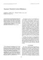
Show Journal of Clinirnl Neuro-ophthalmology 7(3):139-143, 1987. © 1987 Raven Press, Ltd., New York Pituitary Apoplexy Presenting With Light-Near Dissociation of the Pupils Bruce D. Nichols, M.D., and Kenneth G. Romanchuk, M.D., F.R.C.S. (c) We describe a patient with pituitary apoplexy causing sudden visual loss and light-near dissociation of the pupils but minimal ophthalmoplegia. Good visual recovery occurred despite an 8-day delay in neurosurgical treatment. Key Words: Hypophysectomy-Pituitary neoplasmsPupil- Pupillary reflex. From the Department of Ophthalmology, University of Saskatchewan, Saskatoon, Saskatchewan, Canada. Address correspondence and reprint requests to K. G. Romanchuk, M.D., Department of Ophthalmology, University of Saskatchewan, Saskatoon, Saskatchewan, Canada S7N DXD. 139 Pituitary apoplexy is a sudden hemorrhage or infarct in the pituitary gland. Patients usually present with severe headache, sudden loss of vision, ophthalmoplegia, alteration in the level of consciousness, and evidence of subarachnoid hemorrhage, but unusual presentations have been reported. Swift neurosurgical decompression is recommended, although good visual recovery has been reported despite delay in such treatment. We report a patient who suffered pituitary apoplexy due to pituitary tumor causing loss of vision and light-near dissociation of her pupils and yet had good visual recovery despite an 8-day delay in treatment. CASE REPORT A 53-year-old woman complained of sudden onset of severe bitemporal headache, marked loss of vision, and oblique distortions of images but no frank diplopia. She was admitted to her local hospital with suspected "nervous breakdown." Her condition did not improve, and after 7 days she was transferred to the neurological service here for further investigation of unremitting headache. Elective ophthalmological consultation was obtained 1 day after this, at which time her best corrected distance vision was counting fingers at 1 ft in the nasal field of each eye. Near acuity through her bifocals was 20/200 equivalent print left, but she could not read even 20/400 equivalent print right. Visual fields by confrontation showed a hemianopic temporal defect in the left eye and only a small nasal island of vision in the right eye. The pupils were mid-dilated and showed only very slight constriction when intense light was shone directly into them at the slit lamp, but they constricted briskly when the patient looked at an accommodative stimulus at near. Slight anisocoria 140 B. D. NICHOLS AND K. G. ROMANCHUK AG. t. Lateral skull x-ray showing enlargement of the sella. was present in room light with the right pupil measuring 5 mm and the left 6 mm, with no change in dim light. The left upper eyelid showed 1 mm of ptosis. Corneal touch reflex was normal bilaterally. Extraocular movements were full, but perhaps a slight right exotropia was present by - T ··<:n showing pituitary tumor I Clill Nellro-ophthalmol, Vol. 7, No.3, 1987 FIG. 3. Coronal (A) and sagittal (B) reconstructions from CT scan showing midline lesion extending anterosuperiorly from the sella to below the third ventricle. Hirschberg corneal light reflex testing, although fixation was poor. Ophthalmoscopy showed mild temporal pallor of the right optic disc only. Physical examination was unremarkable and, in particular, there was no evidence of endocrine dysfunction. The patient was afebrile. A lateral skull x-ray ordered immediately after the ophthalmological examination showed enlargement of the sella, dorsum sella, and posterior clinoids (Fig. 1). Emergency neurosurgical consultation was requested; computerized tomography (CT), done on an emergency basis, showed an enlarged sella with a hypodense mass in the pituitary fossa but with an area of attenuation greaterthan- normal brain tissue at the anterosuperior margin (Fig. 2), Sagittal and coronal reconstruction showed a midline lesion extending anterosuperiody from the sella to below the third ventricle (Fig. 3). The patient underwent emergency transsphenoidal hypophysectomy that day; necrotic tissue and old blood were evacuated from the pituitary fossa, Pathological diagnosis was difficult due to necrosis but was reported to be compatible with hemorrhagic tissue of chromophobe adenoma type, Two days after surgery, the patient's best corrected distance vision had recovered to 20/200 + right and 20/200 left, and both pupils now reacted briskly to light; no relative afferent defect was detected, The pupils were now equal in size, and the left ptosis was gone. Ocular versions were full (Fig. 4). Four days after surgery, best corrected distance vision had recovered to 20/20 - right and 20/60left. Automated perimetry showed bitemporal field loss (with small islands of vision remaining lateral to fixation, more so on the left than the right) and some inferior nasal field loss bilaterally (Fig, 5). The eyes were now orthophoric on cover PITUITARY APOPLEXY FIG. 4. Normal extraocular movements 2 days postoperatively. 141 testing when looking in the distance, and only a slight exophoria was present at near when looking through bifocals. Seven months after surgery, the patient's best corrected distance vision was 20/20 in each eye, and ocular examination was normal apart from a temporal pallor of each optic disc (right more so than left) and bitemporal visual field depression, especially superotemporally on automated perimetry (Fig. 6). DISCUSSION The sudden expansion of a pituitary adenoma due to spontaneous hemorrhage can cause the acute clinical syndrome known as pituitary apoplexy. The distinctive symptoms are sudden onset of severe headache, sudden loss of vision, partial or complete ophthalmoplegia, evidence of subarachnoid hemorrhage, and sometimes alteration in the level of consciousness. The diagnosis is strengthened if there is sellar enlargement on lateral skull x-ray film, defects of the visual fields, or clinical hypopituitarism. Pituitary apoplexy may be precipitated by some factors such as head trauma, estrogen therapy, anticoagulant therapy, angiography, radiotherapy, bromocriptine therapy, pregnancy, oral contraceptive therapy, diabetes with cerebrovascular disease, and sickle cell disease (1,2). Other causes of abrupt loss of vision must be ruled out such as ocular causes, orbital causes, and intracranial lesions other than pituitary adenoma such as tumors of the optic nerve and chiasm, supraclinoid aneurysm, parasellar lesions, thrombosis of the carotid artery, hydrocephalus of the third ventricle, chiasmal arachnoiditis, fracture of the anterior cranial fossa, basofrontal tumor of the skull, and pseudotumor cerebri (3). Unusual presentations of pituitary apoplexy, such as epistaxis, have been reported (4). The reported incidence of hemorrhage in pituitary adenomas varies widely from 1.5 to 27.7% (1). The types of pituitary tumors that undergo apoplexy are reported as being chromophobe and eosinophilic adenomas but not basophilic, fetal cell, or transitional (5). Apoplexy has been reported in nonfunctioning tumors, prolactinomas, and tumors associated with the production of growth hormone (2). I Clill Nellro-ophthalmol. Vol. 7, No.3, 1987 142 A B. D. NICHOLS AND K. G. ROMANCHUK ., >' .. FIG. 5. Automated perimetry 4 days postoperatively showing bitemporal field loss. (A) Left visual field. (B) Right visual field. Treatment of pituitary apoplexy is swift chiasmal decompression (especially when vision is deteriorating rapidly) and corticosteroid replacement. Delay in treatment, although not recommended, sometimes does not interfere with recovery of vision; our patient had a good visual recovery despite an 8-day delay in treatment, and similar recovery has been reported with a delay of up to 10 days (2,6). Medical treatment has been suggested to sometimes suffice when there is limited suprasellar extension and intact or improving vision (7). A The sudden expansion due to ischemic necrosis often takes place in an intrasellar secretory adenoma confined by a competent diaphragma sella (7). Compression of adjacent structures in the cavernous sinus, such as the optic chiasm, optic nerves, and the third, fourth, and sixth cranial nerves, may cause a rapidly progressive ophthalmoplegia. It is said that third (oculomotor) nerve palsy occurs more often than sixth (abducens) nerve palsy, and partial third nerve palsy has also been reported (5,8). Our patient presented with most of the features -C'c"t0cf n0r imetry 7 months postoperatively showing further recovery of the visual fields but still superc , C,' : eft visual field. (B) Right visual field. PITUITARY APOPLEXY 143 of pituitary apoplexy: severe headache, loss of vision, and typical field changes. Perhaps she had minimal ophthalmoplegia consisting of a partial left third (oculomotor) nerve palsy (minimal left ptosis, slight dilation of the left pupil, and an exotropia with the patient preferring to fix with her left eye as near visual acuity and remaining field were better on the left than the right). She definitely had light-near dissociation of the pupils. The light-near dissociation can easiy be explained by postulating that the afferent limb of the pupillary light reflex arc was sufficiently damaged by rapid expansion of the tumor to cause the poor pupillary reaction to direct light stimulation while the near response remained intact because it does not depend on the afferent light impulses. In those cases of pituitary apoplexy with total bilateral ophthalmoplegia, pupillary constriction to an accommodative stimulus at near could disappear because the efferent pathway for constriction in the third (oculomotor) nerves would be affected. Light-near dissociation of the pupils, when certain other criteria are met, is characteristic of the Argyll Robertson pupil; this is diagnostic of neurosyphillis. Other clinical syndromes can show light-near dissociation: the tonic pupil of Adie's syndrome; the "midbrain" pupils of the periaqueductal (Parinaud's) syndrome; and gaze-evoked pupillary constriction following aberrant regeneration in third (oculomotor) nerve palsies (9). As Walsh and Hoyt wrote, one should avoid calling light-near dissociation in these cases, and in our patient, a "pseudo-Argyll Robertson pupiV' as the pathology and etiology are clearly dissimilar (10). REFERENCES 1. Wakai S, Fukushima T. Teramoto A, Sano K. Pituitary apoplexy: its incidence and clinical significance. J Neurosurg 1981;55:187-93. 2. Fong LP, Fabrinyi GCA. Ophthalmic manifestations of pituitary apoplexy. Med JAust 1985;142:142-3. 3. Glew WB. Simulated pituitary apoplexy: report of an unusual case due to hemorrhage into hypothalamic astrocytoma. Ann OphthalmoI1977;9:139-42. 4. Keane JR. Pituitary apoplexy presenting with epistaxis. J Clin Neuro-ophthalmol 1984;4:7-8. 5. Epstein S, Pimstone BL, de Villiers Je. Jackson WPU. Pituitary apoplexy in five patients with pituitary tumours. Br Med J1971;2:267-70. 6. Robinson JL. Sudden blindness with pituitary tumours. J Neurosurg 1972;36:83-5. 7. Glaser JS. Topical diagnosis: the optic chiasm. In: Duane TO, Jaeger EA, eds. Clinical ophthalmology (vol. 2). New York: Harper & Row, 1986. 8. Wray SH. Neuroophthalmologic manifestations of pituitary and parasellar lesions. Clin Neurosurg 1976;24:86-117. 9. Thompson HS. Light-near dissociation of the pupil. Ophthalmologica 1984;189:21-3. 10. Walsh FB, Hoyt WF. Afferent impairment of pupillary light reflex: lesions of the retinomesencephalic pathway to the Edinger-Westphal nucleus. In: Walsh FB, Hoyt WF, eds. Clinical neuroophthalmology (vall). Baltimore: Williams & Wilkins, 1969:502-3. I Gin Neuro-ophthalmol, Vol. 7, No.3, 1987 |