| Title |
Giant Cavernous Malformation of the Occipital Lobe |
| Creator |
Chicani, CF; Miller, NR; Tamargo, RJ |
| Affiliation |
Department of Ophthalmology, The Johns Hopkins Hospital, Baltimore, Maryland, USA. |
| Abstract |
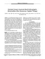
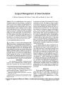
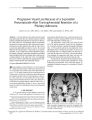
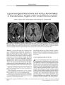
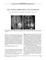
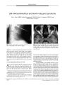
A 15-year-old boy who developed severe headaches and an incomplete homonymous hemianopia was found to have a large, well-circumscribed, multilobulated intracranial mass in the contralateral occipital lobe. The initial impression was that of a low-grade glioma or a vascular malformation. When the lesion increased in size and complexity, concern arose about the possibility of a malignant glioma. Upon craniotomy, it proved to be a giant cerebral cavernous malformation. This case is remarkable in that most cavernous malformations do not become symptomatic before the third decade of life and rarely attain such a large size. |
| Subject |
Adolescent; Brain Neoplasms/complications; Brain Neoplasms/diagnosis; Brain Neoplasms/physiopathology; Brain Neoplasms/surgery; Cerebral Hemorrhage/etiology; Hemangioma, Cavernous, Central Nervous System/complications; Hemangioma, Cavernous, Central Nervous System/diagnosis; Hemangioma, Cavernous, Central Nervous System/physiopathology; Hemangioma, Cavernous, Central Nervous System/surgery; Hemianopsia/etiology; Hemianopsia/physiopathology; Humans; Magnetic Resonance Imaging; Male; Neurosurgical Procedures; Occipital Lobe/pathology; Occipital Lobe/radiography; Tomography, X-Ray Computed; Visual Fields |
| Format |
application/pdf |
| Publication Type |
Journal Article |
| Collection |
Neuro-Ophthalmology Virtual Education Library: Journal of Neuro-Ophthalmology Archives: https://novel.utah.edu/jno/ |
| Publisher |
Lippincott, Williams & Wilkins |
| Holding Institution |
Spencer S. Eccles Health Sciences Library, University of Utah |
| Rights Management |
© North American Neuro-Ophthalmology Society |
| Setname |
ehsl_novel_jno |
| ID |
225159 |
| Reference URL |
https://collections.lib.utah.edu/ark:/87278/s63f7vpw/225159 |