| Identifier |
walsh_2021_s1_c1-slides |
| Title |
A 4-Year Wait |
| Creator |
Dmitry Balian, Sachin Kedar, Michaelyn Everhart, Aleh Bobr, Liudmila Muinov |
| Subject |
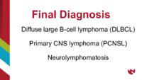
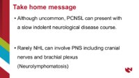
Orbital Lymphoma; 3rd Nerve Palsy; 4th Nerve Palsy; 7th Nerve Palsy; Trigeminal Neuralgia |
| History |
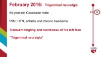
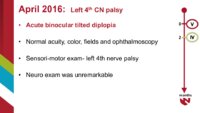
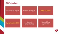
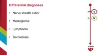
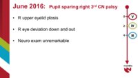
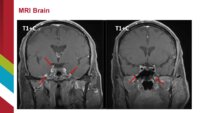
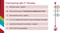
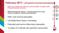
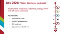
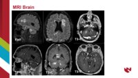
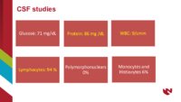
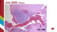
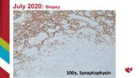
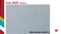
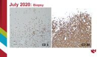
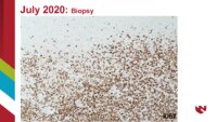
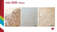
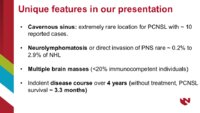
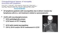
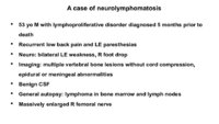
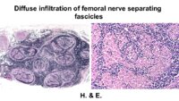
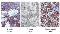
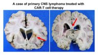
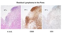
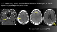
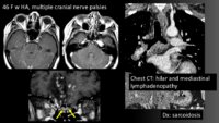
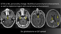
A 64 year old male with past history of hypertension, arthritis and chronic headache developed transient tingling and numbness of his left face in February 2016 and was diagnosed to have "trigeminal neuralgia". Two months later, he presented to the ED with sudden onset of binocular tilted diplopia. Clinical examination was consistent with the 4th nerve palsy with unremarkable neurological exam. VA OD was 20/30 and OS 20/20, ophthalmoscopic exam unremarkable. MRI brain showed a contrast enhancing lesion adjacent to left cavernous sinus extending into the foramen rotundum involving left V2 trigeminal nerve division. Nerve sheath tumor, meningioma, lymphoma and sarcoidosis were suspected. CSF showed mild pleocytosis (11 WBC with 82% of lymphocytes), normal glucose and protein. His symptoms improved but never completely resolved. Three months later he presented to the ED with pupil sparing right third nerve palsy. Infectious, inflammatory, paraneoplastic panel labs were negative except for elevated levels (311 U/ml) of Anti-Dnase B antibody. CSF again showed mild pleocytosis but normal protein and glucose. Additional work up including conjunctival biopsy (for sarcoidosis) and PET/CT SPECT imaging was unrevealing. Six months later (February 2017) he developed left painful brachial plexopathy 2 weeks after Pneumococcus vaccine. MRI left brachial plexus showed enhancement of the roots, trunks, divisions, cords and branches. He underwent 3 months of IV steroids for "presumed post-vaccine inflammatory plexopathy" with significant improvement. He was doing well until July 2020 when he presented with loss of balance, dizziness, droopy eyelid and left facial weakness. Examination showed ptosis of the right eye, weakness of left frontalis and orbicularis oris muscles, flattening of the left nasolabial fold, decreased motor strength and hyperreflexia. MRI brain now showed multiple supratentorial and infratentorial contrast enhancing lesions. A procedure was performed. |
| Date |
2021-02 |
| Language |
eng |
| Format |
application/pdf |
| Type |
Text |
| Source |
53rd Annual Frank Walsh Society Meeting |
| Relation is Part of |
NANOS Annual Meeting 2021: Walsh Session I |
| Collection |
Neuro-ophthalmology Virtual Education Library: NOVEL http://NOVEL.utah.edu |
| Publisher |
Spencer S. Eccles Health Sciences Library, University of Utah |
| Holding Institution |
North American Neuro-Ophthalmology Association. NANOS Executive Office 5841 Cedar Lake Road, Suite 204, Minneapolis, MN 55416 |
| Rights Management |
Copyright 2021. For further information regarding the rights to this collection, please visit: https://NOVEL.utah.edu/about/copyright |
| ARK |
ark:/87278/s6ms9rn2 |
| Contributor Primary |
Dmitry Balian, MD |
| Contributor Secondary |
Sachin Kedar, Michaelyn Everhart, Aleh Bobr, Liudmila Muinov |
| Setname |
ehsl_novel_fbw |
| ID |
1694272 |
| Reference URL |
https://collections.lib.utah.edu/ark:/87278/s6ms9rn2 |