| OCR Text |
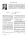
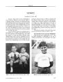
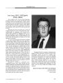
Show THE FOURTH HOYT LECTURE William F. Hoyt, MD William Fletcher Hoyt, MD, professor emeritus of Ophthalmology, Neurology, and Neurosurgery, University of California, San Francisco, was born and raised in Berkeley, California. He took his undergraduate education at the University of California, Berkeley and his medical education at the University of California San Francisco ( UCSF). After a year's study at the Wilmer Institute, Johns Hopkins University, under the mentorship of Frank B, Walsh, MD, he returned to UCSF in 1958 to found the neuro- ophthalmology service. During a 36- year academic career- all of it at UCSF- he authored 266 journal articles, co- authored ( with Frank B. Walsh, MD) the biblical third edition of Clinical Neuro- ophthalmology, and trained 71 neuro- ophthalmology fellows. In 1983, he received the title of Honorary Doctor of Medicine from the Karolinska Institute. He is widely acknowledge as one of the titans of twentieth century neuro- ophthalmology. In recognition of his contributions, the North American Neuro- Ophthalmology Society ( NANOS), in conjunction with the American Academy of Ophthalmology, in 2001 initiated the Hoyt lecture to be delivered each year at the Annual Meeting of the American Academy of Ophthalmology. Amblyopia: A Neuro- Ophthalmic View Creig S. Hoyt, MD Abstract: Amblyopia designates monocular or binocular visual loss associated with form deprivation caused by obstruction or deformation of a light stimulus, strabismus, or uncorrected refractive error. Elegant neuro scientific studies have identified a critical period early in life in which the visual system is vulnerable to amblyopia and the site of pathology as the primary visual cortex. However, recent observations have challenged these concepts. A broader understanding of the pathophysiology of amblyopia will be critical to refining therapy for this condition. ( J Neuro- Ophthalmol 2005; 25: 227- 231) The term " amblyopia," comes from the Greek words " amblyos" ( blunt) and " opia" ( vision). Amblyopia has been recognized as a clinical disorder for more than 300 years. The term has been used to describe the potentially reversible monocular or binocular visual loss associated with occlusion of the visual axis ( form deprivation), strabismus, or anisometropia/ ametropia. Occlusion of the non-affected eye has remained the primary form of therapy since the 1740s. Recently, brave voices have challenged the notion that screening for amblyopia is worthwhile, arguing that proof of efficacy for amblyopia therapy is wanting ( 1). But carefully controlled prospective treatment trials have provided provocative results yet irrefutable proof of the benefit of amblyopia therapy ( 2- 4). A prospective study, Department of Opthalmology, University of California, San Francisco, CA. Address correspondence to Creig S. Hoyt, Department of Opthalmology, University of California, San Francisco, 10 Kirkham Street, San Francisco, CA 94143- 0730; E- mail: cshoyt@ itsa. ucsf. edu however, did suggest that there may be no clinical benefit to treating mild forms of amblyopia ( 5). I will not address the practical treatment issues surrounding these recent studies of amblyopia. Rather, I will review our understanding of the neuro- anatomical and neurophysiological correlates of amblyopia. EXPERIMENTAL EFFECTS OF VISUAL FORM DEPRIVATION With the development of single- neuron extracellular recording techniques, the visual pathways of the brain in experimental visual deprivation states began to be probed by Wiesel, Hubel, von Noorden, Crawford, and others ( 6- 9). An experimental model of form deprivation amblyopia ( occlusion) was quickly established, ( 10- 13) and the results had a profound effect on the clinical management of infants with congenital cataracts ( 14- 19). Some of the details of these experimental models of form deprivation were appropriately challenged as quantitatively insufficient to be applied directly to clinical problems ( 20). However, the identification of the early profound alteration in the neural visual pathways convinced clinicians of the importance of early surgery in the management of congenital cataracts ( 15- 19). The specific conclusions drawn from these experimental models of form deprivation were: 1) The importance of critical periods in normal visual development and in the treatment protocols for amblyopia; 2) The role of competition between the " normal" eye and the deprived eye in the genesis of amblyopia; and 3) The discovery that the primary site of neurologic pathology associated with experimental form deprivation is Layer IVc of Vi, the primary J Neuro- Ophthalmol, Vol. 25, No. 3, 2005 227 J Neuro- Ophthalmol, Vol. 25, No. 3, 2005 Hoyt Creig S. Hoyt, MD is the Theresa and Wayne Caygill Professor of Ophthalmology and chair of the Department of Ophthalmology, University of California, San Francisco ( UCSF). Born in Pittsburgh, PA, he graduated from Amherst College in 1964 and from Cornell University Medical College in 1968. After an internship at Stanford University Medical Center, he undertook two years of residency in neurology at UCSF and then switched to ophthalmology, completing his residency at UCSF in 1976. From 1976 to 1977, he spent a fellowship year at the Royal Children's Hospital in Melbourne, Australia, which formed the basis of an outstanding career in pediatric ophthalmology with an accent on neurodevelopmental aspects of vision. From his houseboat docked off the coast of Sausalito, California, he has published and edited widely, and in 2000, became the first Yankee to be named editor of the British Journal of Ophthalmology. In 2003, he assumed the position of chair of the UCSF Department of Ophthalmology. visual cortex ( 10- 13). The neuropathological model of amblyopia based on occlusion experiments was not only elegant but appeared to be complete. However, subsequent observations have made us less confident that current neu-ropathologic models of amblyopia are completely applicable to the problems of the amblyopic patient. What are the difficulties of these experimental models of visual deprivation states? First, a rigidly denned critical period for the development and effective treatment of form deprivation amblyopia seems justified by the data obtained about visual outcomes and the timing of surgical intervention/ visual rehabilitation in congenital cataracts ( 15- 19). However, in the case of patients with strabismic and/ or amsometropic amblyopia, strictly limited critical periods cannot be defined. Recent important clinical observations include: 1) Treatment success reported in strabismic and amsometropic patients who are older than the usual periods denned for therapeutic success ( 21- 23); 2) Spontaneous deterioration (" slippage") of visual acuity in the strabismic amblyopic eye even after the patient is older than the defined critical period ( 24,25); and 3) Significant spontaneous improvement in the visual acuity of the amblyopic eye ( often in only a few days) when visual function is lost in the fixing " normal" eye in at least 10% of all adult patients with strabismic and/ or anisometropic amblyopia ( 26,27). Second, there are reasons to question the proposed site of neurologic alterations in amblyopia. The notion that alterations in the ocular dominance columns of layer IVc in Vi occur in form deprivation seems well established. However, there is reason to doubt that these alterations occur in human strabismic and/ or anisometropic amblyopes. Horton et al ( 28,29) have shown in human necropsy studies that layer IVc in Vi is unaffected in anisometropic or strabismic amblyopia. These investigators point out that this feature might have been anticipated since layer IVc in Vi matures quickly ( in a matter of months) before anisometropia occurs or strong fixation preference in strabismus is established ( 29). So, further study is needed on the question of where in the visual neural pathways anisometropic or strabismic amblyopia has its primary effect. The potential for the development of rational effective pharmacological therapies for amblyopia is at stake. SITES OF FUNCTIONAL ALTERATION IN VISUAL DEPRIVATION Let us review what is known about alterations at various levels of the visual pathways in experimental visual deprivation models. Is there any abnormality in the retina in amblyopia? This controversial question seemed to be resolved until recently. Ikeda's ( 30- 32) recordings of retinal dysfunction in amblyopia were effectively challenged by Sherman and Stone and others ( 33- 34). Arden's proposal ( 35) that the pattern ERG was abnormal in amblyopia was countered by Hess' work ( 36,37) that accounted for fixation error and its effect on the recordings. Despite some early studies indicating that a primary site of pathology in amblyopia might be the retina, the conclusions from a wide range of animal studies and human electrophysiological investigations are that the retina demonstrates essentially normal physiologic function in the presence of amblyopia ( 38). However, recent multifocal ERG studies in amblyopic eyes have reopened the issue that there is retinal dysfunction in amblyopia ( 38). Moreover, the clinician is left to ponder what alterations in the visual pathways account for the afferent pupillary defect that is detected in many amblyopic eyes. The six laminae of the lateral geniculate nucleus with their distinctive patterns of inputs from the ipsilateral and contralateral eye made it an early target of neurophysio-logical studies of amblyopia. Ikeda and Tremain ( 39) found lowered acuity of the sustained x- cells in the lateral geniculate nucleus. This finding was refuted by the work of Gillard- Crewther and Crewther ( 40). Von Noorden and Crawford ( 41) examined the anatomic structure of the lateral geniculate nuclei from a human strabismic amblyopia. In the lateral geniculate nucleus ipsilateral to the amblyopic eye, they found that the layers supplied by the amblyopic 228 © 2005 Lippincott Williams & Wilkins Fourth Hoyt Lecture J Neuro- Ophthalmol, Vol. 25, No. 3, 2005 eye were shrunken. A question remains whether this cell shrinkage in the lateral geniculate nucleus was primary or secondary to influences from the cortex on the lateral geniculate nucleus. That cell shrinkage is secondary to cortical influences seems more likely in view of recent models of lateral geniculate function. The simplistic view that the lateral geniculate nucleus functions primarily as a relay station for retinal ganglion cell projections to the visual cortex has been overturned. It is now clear that only 20% of the input to the lateral geniculate nucleus is retinal in origin ( 42). The greatest inputs to the lateral geniculate nucleus are from the visual cortex ( 43). An additional source of significant input is from the brainstem reticular formation ( 44). These inputs appear to be involved primarily in ocular motility and visual attention mechanisms ( 42). Indeed, the lateral geniculate nucleus is now seen to serve primarily a gating function related to selective visual attention inputs ( 42). The anatomical and physiological changes seen in the lateral geniculate nucleus in experimental models of amblyopia probably reflect changes in the visual cortex rather than primary changes resulting from the visual deprivation itself. Thus, by exclusion we return to the visual cortex as the likely sight of the essential neuropathology of amblyopia. Yet we must remind ourselves of Horton's necropsy studies of anisometropia and strabismic amblyopia ( 28,29). At least in these forms of amblyopia we must look beyond layer IVc of W What is the evidence for visual cortex dysfunction associated with anisometropic and strabismic amblyopia in man? Although visual evoked potential studies routinely reported reduced and distorted VEP's in human amblyopes, ( 43) the most compelling data are provided by neuro- imaging techniques. Recent advances in neuro- imaging allow functional as well as structural assessment of specific areas of the cortex in adult human amblyopes. Using positron emission tomography ( PET), Demer et al ( 44) have reported reduced blood flow in the primary visual cortex in two patients with form deprivation amblyopia. They have also reported reduced glucose metabolism in the primary visual cortex in one anisometropic and in one strabismic amblyope. In contrast, Imamura et al ( 45) studied a small number of strabismic amblyopes with PET scans and argued that there is normal striate function but abnormal extrastriate cortex function. Functional MRI ( fMRI) offers better image resolution and quantitative capabilities for this type of study. In four adult amblyopes studied with fMRI, Barnes et al ( 46) found that three showed marked reduction in striate cortex activity associated with viewing by the amblyopic eye. However, in an fMRI study of a small number of strabismic and anisometropic amblyopes, Sireteanu et al ( 47) found striate cortex activity to be normal. Extrastriate cortex activity was adversely affected. Similar specific pathologic reduction of activity in the extrastriate cortex has been reported by Lerner et al ( 48) in their fMRI studies. This suggestion that extrastriate abnormalities occur in human amblyopes gains additional support from psychophysiological studies that find that global motion processing and detection of contrast- defined contours are abnormal in human amblyopes ( 49,50). Both of these functions are thought to be mediated by extrastriate systems ( 51). That the extrastriate cortex is affected in some human amblyopes appears clear. However, it remains to be seen if this is due to an intrinsic defect or is merely a consequence of alterations in the striate cortex. AMBLYOPIA AND BINOCULAR COMPETITION Although the exact location and precise extent of the pathology in the cortex will require further studies, a compelling argument can be made that amblyopia is primarily a cortical disorder. Were Hubel and Wiesel correct in suggesting that this cortical pathology comes about as the result of competition between the unaffected and the amblyopic eye ( 52)? It now appears that neural mechanisms of amblyopia may not be due to a simple loss of cortical binocularity. There is strong evidence to suggest that residual binocular interactions remain present in the amblyopic cortex. The model of competition between geniculocortical afferents in amblyopia appears less certain now than at the time of the original proposal of Hubel and Wiesel. IMPLICATIONS FOR AMBLYOPIA TREATMENT What does this search for the neurologic site of amblyopia imply about the potential for direct pharmacological treatments of human amblyopia? Let us assume that striate cortex and the extrastriate cortex are involved in the pathophysiology of amblyopia. Let us further assume that maintaining synaptic plasticity in these cortical areas is essential in order to reverse visual loss associated with amblyopia. It is now clear that maintaining synaptic plasticity in the visual cortex depends on a complex sequence of events involving a number of different classes of molecules ( 53). N- methyl- D- aspartate receptors ( NMDA), neurotrophic factors, nitric oxide, and inhibition all seem to play important roles in the neural changes that occur in the striate cortex in experimental deprivation models ( 54). Yet the complexity of the interactions is even more striking if one considers that NMDA, for example, is now known to exist as a complex of at least 75 proteins that can be grouped into at least five classes ( 55). The additional possibility that apoptosis may be important in maintaining neuronal plasticity in the developing visual system ( 56) adds even more uncertainty about whether a rational 229 J Neuro- Ophthalmol, Vol. 25, No. 3, 2005 Hoyt effective pharmacological treatment for amblyopia can be developed. Those who embraced the original model of occlusion amblyopia and extrapolated from it a rationale for early surgery in congenital cataract patients now realize that much is still to be learned of the neurologic substrates responsible for amblyopia, especially anisometropic and strabismic amblyopia. Further studies are necessary to define the exact extent of neurologic changes in the cortex in these milder forms of amblyopia. Only then, perhaps, will the molecular underpinnings of synaptic plasticity in the visual cortex be detailed and appropriate pharmacologic therapies developed for treating amblyopia. Much remains to be done and many questions need to be answered. REFERENCES 1. Stewart- Brown SL, Haslum MN, Howlett B. Pre- school vision screening: A service in need of rationalisation. Arch Dis Child 1988; 63: 356- 9. 2. Holmes JM, Kraker RT, Beck RW et al. A randomized trial of prescribed patching regimens for treatment of severe amblyopia in children. Ophthalmology 2003; 110: 2075- 87. 3. Pediatric Eye Disease Investigator Group. The course of moderate amblyopia treatment with atropine in children: experience of the amblyopia treatment study. Am J Ophthalmol 2003; 136: 630- 9. 4. Pediatric Eye Disease Investigator Group. The course of moderate amblyopia treated with patching in children: experience of the amblyopia treatment study. Am J Ophthalmol 2003; 136: 620- 9. 5. Clarke MP, Wright CM, Hrisos S et al. Randomised controlled trial of treatment of unilateral visual impairment detected at preschool vision screening. 5M/ 2003; 327: 1251^ k 6. Hubel DH, Wiesel TN. Binocular interaction in striate cortex of kittens reared with artificial squint. JNeurophysiol 1965; 28: 1041- 59. 7. Baker FH, Grigg P, von Noorden GK. Effects of visual deprivation and strabismus on the response of neurons in the visual cortex of the monkey including studies on the striate and prestriate cortex in the normal animal. Brain Res 1974; 66: 185- 208. 8. von Noorden GK, Crawford ML. Form deprivation without light deprivation produces the visual deprivation syndrome in macaca mulatta. Brain Res 1977; 129: 37^ 4. 9. Blakemore C, Garey LJ, Vital- Durand F The physiological effects of monocular deprivation and their reversal in the monkey's visual cortex. J Physiol 1978; 283: 223- 62. 10. Hubel DH, Wiesel TN. The period of susceptibility to the physiological effects of unilateral eye closure in kittens. J Physiol 1970; 206: 419- 36. 11. Daw NW, Wyatt HI Kittens reared in a unidirectional environment: Evidence for a critical period. J Physiol 1976; 257: 155- 70. 12. Daw NW, Bernan NE, Ariel M. Interaction of critical periods in the visual cortex of kittens. Science 1978; 199: 565- 7. 13. Harwerth RS, Smith EL, Duncan GC et al. Multiple sensitive periods in the development of the primate visual system. Science 1986; 232: 235- 8. 14. Vaegan, Taylor D. Critical period for deprivation amblyopia in children. Trans Ophthalmol Soc UK 1979; 99: 432- 9. 15. Beller R, Hoyt HC, Marg E et al. Good visual function after neonatal surgery for congenital monocular cataracts. An J Ophthalmol 1981; 91: 559- 65. 16. Pratt- Johnson JA, Tillson G. Visual results after removal of congenital cataracts before the age of one year. Can J Ophthalmol 1981; 16: 19- 21. 17. Birch EE, Stager DR. Prevalence of good visual acuity following surgery for congenital unilateral cataract. Arch Ophthlamol 1988; 106: 40- 3. 18. Robb RM, Mayer DL, Moore BD. Results of early treatment of unilateral congenital cataracts. JPediatr Ophthalmol Strab 1987; 24: 178- 81. 19. Birch EE, Stager DR. The critical period for surgical treatment of dense congenital unilateral cataract. Invest Ophthalmol Vis Sci 1996; 36: 1532- 8. 20. Marg E. Prentice Memorial Lecture: Is the animal model for stimulus deprivation amblyopia in children valid or useful? Am J Optom Physiol Opt 1982; 59: 451- 64. 21. Franceschetti A. Efficacy of amblyopia therapy initiated after nine years of age. Eye 2005; 19:[ Epub ahead of print]. 22. Hunter DG. Treatment of amblyopia in older children. Arch Ophthlamol 2005; 123: 557- 8. 23. Scheiman MM, Hertle RW, Beck RW, et al. Randomized trial of treatment of amblyopia in children aged 7 to 17 years. Arch Ophthalmol 2005; 123- 437- 47. 24. Scott WE, Dickey CF Stability of visual acuity in amblyopic patients after visual maturity. Graefes Arch Clin Exp Ophthalmol 1988; 226: 154- 7. 25. Gurovich L, Ciancia A, Herrera MC. Estabilidad a largo plazo de los resuctados obtenidos en la ambliopia nediante tratamiento occlusivo. In Prieto- Diaz J, ed. Transactions of XII Congreso Del Consejo Latin Americano de Estrab 1996; 49- 59. 26. Hoyt CS. Why is the adult amblyopic eye unstable? Br J Ophthalmol 2004; 88: 1105- 6. 27. Chua B, Mitchell P. Consequences of amblyopia on education, occupation and long- term visual loss. Br J Ophthalmol 2004; 1119- 21. 28. Horton JC, Hocking DR. Pattern of ocular dominance columns in human striate cortex in strabismic amblyopia. Vis Neurosci 1996; 13: 787- 95. 29. Horton JC; Stryker MP. Amblyopia induced by anisometropia without shrinkage of ocular dominance columns in human striate cortex. Proc Natl Acd Sci USA 1993; 90: 5494- 8. 30. Ikeda H, Wright MJ. Is amblyopia due to inappropriate stimulation of the sustained pathway during development? Br J Ophthalmol 1974; 58: 165- 75. 31. Ikeda H, Wright MJ. Properties of LGN cells in kittens reared with convergent squint: A neurophysiological demonstration of amblyopia. Exp Brain Res 1976; 25: 63- 77. 32. Ikeda H, Tremain KE. Amblyopia occurs in retinal ganglion cells in cats reared with convergent squint without alternating fixation. Exp Brain Res 1979; 35: 559- 82. 33. Sherman SM, Stone J. Physiological normality of the retina in visually deprived cats. Brain Res 1973; 60: 224- 30. 34. Kratz NE, Mangel SC, S Lehmkuhle et al. Retinal X- and Y- cells in monocularly lid- sutured cats: Normality of spatial and temporal properties. Brain 1979; 172: 545- 51. 35. Arden GB, Vaegan, Hogg CR et al. Pattern ERGs are abnormal in most amblyopes. Trans Ophthalmol Soc UK 1980; 100: 453- 60. 36. Hess RF, Baker CL Jr. Assessment of retinal function in severely amblyopic individuals. Vision Res \ 9% A; 2A:\?>(> 1- 1(>. 37. Hess RF, Baker CL, Jr. Verhoeve JN et al. The pattern evoked electroretinogram: Its variability in normals and its relationship to amblyopia. Invest Ophthalmol Vis Sci 1985; 26: 1610- 23. 38. Hess RF. Amblyopia: Sight unseen. Clin Exp Optom 2001; 84: 321- 36. 39. Ikeda H, Tremain K. Amblyopia and cortical binocularity. Trans Ophthalmol Soc UK 1980; 100: 450- 2. 40. Gillard- Crewther S, Crewther DP. Neural site of strabismic amblyopia in cats: X- cell acuities in the LGN. Exp Brain Res 1988; 72: 503- 9. 41. von Noorden GK, Crawford ML. The lateral geniculate nucleus in human strabismic amblyopia. Invest Ophthalmol Vis Sci 1992; 33: 2729- 32. 42. Asper L, Crewther D, Crewther SG. Strabismic amblyopia: Part 2. Neural processing Clin Exp Optom 2000; 83( 4): 200- ll. 43. Levi DM, Harwerth RS. Contrast evoked potentials in strabismic and anisometropic amblyopia. Invest Ophthalmol Vis Sci 1978; 17: 571- 5. 230 © 2005 Lippincott Williams & Wilkins Fourth Hoyt Lecture J Neuro- Ophthalmol, Vol. 25, No. 3, 2005 44. Demer XL, von Noorden GK, Volkow ND et al. Imaging of cerebral blood flow and metabolism in amblyopia by positron emission tomography. Am J Ophthalmol 1988; 105: 337^ 7. 45. Imamura K, Richter H, Fischer H et al. Reduced activity in the extrastriate visual cortex of individuals with strabismic amblyopia. NeurosciLett 1997; 225: 173- 6. 46. Barnes GR, Hess RF, Dumoulin SO et al. The cortical deficit in humans with strabismic amblyopia. J Physiol 2001; 523: 281- 97. 47. Sireteanu R, Tonhausen N, Muckli L et al. Cortical site of amblyopic deficit in strabismic and anisometropic subjects investigated with MRI. Invest Ophthalmol Vis Sci ( Suppl) 1998; 39: S909. 48. Lerner Y, Pianka P, Asmon B et al. Area- specific amblyopic effects in human occipitotemporal object representation. Neuron 2003; 40: 1023- 9. 49. Wong EH, Levi DM, Mcgraw PV Is second- order spatial loss in amblyopia explained by the loss of first- order spatial input? Vision Res 2001; 41: 2951- 60. 50. Simmers AJ, Ledgeway T, Hess RF et al. Deficits to global motion processing in human amblyopia. Vision Res 2003; 43: 729- 38. 51. Barrett BT, Bradley A, Mcgraw PV Understanding the neural basis of amblyopia. Neuroscientist 2004; 10: 106- 17. 52. Hubel DH, Wiesel TN, Levay S. Plasticity of ocular dominance columns in monkey striate cortex. Philos Trans R Soc LandB Biol Sci 1977; 278: 377- 409. 53. Shaw CA, Lanius RA, Van den Doel K. The origin of synaptic neuroplasticity: Crucial molecules or a dynamical cascade? Brain Res Brain Res Rev 1994; 19: 241- 63. 54. Berardi N, Pizzorusso T, Ratto GM et al. Molecular basis of plasticity in the visual cortex. Trends Neurosci 2003; 26: 369- 78. 55. Husi H, Grant SG. Proteomics of the nervous system. Trends Neurosci 2001; 24: 259- 66. 56. Nucci C, Piccirilli S, Nistico R et al. Apoptosis in the mechanisms of neuronal plasticity in the developing visual system. Eur J Ophthalmol 2003; 13( Suppl 3): S36^ 3. 231 |